. 2003 Mar 3;22(5):1025-35.
doi: 10.1093/emboj/cdg106.

Intrabodies based on intracellular capture frameworks that bind the RAS protein with high affinity and impair oncogenic transformation

Affiliations

Intrabodies based on intracellular capture frameworks that bind the RAS protein with high affinity and impair oncogenic transformation

Tomoyuki Tanaka et al. EMBO J. .

Abstract

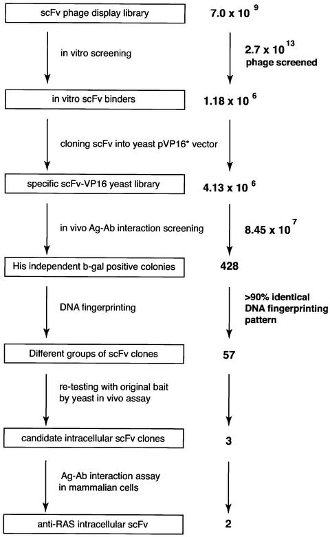
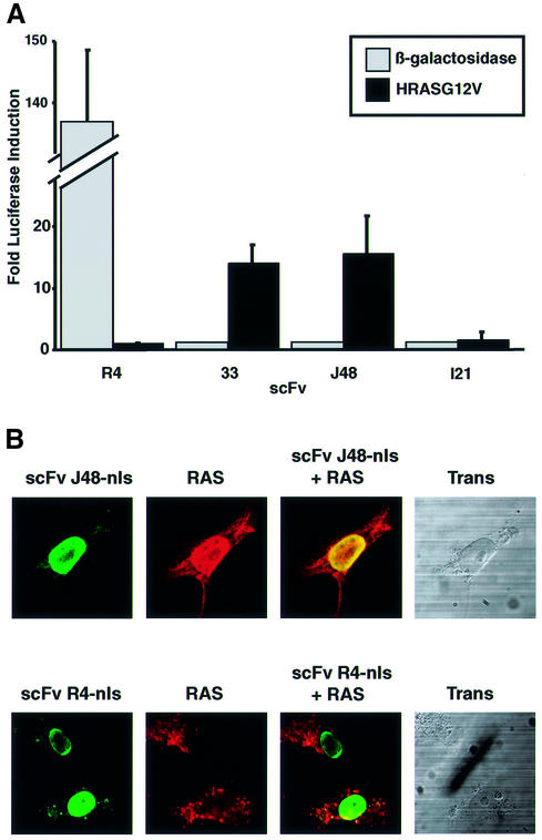
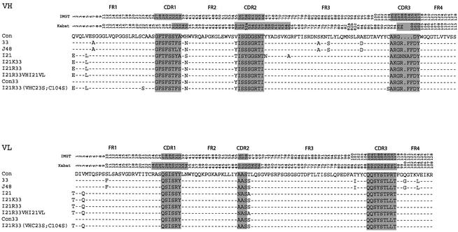
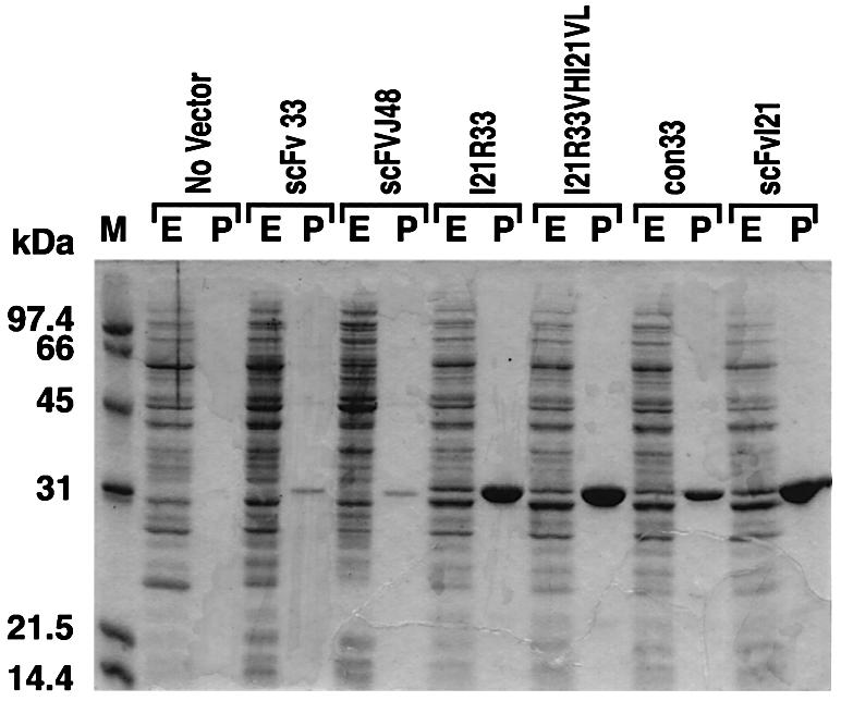
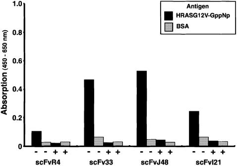
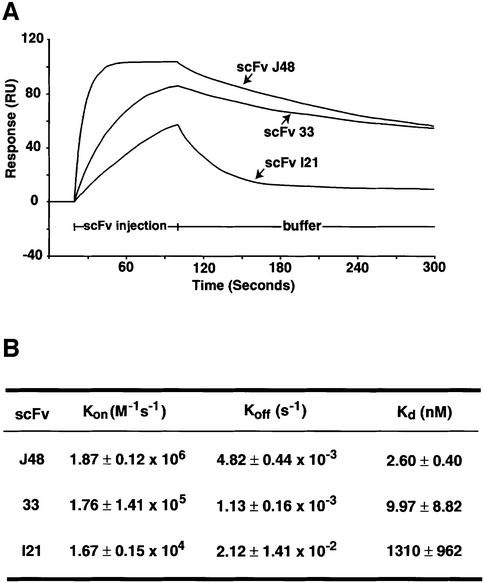
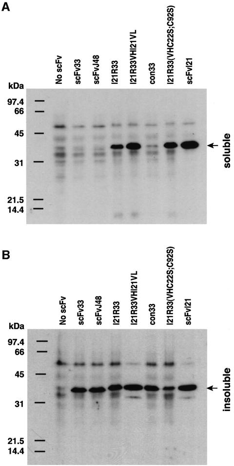
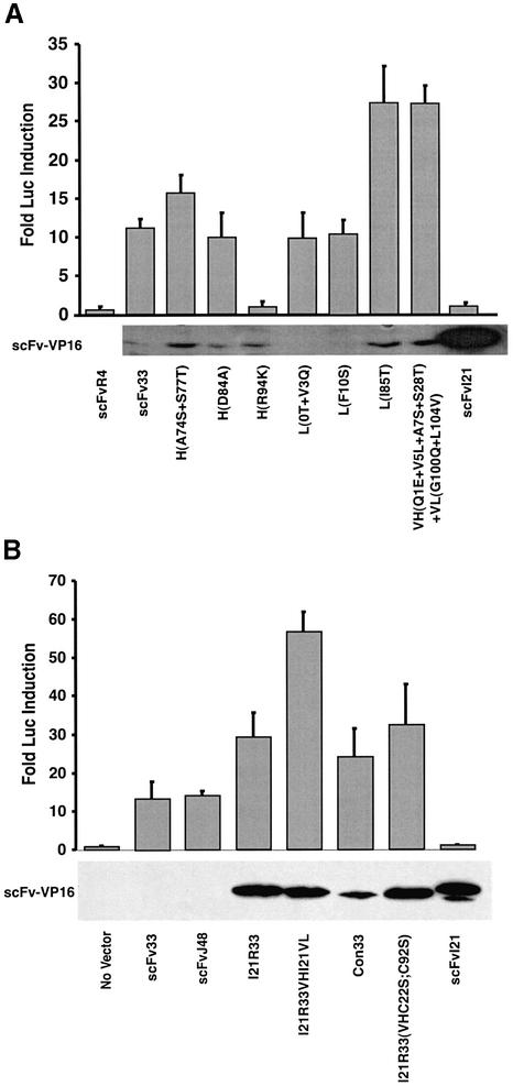
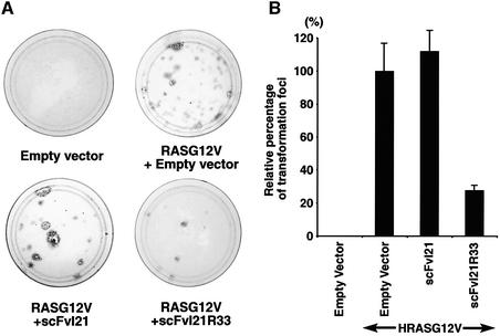
We have applied in vivo intracellular antibody capture (IAC) technology to isolate human intrabodies which bind to the oncogenic RAS protein. IAC facilitates the capture of antibody fragments, in this case single-chain Fvs (scFvs), which tolerate reducing environments, such as the cytoplasm of cancer cells. Three anti-RAS scFvs with different affinity, solubility and intracellular binding activity were characterized. The anti-RAS scFvs with highest affinity were expressed relatively poorly in mammalian cells, and greater soluble expression was achieved by mutating the antibody framework to canonical consensus scaffolds, previously derived from IAC, without losing antigen specificity. Mutagenesis experiments showed that the consensus scaffolds are functional as intrabody fragments without an intra-domain disulfide bond. Furthermore, we could convert an intrabody which does not bind RAS in mammalian cells into a high-affinity reagent capable of inhibiting RAS-mediated NIH 3T3 transformation by exchanging VH and VL complementarity-determining regions onto its consensus scaffold. These data show that the consensus scaffold is a robust framework by which to improve intrabody function.


Figures

None
Fig. 1. Intracellular antibody capture of anti-RAS scFvs. A total of 2.7 × 1013 clones from three different phage libraries (Sheets et al., 1998; de Wildt et al., 2000) (total diversity 7.0 × 109) were screened with purified HRASG12V antigen in vitro. A total of 1.18 × 106 phage were recovered, phagemid DNA was prepared and scFv fragments cloned into the yeast vector pVP16* to make a sublibrary of 4.13 × 106 clones. Yeast clones (8.45 × 107) were screened in the yeast L40 strain expressing the LexA-HRASG12V bait; 428 colonies grew on histidine selective plates and showed strong activation of the lacZ gene, determined by β-gal filter assay. All prey plasmids were isolated from histidine-independent and β-gal-positive yeast colonies and were fingerprinted by digestion with restriction enzymes BstNI, MspI, MboI, RsaI or HinfI to identify the differing scFv clones. Subsequently, 57 scFv clones which had different DNA fingerprinting patterns were re-tested in yeast with LexA-HRASG12V bait, and three scFvs (which originated from different libraries) were isolated. Of these three anti-RAS scFvs, only two detectably bound RAS protein in a mammalian reporter assay.
None
Fig. 2. Interaction of anti-RAS scFv with RAS protein in mammalian cells. (A) Luciferase assay. COS7 cells were transiently co-transfected with various scFv-VP16-AD fusions and the Gal4-DBD bait plasmid pM1-HRASG12V (closed boxes) or pM1-lacZ (open boxes), together with the firefly luciferase reporter plasmid pG5-Luc and an internal Renilla luciferase control plasmid pRL-CMV. scFv-VP16 prey vectors were used expressing anti-RAS scFv33, J48 and I21 or anti-β-gal scFvR4 (Martineau et al., 1998). The luciferase activities were measured 48 h after transfection using the Dual Luciferase Assay System (Promega) and a luminometer. The luciferase activities of each assay were normalized to the Renilla luciferase activity (used as internal control for the transfection efficiency). The fold luciferase induction level is shown, with the activity of each scFv-VP16 with non-relevant bait taken as baseline. (BIn situ immunofluorescence study. COS7 cells were transiently co-transfected with pEF/myc/nuc-scFvJ48 (anti-RAS scFv) or scFvR4 (anti-β-gal scFv) and pHM6-HRASG12V vectors expressing the RAS antigen. After 48 h, cells were fixed and stained with 9E10 monoclonal antibody (detecting the myc-tagged scFv) and rabbit anti-HA tag polyclonal serum, followed by secondary fluorescein-conjugated anti-mouse and Cy3-conjugated anti-rabbit antibodies, respectively. The staining patterns were examined using a BioRadiance confocal microscope. Co-location of antigen and intrabody fluorescence was found for scFvJ48 co-expressed with RAS. Green (fluorescein) = fluorescence of scFv; red (Cy3) = fluorescence of antigen.
None
Fig. 3. Sequence of anti-RAS intracellular scFvs. The nucleotide sequences were obtained and the derived protein translations (shown as the single letter code) were aligned. Dashes in the framework regions (FRs) represent identities with the consensus (Con) sequence (derived from anti-BCR and anti-ABL scFvs isolated by the IAC method; Tse et al., 2002). The numbers indicate the reference positions of the residues, according to the system of Lefranc and Lefranc (2001) (top column number, indicated as IMGT) and Kabat et al. (1991) (second column, Kabat). The 15 residues of the linker, (GGGGS)3, between the heavy chain domain (VH) and light chain variable domain (VL) are not shown. The complementarity-determining regions (CDRs) are highlighted on a grey background and demarcated from FRs. The three anti-RAS intracellular scFvs are designated 33, J48 and I21. All anti-RAS scFvs belong to the VH3 subgroup of heavy chains and Vκ1 subgroup of light chains shown in the middle (designed VH3 or Vκ1) from the Kabat database (Kabat et al., 1991) or IGHV3 and IGKV1 from the Lefranc database (Lefranc and Lefranc, 2001). The mutated anti-RAS scFvs are shown designated as I21K33, I21R33, I21R33VHI21VL, con33 and I21R33VH (VHC22S;C92S) or (VHC23S;C104S) (Kabat or IMGT nomenclatures, respectively). I21K33 comprises the six CDRs of scFv33 in the I21 framework, and I21R33 is identical except for a mutation Lys94(106)Arg; I21R33VHI21VL comprises the VH domain of I21R33 fused to the VL domain of I21; con33 has all six CDRs of scFv33 in the canonical consensus framework (Tse et al., 2002b); I21R33 (VHC22S;C92S) or (VHC23S;C104S) is a mutant of clone I21R33 with the mutations Cys22(23)Ser and Cys92(104)Ser of the VH domain. There are only four amino acid differences (at positions H1, H5, L0 and L3) between consensus and I21R framework regions. ScFv 33 and J48 belong to IGHV3-48 with IGHJ4 and IGHD1-26 (VH domain), and to IGKV1-39 with IGKJ4 (VL domain) according to the IMGT database (Lefranc and Lefranc, 2001). ScFvI21 belongs to IGHV3-23 with IGHJ4 and IGHD1-8 (VH domain) and IGKV1D-39 with IGKJ1 (VL domain).
None
Fig. 4. Periplasmic expression and purification of anti-RAS scFvs. The scFvs with the PelB leader sequence at the N-terminus and a His tag and myc tag at the C-terminus were expressed periplasmically from the pHEN2-scFv vector in E.coli HB2151 using 1 mM IPTG for 2 h at 30°C in 1 l of 2× TY medium with 100 µg/ml ampicillin and 0.1% glucose. After induction, the cells were harvested and extracted in 4 ml of ice-cold 1× TES buffer (0.2 M Tris–HCl pH 7.5, 0.5 mM EDTA, 0.5 M sucrose), and a further 6 ml of 1:5 TES buffer were added. The supernatants of cell extracts were used as the soluble periplasmic fraction. The His-tagged scFvs were purified by immobilized Ni2+ ion chromatography and fractionated by 15% SDS–PAGE, and proteins were revealed by Coomassie Blue staining. The approximate yields of purified anti-RAS scFv33 and J48 were <100 µg/l of culture; those of scFvI21R33, I21R33VHI21VL and I21 were >3 mg/l; and that of con33 was 1 mg/l. E = complete periplasmic extracts; P = purified scFv; M = molecular weight markers.
None
Fig. 5. Specific antigen binding and competition ELISA of anti-RAS scFvs. Purified HRASG12V-GppNp (4 µg/ml, ∼200 nM; black boxes) or BSA (30 mg/ml, ∼450 µM; grey boxes) were coated onto ELISA plates for 1.5 h at room temperature. For both sets of wells, 3% BSA in PBS was added for blocking and, subsequently, purified scFv (450 ng per well) was added and incubated overnight at 4°C. After washing with PBS–0.1% Tween-20, bound scFv was detected with HRP-conjugated anti-polyhistidine antibody (HIS-1, Sigma) and signals quantitated using an Emax microplate reader (Molecular Devices). For competition assays (indicated in the figure as +), scFvs were pre- incubated with HRASG12V-GppNp (8 µg/ml; ∼400 nM) for 30 min at room temperature before addition to the ELISA well.
None
Fig. 6. Affinity measurements of anti-RAS scFvs using BIAcore. Biosensor measurements were made using the BIAcore2000. Purified scFvs from bacterial cultures were used. (A) Sensograms showing the binding of anti-RAS scFv with HRASG12V-GppNp antigen (immobilized 1500 RU). The purified scFvs (10–2000 nM) were loaded on two channels of the chip, containing either immobilized HRASG12V-GppNp or no antigen. The sensograms of each measurement were normalized by the resonance of the channel without antigen. (B) The table summarizes the values of the association rate (Kon), dissociation rate (Koff) and calculated equilibrium dissociation constants (Kds) by BIAevaluation 2.1 software.
None
Fig. 7. Influence of framework residues on the solubility of expressed scFvs in COS7 cells. COS7 cells were transiently transfected with pEF/myc/cyto-scFv expression clones as indicated. Soluble and insoluble proteins were extracted, as described in Materials and methods, and fractionated by 15% SDS–PAGE. After electrophoresis, proteins were transferred to membranes and incubated with the anti-myc tag monoclonal antibody 9E10. The migration molecular weight markers (in kDa) are shown on the left. Arrows on the right indicate the scFv fragment band.
None
Fig. 8. Improvement of the intracellular interaction between anti-RAS intrabodies and RAS antigen by the mutation of framework sequences. Mammalian two-hybrid antibody–antigen interaction assays were performed in COS7 cells. (A) COS7 were transfected with the pEF-scFv-VP16 vectors and pM1-HRASG12V, together with the luciferase reporter clones, and luciferase levels were determined as described in Materials and methods. Upper panel: the normalized fold induction of luciferase signals (zero being taken as the signal from prey plasmid without scFv) for scFv-VP16 binding RAS antigen bait. Lower panel: a western blot of COS7 cell extracts after the expression of scFv-VP16 fusion proteins. ScFv-VP16 fusion proteins were detected by western blot using anti-VP16 (14-5, Santa Cruz Biotechnology) monoclonal antibody and HRP-conjugated anti-mouse IgG antibody. ScFv used as a control was anti-β-gal scFvR4 (Martineau et al., 1998). scFv33 mutants were numbered according to Kabat et al. (1991) and the numbers in parentheses indicate numbering according to Lefranc and Lefranc (2001) (see Figure 33): VH(A74S+S77T), substitutions of Ala74(83)Ser and Ser77(86)Thr of VH; VH(D84A), substitution of Asp84(96)Ala of VH; VH(R94K), substitution of Arg94(106)Lys of VH; VL(0T + V3Q), addition of threonine between the linker and VL domain plus substitution of Val3(3)Gln of VL; VL(F10S), substitution of Phe10(10)Ser of VL; VL(I85T), substitution of Ile85(100)Thr of VL; VH(Q1E + V5L + A7S + S28T) + VL(G100Q + V104L), substitutions of Gln1(1)Glu, Val5(5)Leu, Ala7(7)Ser and Ser28(29)Thr of VH plus Gly100(120)Gln and Val104(124)Leu of VL. (B) COS7 cell two-hybrid antibody– antigen interaction assay using scFvs with framework mutations to convert to consensus sequence scaffolds. The various scFv-VP16 prey constructs shown were transiently transfected with pM1-HRASG12V bait plasmid in COS7 cells, and the luciferase activities were measured 48 h after transfection. The fold luciferase activity levels are shown in the histogram, with the activity of no scFv (prey plasmid without scFv) as baseline. The expression levels of scFv-VP16 in the soluble fraction of COS7 cells are shown in the lower panel. The bands were visualized by western blot using anti-VP16 (14-5) antibody and HRP-conjugated anti-mouse IgG antibody.
None
Fig. 9. Inhibition of RAS-dependent NIH 3T3 cell transformation activity by anti-RAS scFv. Mutant HRASG12V cDNAs were subcloned into the mammalian expression vector pZIPneoSV(X), and anti-RAS scFv into the pEF-FLAG-Memb vector which has a plasma membrane targeting signal at the C-terminus of scFv and a FLAG tag at the N-terminus. A 100 ng aliquot of pZIPneoSV(X)-HRASG12V and 2 µg of pEF-FLAG-Memb-scFv were co-transfected into NIH 3T3 cells clone D4. Two days later, the cells were transferred to 10 cm plates and grown for 14 days in DMEM containing 5% donor calf serum with penicillin and streptomycin. Finally, the plates were stained with crystal violet, and foci of transformed cells were counted. (A) Representative photograph of stained plates. Empty vector in the left top panel indicates co-transfection of pZIPneoSV(X) without HRASG12V, and pEF-FLAG-Memb without scFv as negative control. No foci formation was observed. The right top panel indicates pZIPneoSV(X)-HRASG12V with pEF-FLAG-Memb without scFv as positive control. In the other plates, the HRASG12V vector was co-transfected with either pEF-FLAG-Memb-scFvI21 or pEF-FLAG-Memb-scFvI21R33. (B) The relative percentage of transformed foci was determined as the number of foci normalized to the focus formation induced by pZIPneoSV(X)-HRASG12V and pEF-Memb empty vector, which was set at 100. Results represent one experiment with each transfection performed in duplicate. Two additional experiments yielded similar results.

References

    1. Adjei A.A. (2001) Blocking oncogenic Ras signaling for cancer therapy. J. Natl Cancer Inst., 93, 1062–1074. - PubMed
    1. Biocca S., Pierandrei-Amaldi,P. and Cattaneo,A. (1993) Intracellular expression of anti-p21ras single chain Fv fragments inhibits meiotic maturation of Xenopus oocytes. Biochem. Biophys. Res. Commun., 197, 422–427. - PubMed
    1. Biocca S., Pierandrei-Amaldi,P., Campioni,N. and Cattaneo,A. (1994) Intracellular immunization with cytosolic recombinant antibodies. Biotechnology, 12, 396–399. - PubMed
    1. Biocca S., Ruberti,F., Tafani,M., Pierandrei-Amaldi,P. and Cattaneo,A. (1995) Redox state of single chain Fv fragments targeted to the endoplasmic reticulum, cytosol and mitochondria. Biotechnology, 13, 1110–1115. - PubMed
    1. Bird R.E. et al. (1988) Single-chain antigen-binding proteins. Science, 242, 423–426. - PubMed

Publication types

MeSH terms