. 2003 Mar 3;22(5):1036-46.
doi: 10.1093/emboj/cdg107.

A role for N-glycanase in the cytosolic turnover of glycoproteins

Affiliations

A role for N-glycanase in the cytosolic turnover of glycoproteins

Christian Hirsch et al. EMBO J. .

Abstract

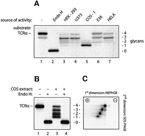
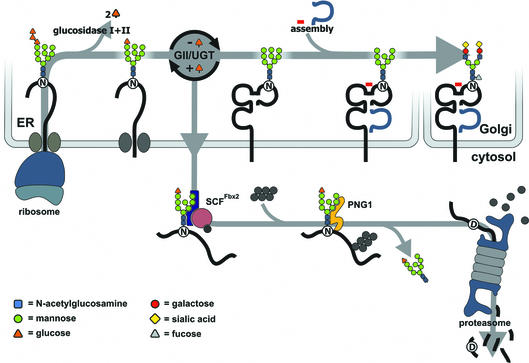
Successful maturation determines the intracellular fate of secretory and membrane proteins in the endoplasmic reticulum (ER). Failure of proteins to fold or assemble properly can lead to their retention in the ER and redirects them to the cytosol for degradation by the proteasome. Proteasome inhibitors can yield deglycosylated cytoplasmic intermediates that are the result of an N-glycanase activity, believed to act prior to destruction of these substrates by the proteasome. A gene encoding a yeast peptide:N-glycanase, PNG1, has been cloned, but this N-glycanase and its mammalian homolog were reported to be incapable of deglycosylating full-length glycoproteins. We show that both the yeast PNG1 enzyme and its mammalian homolog display N-glycanase activity towards intact glycoproteins. As substrates, cytosolic PNGase activity prefers proteins containing high-mannose over those bearing complex type oligosaccharides. Importantly, PNG1 discriminates between non-native and folded glycoproteins, consistent with a role for N-glycanase in cytoplasmic turnover of glycoproteins.


Figures

None
Fig. 1. Mammalian cell extracts contain an N-glycanase activity that deglycosylates TCRα. (A) [35S]methionine-labeled TCRα was immuno precipitated after incubation with buffer alone (lane 1), with Endo H (lane 2) or with detergent extracts containing 750 µg of protein obtained from the indicated cell lines (lanes 3–7). Immunoprecipitates were resolved by SDS–PAGE, and proteins were visualized by fluorography. The number of N-linked glycans attached to TCRα is indicated. The doublet observed occasionally for the TCRα carrying two residual glycans is most probably due to heterogeneous deglycosylation of the polypeptide since the four N-linked attachment sites of TCRα allow six different glycosylation patterns. (B) [35S]methionine-labeled TCRα was obtained by immunoprecipitation after incubation with buffer alone (lanes 1 and 2) or after incubation with extracts from COS-1 cells (lanes 3 and 4). Removal of all N-linked glycans in lanes 2 and 4 by digestion with Endo H resulted in polypeptides with identicalelectrophoretic mobility. The number of N-linked glycans is indicated in (C). To obtain the complete ladder of partially deglycosylated TCRα molecules, only 300 µg of COS cell extracts were used for this deglycosylation. (C) [35S]methionine-labeled TCRα was incubated with COS-1 cell extracts and analyzed by two-dimensional gel electrophoresis (as described in Materials and methods). For each glycan lost, a single negative net charge is acquired.
None
Fig. 2. N-Glycanase is located predominantly in the cytosol. COS-1 cells were subjected to subcellular fractionation (as described in Huppa and Ploegh, 1997). Separation of fractions by SDS–PAGE was followed by immunoblotting with an α-LDH antibody as cytosolic marker (lanes 1–4). In parallel, fractions were tested for N-glycanase activity by incubation with TCRα as a substrate in the described assay (lanes 5–8).
None
Fig. 3. The N-glycanase activity in COS cells is a single enzymatic entity. The purification scheme for N-glycanase from COS cells consisted of seven steps. First, COS-1 cytosol was obtained by differential centrifugation. Then N-glycanase was enriched by ammonium sulfate precipitation. The precipitate was resuspended and separated on a hydroxylapatite column. The flowthrough, which contained the N-glycanase activity, was separated on four chromatographic colums. Individual fractions obtained after each chromatographic separation were assayed for N-glycanase activity using TCRα as the substrate. Fractions containing activity were pooled as indicated and used in the next purification step.
None
Fig. 4. The deglycosylation activity observed in 3T3 and COS-1 cells is encoded by PNG1 mRNA. PNG1 knockdowns were generated in 3T3 (A) and COS-1 (B) cells as described in Materials and methods. Extracts from generated cell lines were tested in the deglycosylation assay, using [35S]methionine-labeled TCRα as a substrate. In (A), 400 µg of 3T3 cell extracts were used, in (B) 600 µg of COS-1 cell extracts. Lane 1: TCRα substrate incubated without cell extracts. Lane 2: TCRα incubated with wild-type cell extracts. Lanes 3–7: TCRα substrate incubated with extracts from PNG1 knockdown cell lines, infected with constructs I–IV (see Materials and methods for details). Note that extracts from cells infected with knockdown construct III were used from two independent DNA isolates, since the complete nucleotide sequence of this construct could not be determined. The N-glycanase activity observed in the wild-type COS extracts is lower than that seen in Figure 1, lane 5, because for the experiment shown here the extracts have been frozen prior to performing the deglycosylation assay.
None
Fig. 5. The yeast and the mammalian PNG1 can deglycosylate full-length TCRα. (A) PNG1 was expressed in BL21 cells and purified in two steps. An aliquot from each step was separated by SDS–PAGE, and protein bands were visualized by silver staining. Lane 1 shows a molecular weight marker, lane 2 contains lysates from uninduced cells, and lane 3 lysates from cells after induction. The bacterial lysates were purified over a Uno Q-12 anion-exchange column (lane 4) and sub sequently separated by gel filtration on a Superdex 75 column (Pharmacia) (lane 5). (B) [35S]methionine-labeled TCRα was incubated with either lysates from uninduced BL21 cells (lane 1) or 10 µg of the purified Png1p (lane 2). Incubation with the purified yeast Png1p yielded a TCRα molecule that had lost either all or three of its four N-linked glycans. (C) [35S]methionine-labeled TCRα was immunoprecipitated after incubation with buffer alone (lane 1), extracts from mock transfected U373 cells (lane 2) or extracts from U373 cells expressing the murine PNG1 (lane 3).
None
Fig. 6. Cys306 is part of a transglutaminase domain that forms the active site of N-glycanase. [35S]methionine-labeled TCRα was incubated either with buffer alone (lane 1) or with [35S]methionine-labeled extracts obtained from HEK-293 cells that were mock transfected, (lane 2), from transfectants expressing PNG1–GFP (lanes 3–5) or from transfectants expressing PNGAla306–GFP (lane 6). NEM or DTT was added to the incubations as indicated. After the incubations, immunoprecipitations with anti-TCRα and anti-GFP antibodies were performed simultaneously.
None
Fig. 7. Only free heavy chains bearing high-mannose type oligosaccharides are a target of N-glycanase. Lymphoblastoid B cells (FH17) were pulse labeled for 10 min. Free class I MHC heavy chain molecules were obtained by immunoprecipitation immediately (0 min) after the pulse using the HC10 antibody (lane 1), and heavy chains complexed with β2m were immunoprecipitated by the conformation-specific W6/32 antibody (lane 2). At 120 min after the pulse, heavy chains were immunoprecipitated by the W6/32 antibody (lane 3). The number of glycans is indicated; the asterisk represents glycans of the complex type. Samples were treated with Endo H (lanes 4–6) or digested with 10 µg of the yeast Png1p (lanes 7–9). Fully assembled class I MHC molecules from the 0 and 120 min time points were denatured by boiling with SDS and incubated with yeast Png1p after re-immunoprecipitation with the HC10 antibody (lanes 10 and 11).
None
Fig. 8. Truncated forms of high-mannose N-glycans are not recognized by N-glycanase. Class I MHC heavy chains were obtained from U373 cells by immunoprecipitation. After digestion with the indicated glycosidases, samples were analyzed either directly by SDS–PAGE (top panel) or IEF (bottom panel), or a subsequent digestion with mammalian PNG1 was performed prior to gel electrophoresis where indicated. Incubation with mannosidase yielded a doublet band, which is presumably due to incomplete digestion of the substrate as indicated by the two structures shown on the left. The faster migrating protein (closed arrowhead for lane 1) is a glycosidase-sensitive proteolytic degradation product (open arrowhead for lanes 2–5) of the heavy chain molecule.
None
Fig. 9. A proposed model for the degradation of MHC heavy chains from the ER. Class I MHC heavy chain molecules are inserted into the ER where the N-linked oligosaccharide is transferred from a dolicholpyrophosphate carrier onto the Asn-X-Ser/Thr acceptor sequence in the nascent chain (Silberstein and Gilmore, 1996). The N-linked oligosaccharide carries three glucose residues that are removed sequentially by ER glucosidases I and II (GI/GII). The concerted action of glucosidase II and UDP-glucose: UGT constitutes a cycle that deglucosylates and reglucosylates the oligosaccharide on the folding polypeptide chain. Calnexin and calreticulin bind to the monoglucosylated form of the oligosaccharide and assist in folding of the polypeptide. Binding of β2m and a peptide allows the heavy chain to exit to the Golgi where the high-mannose type N-linked glycan is converted to a complex type glycan before the fully assembled complex reaches the cell surface. Proteins that fail to fold properly or do not assemble with their appropriate binding partners are retained in the ER and become a substrate of ER mannosidase I (not shown) after a certain lag time, as do properly folded glycoproteins, yielding a Man8 structure. A Man8 structure on a misfolded protein signals that the polypeptide has resided in the ER for some time without acquiring its native structure. Such proteins are extracted from the ER and become ubiquitylated by ubiquitylating enzymes that may reside at the cytosolic face of the ER membrane. A recently discovered E3 ubiquitin ligase, Fbx2, recognizes N-linked glycans in the cytosol and may trigger selective ubiquitylation of glycoproteins upon their arrival in the cytosol (Yoshida et al., 2002). The 26S proteasome degrades the glycoprotein after its deglycosylation by N-glycanase. The deglycosylation results in the conversion of the asparagine that carried the N-linked glycan to an aspartate. It has not yet been proven whether cytosolic oligosaccharides carry a terminal Glc residue.
None
Fig. 10. Structure of class I MHC heavy chain with an N-linked glycan and the proteasome degrading a glycopeptide. (A) Modeling of the HLA-A2–Tax peptide complex with β2m and a high-mannose N-glycan attached to Asn86. The HLA-A2 chain is rendered in red, β2m in green, Tax peptide in white and the oligosaccharide in yellow. Asn86 is blue. (B) Cross-section of the 20S proteasome modeled with the nine-residue N-terminal tail of the α3 subunit deleted to open the outer pore (Groll et al., 2000). Seven α-subunits (green) form the outer pore that leads to the antechamber; the inner cavity is formed by seven β-subunits (blue). The glycopeptide consists of residues 70–95 from the HLA-A2 sequence (red) and an N-glycan (yellow) attached to Asn86 (blue).

References

    1. Bacik I. et al. (1997) Introduction of a glycosylation site into a secreted protein provides evidence for an alternative antigen processing pathway: transport of precursors of major histocompatibility complex class I-restricted peptides from the endoplasmic reticulum to the cytosol. J. Exp. Med., 186, 479–487. - PMC - PubMed
    1. Barber L.D. et al. (1996) Unusual uniformity of the N-linked oligosaccharides of HLA-A, -B and -C glycoproteins. J. Immunol., 156, 3275–3284. - PubMed
    1. Barnstable C.J., Bodmer,W.F., Brown,G., Galfre,G., Milstein,C., Williams,A.F. and Ziegler,A. (1978) Production of monoclonal antibodies to group A erythrocytes, HLA and other human cell surface antigens—new tools for genetic analysis. Cell, 14, 9–20. - PubMed
    1. Baumeister W., Walz,J., Zuhl,F. and Seemuller,E. (1998) The proteasome: paradigm of a self-compartmentalizing protease. Cell, 92, 367–380. - PubMed
    1. Brummelkamp T.R., Bernards,R. and Agami,R. (2002) A system for stable expression of short interfering RNAs in mammalian cells. Science, 296, 550–553. - PubMed

Publication types

MeSH terms

Substances

Associated data