. 2003 Mar 3;22(5):1047-57.
doi: 10.1093/emboj/cdg096.

Phenotypic variation of Plasmodium falciparum merozoite proteins directs receptor targeting for invasion of human erythrocytes

Affiliations

Phenotypic variation of Plasmodium falciparum merozoite proteins directs receptor targeting for invasion of human erythrocytes

Manoj T Duraisingh et al. EMBO J. .

Abstract

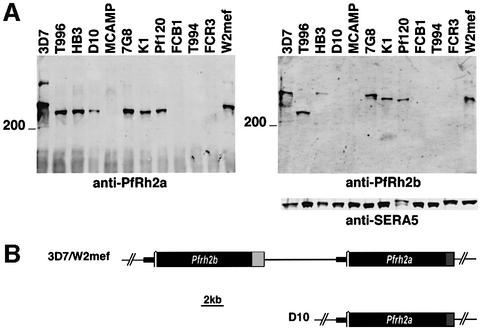
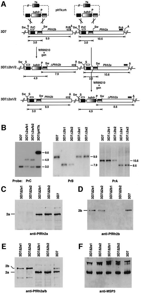
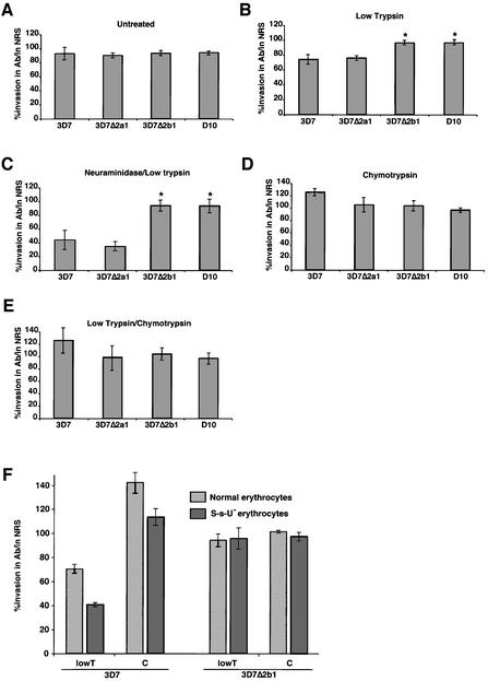
The members of the phylum Apicomplexa parasitize a wide range of eukaryotic host cells. Plasmodium falciparum, responsible for the most virulent form of malaria, invades human erythrocytes using several specific and high affinity ligand-receptor interactions that define invasion pathways. We find that members of the P. falciparum reticulocyte-binding homolog protein family, PfRh2a and PfRh2b, are expressed variantly in different lines. Targeted gene disruption shows that PfRh2b mediates a novel invasion pathway and that it functions independently of other related proteins. Phenotypic variation of the PfRh protein family allows P. falciparum to exploit different patterns of receptors on the erythrocyte surface and thereby respond to polymorphisms in erythrocyte receptors and to evade the host immune system.


Figures

None
Fig. 1. Variant expression, subcellular localization and structure of the PfRh2a and PfRh2b genes and proteins in P.falciparum. (A) Supernatants from 3D7, T996, HB3, D10, MCAMP, 7G8, K1, Pf120, FCB1, T994, FCR3 and W2mef were probed with anti-PfRh2a, anti-PfRh2b or anti-SERA5 antibodies. The position of the 200 kDa molecular weight marker is shown. (B) Structure of the PfRh2a and PfRh2b locus in 3D7, W2mef and D10. The regions in black are identical for the PfRh2b and PfRh2a genes. The light gray (PfRh2b) and darker gray (PfRh2a) sections are non-homologous.
None
Fig. 2. Subcellular localization of PfRh2a/b in 3D7 parasites by immuno electron microscopy. (AC) Cross-sections through merozoites. Scale bars are 200 nm in all panels.
None
Fig. 3. Disruption of the PfRh2b and PfRh2a genes in 3D7 parasites. (A) The pHTkΔrh plasmid has the human dhfr gene (hdhfr) flanked by two target sequences for homologous recombination within the PfRh2a and PfRh2b genes. It also includes the thymidine kinase gene (Tk) for negative selection with ganciclovir. 3D7Δ2b1/2 and 3D7Δ2a1/2 are the cloned transfected lines showing the structure of the double recombination crossovers. The PfRh2a and PfRh2b genes are shown with relevant restriction enzyme sites: Sw, SwaI; S, SacI; A, AvaII; P, PvuII. The sizes are in kbp. (B) Southern blotting of genomic DNA from 3D7, 3D7Δ2a/b1 and 3D7Δ2a/b2 (uncloned population transfected with pHTkΔrh), 3D7/pHTk (uncycled parasites transfected with pHTkΔrh), 3D7Δ2b1 and 3D7Δ2b2 (cloned lines), 3D7Δ2a1 and 3D7Δ2a1/a2 (cloned lines). The left panel was digested with PstI and SwaI, whilst the other panels were digested with SacI, AvaII and PvuII. (CF) PfRh2a and PfRh2b are not expressed in the parasites with the PfRh2a and PfRh2b genes, respectively, disrupted. Supernatants from 3D7Δ2a1, 3D7Δ2a2, 3D7Δ2b1, 3D7Δ2b2 and 3D7 were probed with the relevant antisera: (C) anti-PfRh2a sera; (D) anti-PfRh2b sera; (E) affinity-purified anti-PfRh2a/2b antibodies; (F) anti-MSP3 antibodies.
None
Fig. 4. Specific antibody inhibition of PfRh2b function in 3D7, 3D7Δ2a1, 3D7Δ2b1 and D10. Assays measure the percentage merozoite invasion in the presence or absence of anti-PfRh2a/b IgG antibodies relative to controls with IgG from normal rabbit sera (NRS). The erythrocytes in each panel are: (A) untreated; (B) trypsin treated; (C) neuraminidase/trypsin treated; (D) chymotrypsin treated; and (E) trypsin/chymotrypsin treated. (F) Assays measure the percentage merozoite invasion into S-s-U erythrocytes that lack glycophorin B. Error bars in all cases show 95% confidence limits from 4–10 experiments in triplicate.
None
Fig. 5. Disruption of the PfRh1 gene in 3D7. (A) Pr represents the probe used for Southern hybridization in (B). The restriction enzyme shown as N is NsiI. pHTkΔrh1 has integrated into PfRh1 by a double recombination crossover event to yield 3D7ΔRh1. (B) Southern hybridization of genomic DNA digested with NsiI from 3D7, 3D7ΔRh1/1 and 3D7ΔRh1/2. Sizes on the right are in kb. (C) Western blot of supernatants from W2mef, 3D7, 3D7ΔRh1/1, 3D7ΔRh1/2 with antibodies to PfRh1 (first panel) and EBA175 (second panel).
None
Fig. 6. A simplified model of merozoite invasion into erythrocytes by 3D7 and 3D7Δ2b parasites and the role of the PfRh protein family. (A) A model explaining the enzyme sensitivity of receptor Z as trypsin resistant, neuraminidase resistant and chymotrypsin sensitive. The invasion data are from Table I. Top panels: both 3D7 and 3D7Δ2b parasites show 100% invasion of untreated erythrocytes. The PfRh proteins (shown at the apical end of the merozoites and in the neck of the rhoptries) are responsible for sensing the apical end interaction and signaling to the micronemes for release of high affinity ligands (see B). The eythrocyte-binding antigens (EBAs) and their receptors are shown as green symbols. In 3D7, PfRh2b is responsible for the dominant sensing pathway for invasion via the chymotrypsin-sensitive receptor Z (brown). Disruption of the PfRh2b gene in 3D7 results in replacement of PfRh2b with an alternative pathway specified by another protein [shown as a blue ligand in the neck of the 3D7 rhoptries and interacting with a receptor (blue) on the erythrocyte in 3D7Δ2b] that binds to a chymotrypsin-resistant receptor. Middle panels: following erythrocyte treatment with trypsin and neuraminidase, 3D7 and 3D7Δ2b give 23 and 3% invasion, respectively. The trypsin and neuraminidase resistance of receptor Z is shown. Bottom panels: following erythrocyte treatment with chymotrypsin, 3D7 and 3D7Δ2b give 67 and 100% invasion, respectively. Some invasion can still be achieved using the alternative parasite ligand shown in the rhoptries that binds to a chymotrypsin-resistant receptor. The chymotrypsin sensitivity of receptor Z is shown. (B) Model for the role of the PfRh proteins in sensing the erythrocyte. This would activate release of proteins from micronemes and recruitment at the tight junction, followed by invasion.

References

    1. Adams J.H., Blair,P.L., Kaneko,O. and Peterson,D.S. (2001) An expanding ebl family of Plasmodium falciparum. Trends Parasitol., 17, 297–299. - PubMed
    1. Baldi D.L., Andrews,K.T., Waller,R.S., Roos,D., Crabb,B.S. and Cowman,A.F. (2000) RAP1 controls rhoptry targeting of RAP2 in the malaria parasite Plasmodium falciparum. EMBO J., 19, 1–9. - PMC - PubMed
    1. Barnwell J.W. and Galinski,M.R. (1998) Invasion of vertebrate cells: erythrocytes. In Sherman,I.W. (ed.), Malaria: Parasite Biology, Pathogenesis and Protection. ASM Press, Washington, DC, pp. 193–120.
    1. Butcher G.A., Mitchell,G.H. and Cohen,S. (1973) Mechanism of host specificity in malarial infection. Nature, 244, 40–41. - PubMed
    1. Dolan S.A., Miller,L.H. and Wellems,T.E. (1990) Evidence for a switching mechanism in the invasion of erythrocytes by Plasmodium falciparum. J. Clin. Invest., 86, 618–624. - PMC - PubMed

Publication types

MeSH terms