. 2003 Mar 3;22(5):1114-24.
doi: 10.1093/emboj/cdg111.

TACE is required for the activation of the EGFR by TGF-alpha in tumors

Affiliations

TACE is required for the activation of the EGFR by TGF-alpha in tumors

Maria Borrell-Pagès et al. EMBO J. .

Abstract

The factors and mechanisms that transduce the intracellular signals sent upon activation of the receptor for the epidermal growth factor (EGFR) and related receptors are reasonably well understood and, in fact, are the targets of anti-tumor drugs. In contrast, less is known about the mechanisms implicated in sending the signals that activate these receptors. Here we show that when its proteolytic shedding is prevented, the transmembrane form of the transforming growth factor-alpha (proTGF-alpha) interacts with, but does not activate, the EGFR. Thus, shedding seems to control not only the availability of the soluble form of the growth factor (TGF-alpha) but also the activity of the transmembrane form. The activity of the protease responsible for the shedding of proTGF-alpha, tumor necrosis factor-alpha converting enzyme (TACE), is required for the activation of the EGFR in vivo and for the development of tumors in nude mice, indicating a crucial role of TACE in tumorigenesis. In agreement with this view, TACE is dramatically overexpressed in the majority of mammary tumors analyzed. Collectively, this evidence points to TACE as a promising target of anti-tumor therapy.


Figures
None
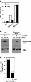
Fig. 1. Effect of BB-94 on the activation of EGFR by transmembrane proTGF-α or soluble TGF-α. (A) Subconfluent A431 cells were incubated with parental CHO cells, CHO/proHA/TGF-α cells or the conditioned media of these cells obtained in the absence or presence of BB-94 as indicated. Then, A431 cells were washed, lysed and the cell lysates analyzed by western blotting with monoclonal antibodies against phosphorylated EGFR or against total EGFR. (B) The results of three independent experiments performed as in (A) were quantified; the averages ± SD are shown. (C) Interaction between A431 cells and parental CHO or CHO/proHA/TGF-α cells. Confluent A431 cells were incubated with parental CHO or CHO/proHA/TGF-α cells with or without BB-94 and the monoclonal antibody C225 (which blocks the interaction between TGF-α and the EGFR) as indicated, and then cells not bound were counted. The percentage of cells bound to A431 was calculated by subtracting the number of cells recovered after the incubation from the number of cells added. The results are expressed as the medians of five independent determinations. (D) Confluent A431 cells were incubated with CHO/proHA/TGF-α in the presence of BB-94 with or without C225 as in (C) and gently washed with PBS. Photomicrographs of representative fields are shown. Bar = 0.1 mm.
None
Fig. 2. Effect of proTGF-α expressed in shedding-defective mutant CHO cells (M2 cells) on the activation of the EGFR. (A) Flow cytometry analysis of parental CHO, CHO/proHA/TGF-α and M2 proHA/TGF-α cells immunostained with anti-HA antibodies. Cells were treated with or without PMA and the level of proHA/TGF-α was determined by flow cytometry. The quantitative results presented are the average of duplicate determinations. (B) Subconfluent A431 cells were incubated with CHO cells, CHO/proHA/TGF-α cells, M2/proHA/TGF-α or the corresponding conditioned media. Then, A431 cells were washed, lysed and the cell lysates analyzed by western blotting with monoclonal antibodies against phosphorylated EGFR or against total EGFR. (C) Interaction between A431 cells and M2/proHA/TGF-α cells. Confluent A431 cells were incubated with parental M2/proHA/TGF-α cells in the presence or absence of the monoclonal antibody C225 and cells not bound were counted. The percentage of cells bound to A431 was calculated by subtracting the number of cells recovered after the incubation from the number of cells added. The results are expressed as the medians of five independent determinations.
None
Fig. 3. Effect of proTGF-α deletion constructs on the activation of the EGFR. (A) Schematic showing the juxtamembrane region of proTGF-α indicating the cleavage site (arrow) and different deletion constructs. The transmembrane domain is shown as a hatched box and the C-terminal cysteine that participates in the EGF motif is shown in bold. (B) Flow cytometry analysis of parental CHO/proHA/TGF-α and CHO cells permanently transfected with the deletion constructs shown in (A). Cells were treated with or without PMA and the level of cell surface anti-HA immunreactivity was determined by flow cytometry. The quantitative results presented are the average of triplicate determinations ± SD. (C) Subconfluent A431 cells were incubated with CHO cells, CHO/proHA/TGF-α cells or CHO cells expressing the indicated deletion constructs. Then, A431 cells were washed, lysed and the cell lysates analyzed by western blotting with monoclonal antibodies against phosphorylated EGFR or against total EGFR. (D) Interaction between A431 cells and cells expressing proTGF-α deletion constructs. Confluent A431 cells were incubated with CHO cells permanently transfected with the indicated deletion constructs in the presence or absence of the monoclonal antibody C225 and cells not bound were counted. The percentage of cells bound to A431 was calculated by subtracting the number of cells recovered after the incubation from the number of cells added. The results are expressed as the medians of five independent determinations.
None
Fig. 4. Analysis of the tumors induced by parental CHO, CHO/proTGF-α, CHO/Δ90–105 and M2/proTGF-α in nude mice. The indicated cells were subcutaneously injected into nude mice. Tumor volumes were measured at the indicated days after injection. Each point represents the mean of six individual determinations ± SD [**Student’s t-test values (P < 0.01) comparing tumors induced by CHO/proHA/TGF-α to those induced by CHO/Δ90–105 or M2/proHA/TGF-α].
None
Fig. 5. Analysis of the EGFR in tumor xenografts. Immunocytochemical staining with anti-HA, anti-total EGFR or anti-phosphorylated EGFR. The number of cells positively stained with the anti-phosphorylated EGFR were counted in 10 different fields, the data shown are the average ± SD.
None
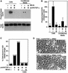
Fig. 6. Characterization of CHO/proTGF-α and M2/proTGF-α cells recovered from tumor xenografts. (A) CHO/proHA/TGF-α, M2/proHA/TGF-α (inset) or Mo1-M2/proHA/TGF-α cells, obtained from a tumor induced by M2/proHA/TGF-α cells, were treated with or without PMA and analyzed by flow cytometry using anti-HA antibodies. (B) The results of three independent experiments performed as in (A) using CHO/proHA/TGF-α, Mo1-CHO/proHA/TGF-α cells, M2/proHA/TGF-α and Mo1-M2/proHA/TGF-α cells were quantified, the average ± SD are shown. (C) CHO/proHA/TGF-α cells were pulsed for 1 h with S35-translabel and chased in complete medium for the indicated times. Cell lysates were immunoprecipitated with antibodies against the cytoplasmic domain of TACE or preimmune serum (P) and immunoprecipitates were resuspended in sample buffer and analyzed by SDS–PAGE and fluorography as described in the Materials and methods. (D) Cells obtained from individual xenografts (Mo5-CHO/proHATGF-α and Mo5-M2/proHATGF-α) were lysed and cell lysates were analyzed by western blot with antibodies directed against the cytoplasmic tail of TACE. CHO/proHA/TGF-α and M2/proHA/TGF-α were used as a control. (E) Activation of EGFR by Mo5-CHO/proHA/TGF-α and Mo5- M2/proHA/TGF-α cells. The assay was performed as described in Figure 1. CHO/proHA/TGF-α and M2/proHA/TGF-α were used as a control.
None
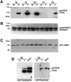
Fig. 7. Analysis of the expression of TACE (A), ADAM10 (B) and MT1-MMP (C) in tumor samples and paired normal tissue. Clarified homogenates of the corresponding tissues were analyzed by western blot with antibodies against the cytoplasmic tail of TACE or ADAM10 or monoclonal antibodies against the metalloprotease domain of MT1-MMP as indicated. (D) Samples from tumor number 20, paired normal tissue and cell lysates from HeLa cells were subjected to western blot analysis with polyclonal antibodies against the prodomain of TACE and against the cytoplasmic domain of TACE, respectively.

Similar articles

Cited by

References

    1. Albanell J. et al. (2001) Activated extracellular signal-regulated kinases: association with epidermal growth factor receptor/transforming growth factor-α expression in head and neck squamous carcinoma and inhibition by anti-epidermal growth factor receptor treatments. Cancer Res., 61, 6500–6510. - PubMed
    1. Albanell J. et al. (2002) Pharmacodynamic studies of the epidermal growth factor receptor inhibitor ZD1839 in skin from cancer patients: histopathologic and molecular consequences of receptor inhibition. J. Clin. Oncol., 20, 110–124. - PubMed
    1. Arribas J. and Massagué,J. (1995) Transforming growth factor α and β amyloid precursor protein share a secretory mechanism. J. Cell Biol., 128, 433–441. - PMC - PubMed
    1. Arribas J. and Merlos-Suárez,A. (2002) Shedding of plasma membrane proteins. Curr. Top. Dev. Biol., in press. - PubMed
    1. Arribas J., Coodly,L., Vollmer,P., Kishimoto,T.K., Rosejohn,S. and Massagué,J. (1996) Diverse cell surface protein ectodomains are shed by a system sensitive to metalloprotease inhibitors. J. Biol. Chem., 271, 11376–11382. - PubMed

Publication types

MeSH terms