. 2003 Mar 3;22(5):1147-57.
doi: 10.1093/emboj/cdg116.

FKHR (FOXO1a) is required for myotube fusion of primary mouse myoblasts

Affiliations

FKHR (FOXO1a) is required for myotube fusion of primary mouse myoblasts

Philippe R J Bois et al. EMBO J. .

Abstract

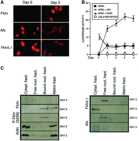
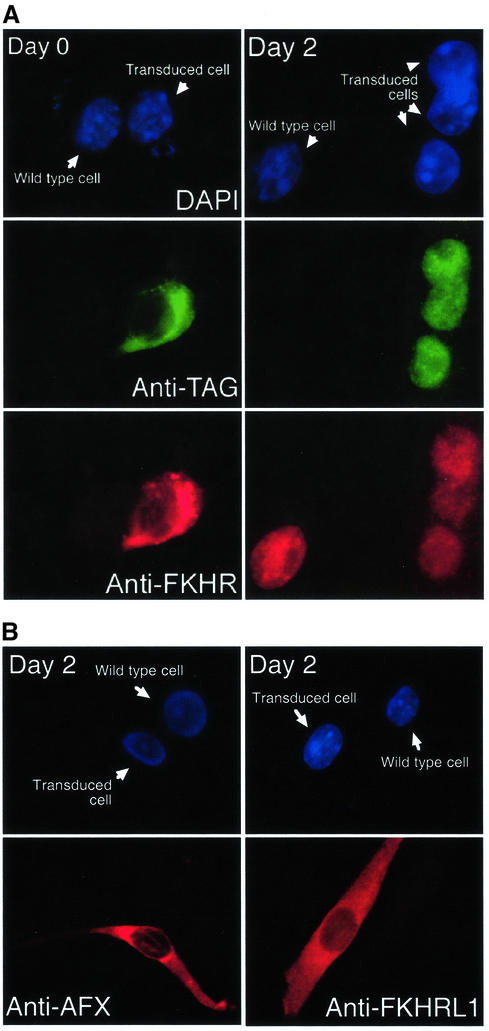
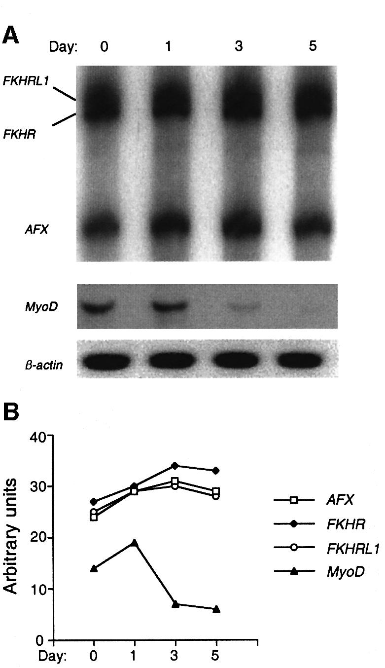
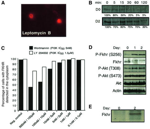
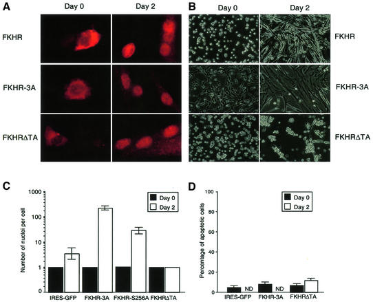
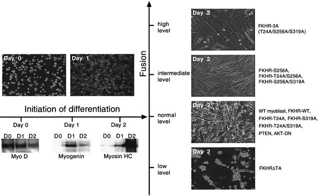
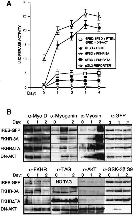
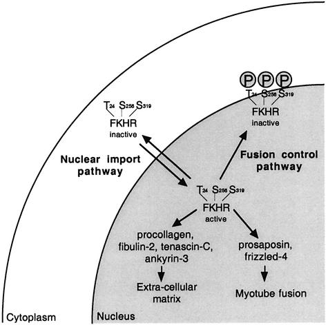
Activation of the transcription factor FKHR (Forkhead in human rhabdomyosarcoma, FOXO1a) in various established cell lines induces cell cycle arrest followed by apoptosis. These effects are inhibited through activation of the phosphatidylinositol 3-kinase/Akt pathway, resulting in FKHR phosphorylation and its export from the nucleus, thus blocking its pro-apoptotic activity. Here we report that FKHR regulates fusion of differentiating primary myoblasts. We demonstrate that FKHR is localized in the cytoplasm of proliferating myoblasts, yet translocates to the nucleus by a phosphorylation-independent pathway following serum starvation, a condition that induces myoblast differentiation. FKHR phosphorylation during terminal differentiation appears to downregulate its fusion activity, as a dominant-active non-phosphorylatable FKHR mutant dramatically augments the rate and extent of myotube fusion. However, this FKHR mutant exerts its effects only after other events initiated the differentiation pro cess. Conversely, enforced expression of a dominant-negative FKHR mutant blocks myotube formation whereas wild-type FKHR has no effect. We conclude that in addition to the role of FoxO proteins in regulating cell cycle progress and apoptosis, FKHR controls the rate of myotube fusion during myogenic differentiation.


Figures

None
Fig. 1. FKHR subcellular localization and transcriptional activity are regulated during muscle cell differentiation. (A) Immunofluorescence analysis of endogenous Fkhr, Afx and FkhrL1 expression was performed on proliferating (day 0, high serum) and differentiating (day 2, low serum) myoblasts. (B) Transient transfection assays were used to determine the FKHR transcriptional activity during myoblast differentiation, using the 6FBD luciferase reporter. Myoblasts were transfected with the pGL3-basic reporter (pGL3-reporter), the 6FBD reporter (6FBD) or the 6FBD reporter with AFX (6FBD + AFX) or wild-type FKHR (6FBD + FKHR). The activation of the 6FBD reporter in the presence of co-transfected FKHRL1 was indistinguishable from that of the 6FBD reporter alone (data not shown). In proliferating myoblasts, the reporter was also partially responsive to transfected AFX. Means of three independent experiments are shown for each transfection. Error bars show the variance at each data point. (C) Immunoblot analysis of Fkhr, Afx and FkhrL1 localization in proliferating (day 0) versus differentiating (day 2) myoblasts. Endogenous Fkhr, Afx and FkhrL1 were detected by western blotting. Cytoplasmic and matrix-bound actin served as a control.
None
Fig. 2. Localization of FKHR, AFX and FKHRL1 in proliferating versus differentiating mouse primary myoblasts. (A) Immunofluorescence of myoblasts transfected with FLAG-tagged FKHR using a DNA stain (DAPI), a FLAG antibody (anti-TAG) or the FKHR antibody. (B) Immunofluorescence of myoblasts transfected with AFX or FKHRL1 using DAPI or indirect immunufluorescence with the respective antibodies. Transduced and untransduced cells are indicated.
None
Fig. 3. Hot-stop quantitative RT–PCR analysis of Fkhr, FkhrL1 and Afx expression during myoblast differentiation. (A) Detection of Fkhr, FkhrL1 and Afx transcripts following RT–PCR using a 4% polyacrylamide gel. MyoD was used as a control to validate the sensitivity of this technique. β-actin was used as a loading control. (B) Relative amounts of Fkhr, FkhrL1, Afx and MyoD mRNA after normalization for the amount of β-actin mRNA. While the levels of MyoD transcripts dropped dramatically during myoblast differentiation, levels of Fkhr, FkhrL1 and Afx transcripts only increased marginally (10–20%).
None
Fig. 4. FKHR shuttling, half-life and phosphorylation are regulated during the differentiation of primary mouse myoblasts. (A) Inhibition of Fkhr nuclear export by Crm1 using leptomycin B (400 nM) in proliferating myoblasts. Cells were incubated for 2 h with leptomycin B, and FKHR was detected by immunofluorescence. (B) The half-life of a His6-tagged FKHR in proliferating (day 0) and differentiating (day 2) myoblasts was determined by immunoprecipitation analyses after a 15 min pulse labeling with [35S]methonine, followed by a 5–120 min chase with cold methionine. (C) Myoblasts treated with the LY294002 (PI3K IC50 = 1.5 µM) inhibitor did not show significant nuclear accumulation of Fkhr even at high doses. However, high doses of wortmannin (100 nM, PI3K IC50 = 5 nM) inhibited 50% of Fkhr nuclear export in growing myoblasts. The effects of the vehicle dimethylsulfoxide (DMSO) are shown as the negative control. (D) Western blot analysis of phosphorylated and non-phosphorylated Fkhr and Akt in proliferating (day 0) and differentiating (day 1 and day 2) myoblasts. Actin was used as a loading control. (E) The phosphorylation status of a His6-tagged FKHR in proliferating (day 0) and differentiating (day 2) myoblasts was determined by immunoprecipitation after a 2 h labeling with [32P]orthophosphate.
None
Fig. 5. Effects of FKHR and FKHR mutants on primary myoblast fusion. (A) Fluorescent immunohistochemistry of FKHR in proliferating (day 0) versus differentiating (day 2) myoblasts expressing FKHR, FKHR-3A and FKHRΔTA using an FKHR antibody. (B) Morphology of proliferating (day 0) and differentiating (day 2) myoblasts expressing FKHR, FKHR-3A or FKHRΔTA. Empty vector, DN-Akt- and PTEN-transduced myoblasts were morphologically indistinguishable from those expressing FKHR (data not shown). (C) The number of nuclei per cell was counted at day 0 (black column) and day 2 (white column) for myoblasts expressing IRES–GFP, FKHR-3A and FKHRΔTA mutants. Means of at least three independent fields are given for each construct. Error bars show the variance at each data point. The number of nuclei for the FKHR-3A mutant is an underestimate, since these cultures often consisted of large syncytia that covered the entire culture dish. (D) The percentage of apoptotic cells is given following PI staining and FACS analysis on myoblasts expressing various FKHR constructs at day 0 and day 2 of differentiation. ND: not determined, since this type of analysis could not be performed with differentiated myoblasts expressing wild-type FKHR or dominant active FKHR (FKHR-3A), because the size of the cells is too large to pass through the FACS.
None
Fig. 6. Summary of myoblast phenotypes generated by expression of different FKHR mutants. Top left panels show the differentiation of wild-type, FKHR mutants, PTEN- and DN-Akt-expressing myoblasts at 0 and 24 h of differentiation. No difference in the extent of fusion between any of these populations was observed. Lower left panels show the temporal expression of early (MyoD), intermediate (myogenin) and late (myosin heavy chain) myogenic markers during 2 days of differentiation, which was the same for all samples. Right panels show the complete range of fusion phenotypes generated by the different FKHR mutants.
None
Fig. 7. FKHR activity during the differentiation of primary mouse myoblasts. (A) Luciferase reporter-based analyses of FKHR transcriptional activity of myoblasts transfected with pGL3, 6FBD, 6FBD + FKHR, 6FBD + FKHR-3A and 6FBD + FKHRΔTA during 4 days of differentiation. Co-expression of PTEN or DN-Akt produced luciferase responses that were identical to those of myoblasts transfected with 6FBD alone. Means of at least three independent experiments are given for each transfection. Error bars show the variance at each data point. (B) Western blot analyses of primary myoblasts transduced with IRES-GFP, FKHR-3A, FKHRΔTA and DN-Akt retroviral vectors, which have no effect on the levels and timing of expression of early (MyoD), intermediate (myogenin) and late (myosin) markers during muscle cell differentiation. Overexpression of DN-Akt was confirmed using an Akt antibody. The efficacy of DN-Akt in repressing known targets of Akt was assessed by determining the level of phospho-GSK-3b Ser9, which was significantly underphosphorylated in myoblasts overexpressing DN-Akt. GFP levels were used as a loading control. FKHR-3A and FKHRΔTA were detected with an antibody to the N-terminal His6 tag, and DN-Akt with an antibody to the N-terminal HA tag. The expression patterns of these markers in PTEN- and FKHR-transduced myoblasts were indistinguishable from those transduced with the IRES-GFP vector (data not shown). In all lanes, 50 µg of protein lysate was loaded, but 100 µg of FKHR-3A was required for its detection with the His6 antibody. Exposure times for the different samples detected by the same antibody were identical. The exposure time to detect FKHR-3A was 40 times longer than that to detect the TAG of FKHRΔTA and DN-Akt.
None
Fig. 8. Model for regulation of FKHR function in primary myoblasts. Two regulatory events control the localization and function of FKHR prior to (nuclear export pathway) and during (fusion control pathway) primary myoblast differentiation. Once activated, FKHR activates genes that are involved in extracellular matrix remodeling and myotube fusion.

Similar articles

Cited by

References

    1. Alvarez B., Martinez,A.C., Burgering,B.M. and Carrera,A.C. (2001) Forkhead transcription factors contribute to execution of the mitotic programme in mammals. Nature, 413, 744–747. - PubMed
    1. Arnold H.H. and Winter,B. (1998) Muscle differentiation: more complexity to the network of myogenic regulators. Curr. Opin. Genet. Dev., 8, 539–544. - PubMed
    1. Ausubel F.M., Brent,R., Kingston,R.E., Moore,D.D., Smith,J.A. and Struhl,K. (2001) Current Protocols in Molecular Biology. John Wiley and Sons, Harvard.
    1. Barr F.G. (2001) Gene fusions involving PAX and FOX family members in alveolar rhabdomyosarcoma. Oncogene, 20, 5736–5746. - PubMed
    1. Bellacosa A., Chan,T.O., Ahmed,N.N., Datta,K., Malstrom,S., Stokoe,D., McCormick,F., Feng,J. and Tsichlis,P. (1998) Akt activation by growth factors is a multiple-step process: the role of the PH domain. Oncogene, 17, 313–325. - PubMed

Publication types

MeSH terms