. 2003 Mar 3;22(5):1168-79.
doi: 10.1093/emboj/cdg115.

HDAC-6 interacts with and deacetylates tubulin and microtubules in vivo

Affiliations

HDAC-6 interacts with and deacetylates tubulin and microtubules in vivo

Yu Zhang et al. EMBO J. .

Abstract

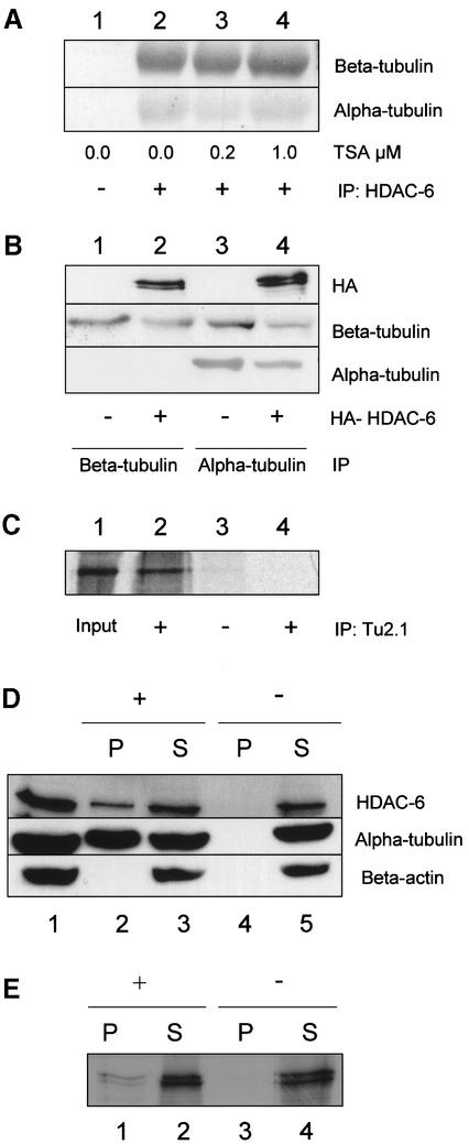
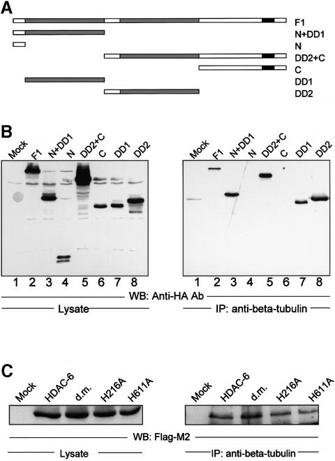
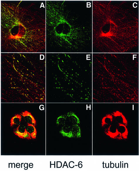
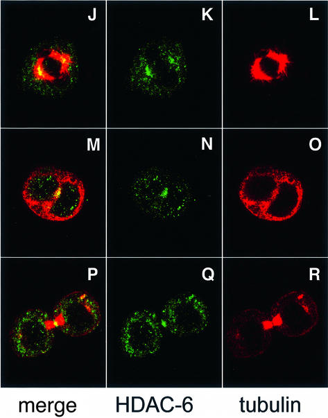
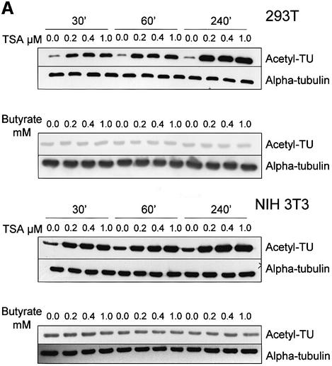
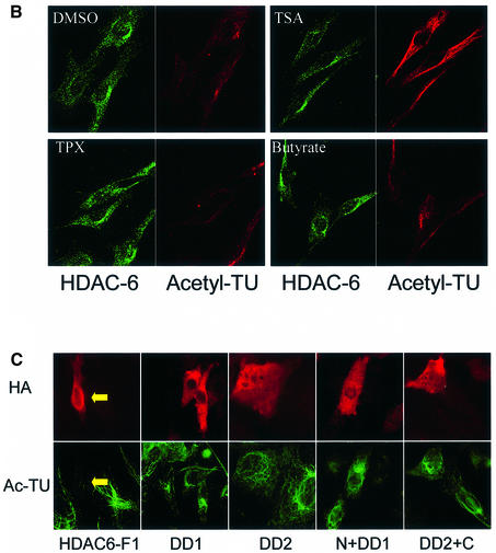
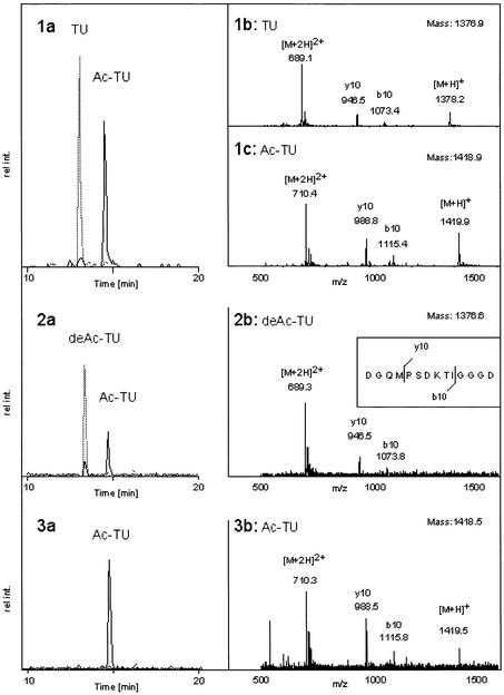
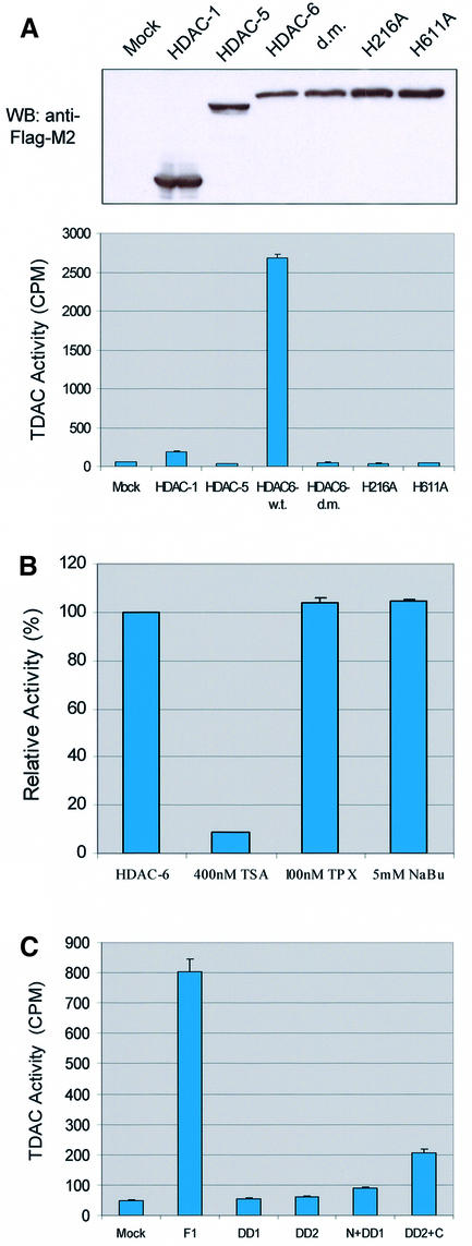
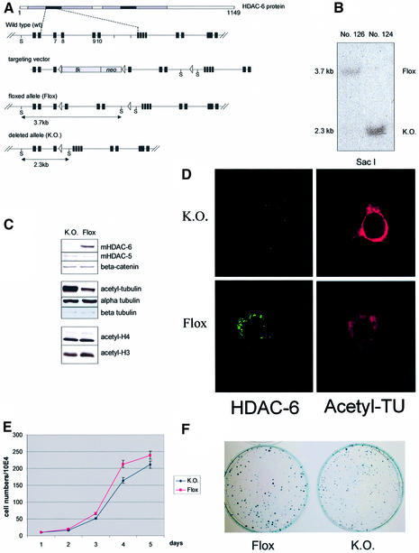
Microtubules are cylindrical cytoskeletal structures found in almost all eukaryotic cell types which are involved in a great variety of cellular processes. Reversible acetylation on the epsilon-amino group of alpha-tubulin Lys40 marks stabilized microtubule structures and may contribute to regulating microtubule dynamics. Yet, the enzymes catalysing this acetylation/deacetylation have remained unidentified until recently. Here we report that beta-tubulin interacts with histone deacetylase-6 (HDAC-6) in a yeast two-hybrid assay and in vitro. We find that HDAC-6 is a micro tubule-associated protein capable of deacetylating alpha-tubulin in vivo and in vitro. HDAC-6's microtubule binding and deacetylation functions both depend on the hdac domains. Overexpression of HDAC-6 in mammalian cells leads to tubulin hypoacetylation. In contrast, inhibition of HDAC-6 function by two independent mechanisms--pharmacological (HDAC inhibitors) or genetic (targeted inactivation of HDAC-6 in embryonic stem cells)--leads to hyperacetylation of tubulin and microtubules. Taken together, our data provide evidence that HDAC-6 might act as a dual deacetylase for tubulin and histones, and suggest the possibility that acetylated non-histone proteins might represent novel targets for pharmacological therapy by HDAC inhibitors.


Figures

None
Fig. 1. HDAC-6 interacts with β-tubulin and microtubules in vivo and in vitro. (A) Co-immunoprecipitation assay. NIH-3T3 cells were treated for 6 h with TSA at the indicated concentration in order to inhibit HDAC activity. After cell extract preparation, HDAC-6 was immunoprecipitated with an anti-mouse HDAC-6-specific antibody (lanes 2–4) or with a control antibody (lane 1). The precipitate was analysed by SDS–PAGE and blotted onto nitrocellulose membranes. The filters were probed with antibodies against β-tubulin (TU 2.1, upper panel) or α-tubulin (DM1A, lower panel). (B) Co-immunoprecipitation assay in 293T cells. In this case, cells were transfected with an expression vector encoding HA-mHDAC-6 (lanes 2 and 4) or an empty expression vector (lanes 1 and 3). Two days after transfection, cell extracts were prepared and β- or α-tubulin was immunoprecipitated with specific antibodies (lanes 1 and 2 with TU2.1, lanes 3 and 4 with DM1A). The precipitates were analysed by western blotting: the filters were probed with antibodies against HA (upper panel), β-tubulin (TU2.1, middle panel) or α-tubulin (DM1A, lower panel). (CIn vitro interaction between HDAC-6 and tubulin. Purified bovine tubulin (Cytoskeleton, Inc.) was incubated with in vitro translated 35S-labelled mHDAC-6 (lanes 2 and 3) or with a control reticulocyte lysate (lane 4). β-tubulin was immunoprecipitated with the TU2.1 antibody (lanes 2 and 4), and the presence of HDAC-6 protein was detected by fluorography. Lane 1 contains 10% of the 35S-labelled mHDAC6 input. (D) HDAC-6 is part of the microtubule-associated proteins in NIH-3T3 cells. Microtubules were purified from NIH-3T3 cell lysate (lane 1) in the presence (lanes 2 and 3) or absence (lanes 4 and 5) of paclitaxel (taxol) and GTP. Microtubules were then pelleted by centrifugation. The proteins present in the pellet (lanes 2 and 4) and supernatant fractions (lanes 3 and 5) were analysed by western blotting using antibodies to detect mHDAC-6 (upper panel), α-tubulin (middle panel) and β-actin (lower panel). (E) Interaction between HDAC-6 and microtubules in vitro. Purified bovine tubulin (Cytoskeleton, Inc.) was assembled into microtubules in the presence (lanes 1 and 2) or absence (lanes 3 and 4) of paclitaxel (taxol) and GTP. Subsequently, in vitro translated 35S-labelled mHDAC6 was added and binding was allowed to proceed. Microtubules were then pelleted by centrifugation. The proteins present in the pellet (lanes 1 and 3) and supernatant (lanes 2 and 4) fractions were analysed by SDS–PAGE, and the presence of HDAC-6 was detected by fluorography.
None
Fig. 2. HDAC-6 interacts with β-tubulin via its HDAC domains. (A) Schematic representation of the N-terminally HA-tagged HDAC-6 deletion constructs used. (B) Co-immunoprecipitation assay. 293T cells were transfected with the HDAC-6 expression vectors (lanes 2–8), and cellular extracts were prepared. HDAC-6 expression was measured by western blot with the anti-HA antibody (left panel). Association with β-tubulin was measured by performing an immunoprecipitation with an anti-β-tubulin antibody, followed by analysis of the precipitate by western blotting with the anti-HA antibody (right panel). (C) 293T cells were transfected with Flag-tagged HDAC-6 constructs and cell extracts were subjected to immunoprecipitation by anti-β-tubulin antibody. In the mutants proteins, the histidine at position 216 or 611 were mutated to alanine. d. m., double mutant protein.
None
Fig. 3. Partial co-localization of HDAC-6 and microtubules in NIH-3T3 cells. (AF) Exponentially growing NIH-3T3 cells were fixed with cold methanol and double-stained for endogenous HDAC-6 (green) and β-tubulin (red). Analysis was performed by confocal microscopy, and individual stainings were merged digitally. In (GI), cells were treated with 10 µM paclitaxel for 4 h to stabilize microtubules, and HDAC-6 and tubulin were immunolocalized as above. In (J–R), mitotic cells were examined: (JL) metaphase; (MO) anaphase; (PR) telophase.
None
Fig. 4. Pharmacological inhibition of HDAC-6 activity leads to increased tubulin acetylation in vivo. (A) Exponentially growing 293T or NIH-3T3 cells were treated for 30 min, 1 h or 4 h with different concentrations of the HDAC inhibitors TSA or sodium butyrate, as indicated. Cell extracts were prepared and analysed by SDS–PAGE followed by western blotting. The membranes were probed with antibodies recognizing α-tubulin, either indiscriminately (DM1A) or only when acetylated (TU6-11). (B) NIH-3T3 cells were treated for 4 h with the indicated chemicals and then fixed. Immunostaining was used to visualize the level of HDAC-6 (green) and of tubulin acetylation (red). (C) Balb/c 3T3 cells were transfected with the indicated expression vectors encoding HDAC-6 proteins tagged at the N-terminus with the HA epitope. Expression of HDAC-6 deletions was monitored with an anti-HA antibody (red), and the state of tubulin acetylation was detected with the TU6-11 antibody (green).
None
Fig. 5. In vitro deacetylation of a tubulin-derived peptide by HDAC-6. A peptide derived from α-tubulin was chemically synthesized as an unacetylated version (TU) or acetylated on Lys40 (Ac-TU). (1a) HPLC separation of a 1:1 mixture of the two peptides. The extracted ion current (XIC) of 689 (grey) and 710 (black) is shown, corresponding to the doubly charged ions of TU and Ac-TU, respectively. (1b) MS of TU with a mass of 1376.9 Da. The Ac-TU has a mass of 1418.9 Da as shown in (1c). Both peptides are partially fragmenting, generating the y10 as well as the b10 ion (insert 2b). (2a) The acetylated peptide was incubated with HDAC-6 and the reaction mix was analysed as described for the standard peptide mixture. The deacetylated TU peptide formed had a mass of 1376.6 Da as shown in (2b). The specific fragment ions y10 and b10 are indicated and explained in the inset of (2b). (3a) Analysis of the products of the incubation of the acetylated peptide with the doubly mutated protein HDAC-6-H216A/H611A shows only the acetylated peptide. This peptide had a mass of 1418.5 Da, and the two specific fragment ions y10 and b10 could be detected again (3b).
None
Fig. 6. Tubulin deacetylase activity is specific to HDAC-6. (A) 293T cells were transfected with expression vectors encoding FLAG-tagged versions of the indicated proteins. Two days later, cell extracts were prepared and immunoprecipitated with the anti-FLAG antibody M2. Upper panel: expression levels of the different HDACs were verified by western blotting using the anti-FLAG antibody. Lower panel: tubulin deacetylase activity (TDAC) was assayed by incubating an [3H]acetyl-labelled peptide from human α-tubulin (amino acids 33–46) and measuring the release of radioactivity. (B) TDAC activity was assayed as above, with immunoprecipitated HDAC-6 alone or in the presence of TSA, TPX or sodium butyrate (NaBu), as indicated. (C) HDAC-6 deletion constructs were transfected into 293T cells, and immunoprecipitated material was used for TDAC assay, as in (A).
None
Fig. 7. Increased tubulin acetylation, but not histone acetylation in HDAC-6-deficient ES cells. (A) Scheme of the floxed mouse HDAC-6 locus and of the deleted (KO) allele (details to be published elsewhere). The position of SacI (S) restriction sites is indicated, as well as the size of the resulting fragments. At the top, a scheme of the HDAC-6 protein is presented showing the two hdac domains (grey) and their core region (black). Cre-mediated recombination between loxP sites 1 and 2 leads to the floxed allele. Cre-mediated recombination between loxP sites 1 and 3 leads to the knockout allele; in this case, exons 7–10 of HDAC-6 are removed and no functional HDAC-6 is made. (B) Southern blot analysis of DNA from a floxed ES cell clone (No. 126) as well as from a knockout clone (No. 124). The position of the floxed or knockout allele is indicated. (C) Analysis of protein expression in floxed or knockout ES cells. Protein extracts were prepared from ES cell clones No. 126 and 124 and analysed by SDS–PAGE followed by western blotting with the indicated antibodies. (D) Immunostaining of HDAC-6 (green) and acetylated tubulin (red) in floxed or knockout ES cells, as indicated. (E) Growth curves of HDAC-6 floxed and knockout ES cell lines. Equal numbers of cells (1 × 105) were seeded in triplicate and aliquots were counted daily during a period of 5 days. (F) Colony formation assay with floxed and HDAC-6 knockout ES cells. A total of 2 × 103 cells were seeded and cultivated for 8 days in triplicate. Cells were then fixed and stained with Methylene Blue. One representative plate of each genotype is shown.

References

    1. Adachi N., Kimura,A. and Horikoshi,M. (2002) A conserved motif common to the histone acetyltransferase Esa1 and the histone deacetylase Rpd3. J. Biol. Chem., 277, 35688–35695. - PubMed
    1. Brown C.E., Lechner,T., Howe,L. and Workman,J.L. (2000) The many HATs of transcription coactivators. Trends Biochem. Sci., 25, 15–19. - PubMed
    1. Cheung W.L., Briggs,S.D. and Allis,C.D. (2000) Acetylation and chromosomal functions. Curr. Opin. Cell Biol., 12, 326–333. - PubMed
    1. Fields S. and Song,O. (1989) A novel genetic system to detect protein–protein interactions. Nature, 340, 245–246. - PubMed
    1. Finnin M.S., Donigian,J.R., Cohen,A., Richon,V.M., Rifkind,R.A., Marks,P.A., Breslow,R. and Pavletich,N.P. (1999) Structures of a histone deacetylase homologue bound to the TSA and SAHA inhibitors. Nature, 401, 188–193. - PubMed

Publication types

MeSH terms