. 2003 Mar 3;22(5):1180-7.
doi: 10.1093/emboj/cdg112.

Stress-induced gene expression requires programmed recovery from translational repression

Affiliations

Stress-induced gene expression requires programmed recovery from translational repression

Isabel Novoa et al. EMBO J. .

Erratum in

  • EMBO J. 2003 May 1;22(9):2307

Abstract

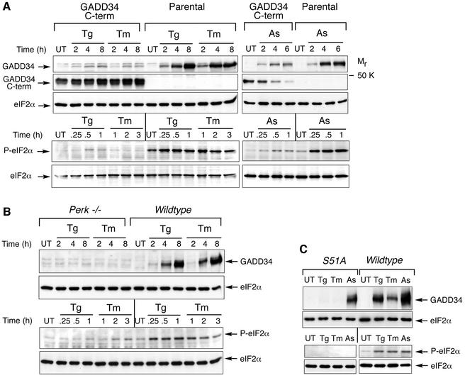
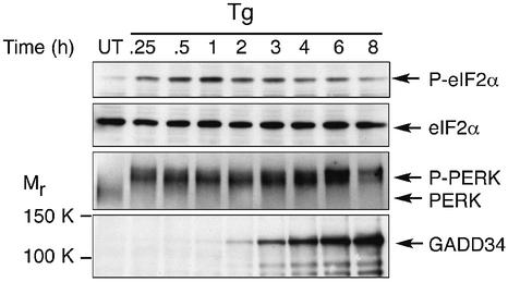
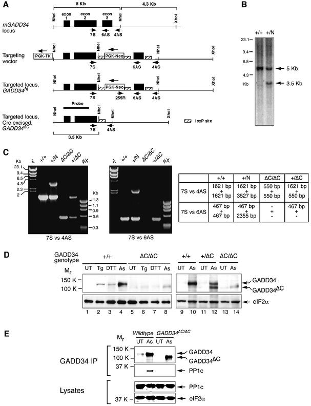
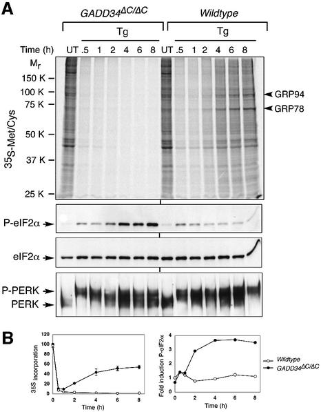
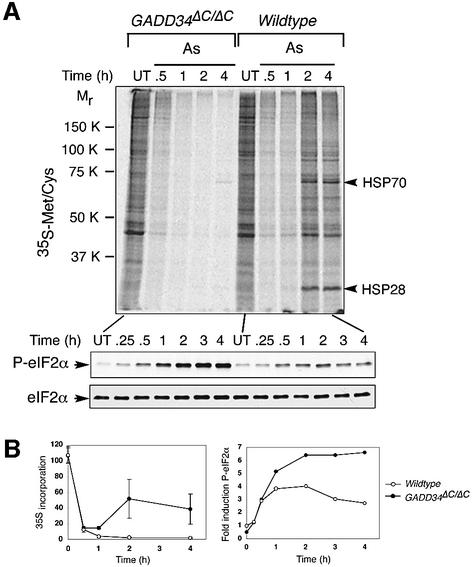
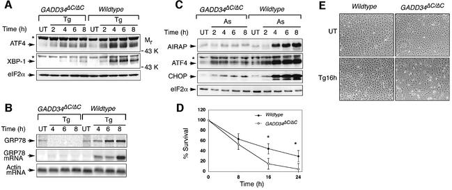
Active repression of protein synthesis protects cells against protein malfolding during endoplasmic reticulum stress, nutrient deprivation and oxidative stress. However, long-term adaptation to these conditions requires synthesis of new stress-induced proteins. Phosphorylation of the alpha-subunit of translation initiation factor 2 (eIF2alpha) represses translation in diverse stressful conditions. GADD34 is a stress-inducible regulatory subunit of a holophosphatase complex that dephosphorylates eIF2alpha, and has been hypothesized to play a role in translational recovery. Here, we report that GADD34 expression correlated temporally with eIF2alpha dephosphorylation late in the stress response. Inactivation of both alleles of GADD34 prevented eIF2alpha dephosphorylation and blocked the recovery of protein synthesis, normally observed late in the stress response. Furthermore, defective recovery of protein synthesis markedly impaired translation of stress-induced proteins and interfered with programmed activation of stress-induced genes in the GADD34 mutant cells. These observations indicate that GADD34 controls a programmed shift from translational repression to stress-induced gene expression, and reconciles the apparent contradiction between the translational and transcriptional arms of cellular stress responses.


Figures

None
Fig. 1. eIF2α phosphorylation promotes GADD34 expression. (A) Immunoblot of GADD34, eIF2α phosphorylated on serine 51 (P-eIF2α) and total eIF2α from untreated (UT), thapsigargin (Tg)-, tunicamycin (Tm)-, or arsenite (As)-treated NIH-3T3 (Parental) cells or cells expressing a C-terminal fragment of GADD34 that constitutively dephosphorylates eIF2α (GADD34 C-term) (Novoa et al., 2001). (B) Immunoblot of GADD34, phosphorylated eIF2α and total eIF2α from untreated, thapsigargin and tunicamycin treated wild-type and PERK–/– mouse fibroblasts. (C) Immunoblot of GADD34 and total eIF2α from untreated (UT), thapsigargin (Tg; 8 h)-, tunicamycin (Tm; 8 h)- and arsenite (As; 4 h)-treated mouse embryonic fibroblasts with the indicated eIF2α genotype (upper panels). eIF2α phosphorylation was measured by immunoblot in the same cells following thapsigargin (Tg; 2 h), tunicamycin (Tm; 2 h) and arsenite (As; 2 h) treatment (lower panels).
None
Fig. 2. Time course of GADD34 expression, eIF2α phosphorylation and PERK activation in ER stressed cells. Shown are immunoblots of P-eIF2α, total eIF2α, activated phosphorylated PERK (P-PERK), inactive PERK and GADD34 from untreated (UT) and thapsigargin (Tg)-treated NIH 3T3 cells.
None
Fig. 3. Targeted mutagenesis of GADD34. (A) Scheme of the genomic organization of mouse GADD34 and targeting strategy to delete exon 3 encoding the PP1c-interacting domain (amino acid residues 549–657). From the top down: the wild-type locus; the targeting vector with the position of the oligonucleotides used to genotype the derivative alleles (arrows) and the loxP-sites (hatched rectangles) showing; the targeted GADD34N locus before Cre-mediated excision of the Neo-cassette and exon 3; and the mutant GADD34ΔC allele after Cre-mediated excision of exon 3 and the Neo cassette. (B) Southern blot analysis of NheI-digested genomic DNA of the indicated GADD34 genotypes. The position of the radiolabeled probe (HinDIII cDNA fragment containing exon 1 and part of exon 2) and the predicted genomic GADD34 NheI fragments (5 and 3.5 Kb) are indicated in (A) above. (C) Detection of the various GADD34 alleles by PCR. The primers 7S versus 4AS, and 7S versus 6AS are shown in (A). The table indicates the expected PCR products for each GADD34 genotype. +, wild-type locus; N, targeted locus; ΔC, targeted locus after CRE-mediated excised of exon 3 and the neo cassette. (D) Immunoblot analysis of GADD34 gene product in untreated (UT), thapsigargin (Tg)-, dithiothreitol (DTT)- and arsenite (As)-treated wild-type (+/+), GADD34ΔC/+ and GADD34ΔC/ΔC fibroblasts. eIF2α immunoblotting serves as a control for loading. (E) GADD34 and protein phosphatase 1 (PP1c) immunoblot of GADD34–PP1c complexes immunoprecipitated with anti-GADD34 antiserum from untreated (UT) and arsenite (As)-treated wild-type and GADD34ΔC/ΔC cells (upper panels). Immunoblot of PP1c and total eIF2α in the lysate that served as the input for the GADD34–PP1c complex immunoprecipitation (two lower panels).
None
Fig. 4. GADD34 is required for eIF2α dephosphorylation and translational recovery during ER stress. (A) Upper panel, autoradiogram of an SDS–PAGE gel showing 35S-methionine/cysteine incorporation into newly synthesized proteins in untreated (UT) and thapsigargin (Tg)-treated wild-type and GADD34ΔC/ΔC fibroblasts. Arrowheads to the right of the autoradiogram indicate the ER stress-inducible chaperones GRP78 and GRP94. Lower panels, immunoblots of P-eIF2α and total eIF2α, and immunoblots of PERK immunoprecipitates from the same cells. (B) Graphic presentation of 35S-methionine/cysteine incorporation into newly synthesized proteins (left graph), and fold induction of phosphorylated eIF2α (right graph) in untreated and thapsigargin-treated wild-type and GADD34ΔC/ΔC fibroblasts. The level of 35S incorporation in untreated cells is set at 100% whereas the signal of phosphorylated eIF2α from untreated wild-type cells is set as 1. Shown are mean ± SEM of experiments performed in duplicate and reproduced twice.
None
Fig. 5. GADD34 is required for eIF2α dephosphorylation and translational recovery during arsenite treatment. (A) Upper panel, autoradiogram of an SDS–PAGE gel showing 35S-methionine/cysteine incorporation into newly synthesized proteins in untreated (UT) and arsenite (As)-treated wild-type and GADD34ΔC/ΔC fibroblasts. Arrowheads to the right of the autoradiogram indicate the arsenite-inducible chaperones HSP70 and HSP28. Lower panels, immunoblots of P-eIF2α and total eIF2α from the same cells. (B) Graphic presentation of 35S-methionine/cysteine incorporation into newly synthesized proteins (left graph), and fold induction of phosphorylated eIF2α (right graph) in untreated and arsenite-treated wild-type and GADD34ΔC/ΔC fibroblasts. The level of 35S incorporation in untreated cells is set at 100% whereas the signal of phosphorylated eIF2α from untreated wild-type cells is set as 1. Shown are mean ± SEM of experiments performed in duplicate and reproduced twice.
None
Fig. 6. GADD34 promotes stress-induced gene expression. (A) Immunoblot of the UPR-induced transcription factors ATF4 and XBP-1 in untreated (UT) and thapsigargin-treated (Tg) wild-type and GADD34ΔC/ΔC fibroblasts. The asterisks mark irrelevant proteins detected by the antisera. (B) Upper panel, autoradiogram of an SDS–PAGE gel of GRP78 immunoprecipitated from untreated (UT) and thapsigargin (Tg)-treated wild-type and GADD34ΔC/ΔC fibroblasts labeled with 35S-methionine/cysteine. Middle and lower panels are northern blots of GRP78 and β-actin mRNA from the same cells. (C) Immunoblot of the arsenite-induced proteins AIRAP (Sok et al., 2001), ATF4 and CHOP from untreated and arsenite-treated wild-type and GADD34ΔC/ΔC fibroblasts. (D) Survival of thapsigargin-treated wild-type and GADD34ΔC/ΔC fibroblasts, measured by their ability to reduce MTT (3-[4,5-dimethylthiazol-2-yl]-2,5-diphenyl-tetrazolium bromide). Shown are the mean ± SEM of measurements carried out in duplicate on two different clones of cells from each genotype and reproduced 3 times. The asterisks denote P < 0.0001 by two-tailed t-test. 100% refers to MTT reduction by the untreated cells. (E) Photomicrographs of untreated and thapsigargin-treated wild-type and GADD34ΔC/ΔC fibroblasts.

References

    1. Brostrom C.O. and Brostrom,M.A. (1998) Regulation of translational initiation during cellular responses to stress. Prog. Nucleic Acid Res. Mol. Biol., 58, 79–125. - PubMed
    1. Brostrom M.A., Lin,X.J., Cade,C., Gmitter,D. and Brostrom,C.O. (1989) Loss of a calcium requirement for protein synthesis in pituitary cells following thermal or chemical stress. J. Biol. Chem., 264, 1638–1643. - PubMed
    1. Calfon M., Zeng,H., Urano,F., Till,J.H., Hubbard,S.R., Harding,H.P., Clark,S.G. and Ron,D. (2002) IRE1 couples endoplasmic reticulum load to secretory capacity by processing the XBP-1 mRNA. Nature, 415, 92–96. - PubMed
    1. Connor J.H., Weiser,D.C., Li,S., Hallenbeck,J.M. and Shenolikar,S. (2001) Growth arrest and DNA damage-inducible protein gadd34 assembles a novel signaling complex containing protein phosphatase 1 and inhibitor 1. Mol. Cell. Biol., 21, 6841–6850. - PMC - PubMed
    1. DeGracia D.J., Kumar,R., Owen,C.R., Krause,G.S. and White,B.C. (2002) Molecular pathways of protein synthesis inhibition during brain reperfusion: implications for neuronal survival or death. J. Cereb. Blood Flow Metab., 22, 127–141. - PubMed

Publication types

MeSH terms