. 2003 Mar 4;100(5):2272-7.
doi: 10.1073/pnas.262791999. Epub 2003 Feb 26.

Integrin beta cytoplasmic domain interactions with phosphotyrosine-binding domains: a structural prototype for diversity in integrin signaling

Affiliations

Integrin beta cytoplasmic domain interactions with phosphotyrosine-binding domains: a structural prototype for diversity in integrin signaling

David A Calderwood et al. Proc Natl Acad Sci U S A. .

Abstract

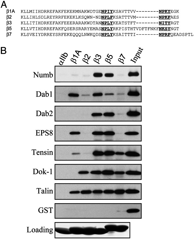
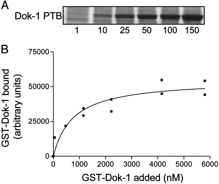
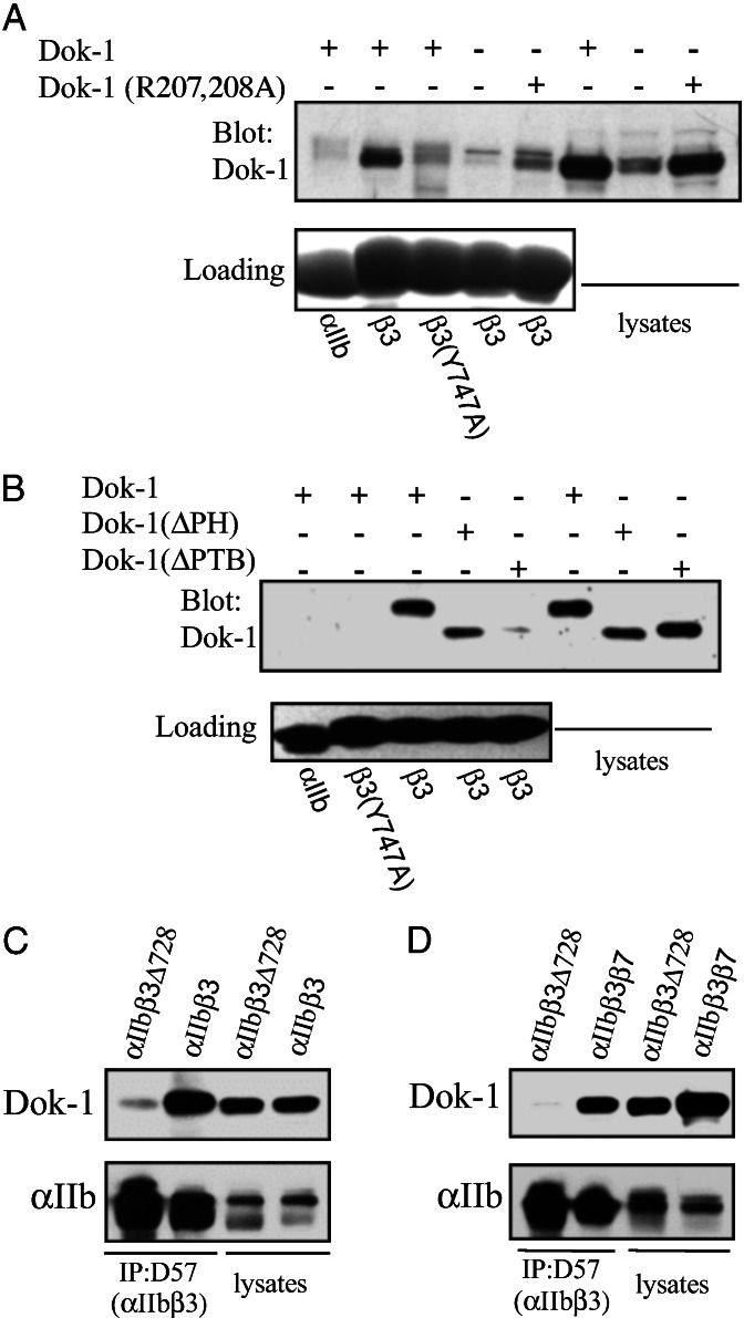
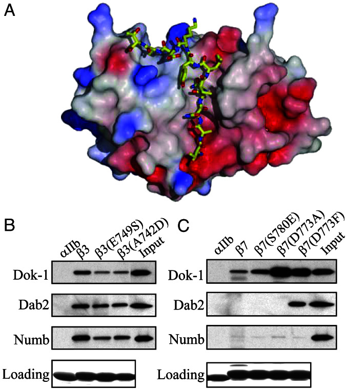
The cytoplasmic domains (tails) of heterodimeric integrin adhesion receptors mediate integrins' biological functions by binding to cytoplasmic proteins. Most integrin beta tails contain one or two NPXYF motifs that can form beta turns. These motifs are part of a canonical recognition sequence for phosphotyrosine-binding (PTB) domains, protein modules that are present in a wide variety of signaling and cytoskeletal proteins. Indeed, talin and ICAP1-alpha bind to integrin beta tails by means of a PTB domain-NPXY ligand interaction. To assess the generality of this interaction we examined the binding of a series of recombinant PTB domains to a panel of short integrin beta tails. In addition to the known integrin-binding proteins, we found that Numb (a negative regulator of Notch signaling) and Dok-1 (a signaling adaptor involved in cell migration) and their isolated PTB domain bound to integrin tails. Furthermore, Dok-1 physically associated with integrin alpha IIb beta 3. Mutations of the integrin beta tails confirmed that these interactions are canonical PTB domain-ligand interactions. First, the interactions were blocked by mutation of an NPXY motif in the integrin tail. Second, integrin class-specific interactions were observed with the PTB domains of Dab, EPS8, and tensin. We used this specificity, and a molecular model of an integrin beta tail-PTB domain interaction to predict critical interacting residues. The importance of these residues was confirmed by generation of gain- and loss-of-function mutations in beta 7 and beta 3 tails. These data establish that short integrin beta tails interact with a large number of PTB domain-containing proteins through a structurally conserved mechanism.


Figures

Figure 1
Figure 1
PTB domains bind to β integrin cytoplasmic tails. (A) An alignment of the amino acid sequences of β integrin cytoplasmic tails. The NPXY or NPXY-like motifs are in bold and underlined. (B) PTB domains, which were expressed and purified as recombinant GST fusion proteins, were incubated with beads coated with recombinant αIIb, β1A, β2, β3, β5, and β7 cytoplasmic tails. Bound proteins were fractionated by SDS/PAGE and GST-PTB domains were detected by Western blotting with anti-GST antibodies. Loading of the recombinant integrin tails on the beads was assessed by Coomassie blue staining.
Figure 2
Figure 2
Tyr to Ala mutations in β integrin cytoplasmic tails inhibit binding of PTB domains. The binding of recombinant GST-PTB domain fusion proteins to recombinant β1A (A), β3 (B), and β7 (C) integrin cytoplasmic tails with Tyr to Ala mutations in their N-terminal NPXY motifs [β1A(Y787A), β3(Y747A), and β7(Y778A)] was assessed as described in the legend to Fig. 1. (D) The binding of GST-PTB domains to recombinant β3 cytoplasmic tails containing mutations in either the N-terminal [β3(Y747A)] or C-terminal [β3(Y759A)] NPXY-like motif was assessed as described in B.
Figure 3
Figure 3
The Dok-1 PTB domain binds integrin β3 tails. (A) Various amounts (1–150 μg as indicated) of recombinant GST-Dok-1 PTB domains were mixed with beads coated with recombinant β3 tails. Bound proteins were fractionated by SDS/PAGE and detected by Coomassie blue staining. (B) Bound GST-Dok-1 fusion protein was quantified by scanning densitometry and the amount bound was plotted against the input concentration.
Figure 4
Figure 4
Intact Dok-1 binds to integrin β tails. (A) Lysates from CHO cells transiently transfected with Dok-1, Dok-1 containing a mutation that reduces PTB domain ligand binding [Dok-1 (R207,208A; ref. 37], or from mock-transfected cells were incubated with beads coated with recombinant αIIb, β3, or β3(Y747A) cytoplasmic domains. Bound proteins were fractionated by SDS/PAGE and Dok-1 was detected by Western blotting with anti-Dok-1 antibodies. Note the specific binding of a band in the untransfected cells, presumably that of endogenous hamster Dok-1. (B) Lysates from CHO cells transiently transfected with epitope-tagged Dok-1, Dok-1 lacking its PH domain [Dok-1(ΔPH)], or Dok-1 lacking its PTB domain [Dok-1(ΔPTB)] were incubated with beads coated with recombinant αIIb, β3, or β3(Y747A) cytoplasmic domains. Bound proteins were fractionated by SDS/PAGE and Dok-1 was detected by Western blotting with antibodies against the epitope tags. (C) CHO cells stably expressing αIIbβ3 or αIIbβ3Δ728 were transfected with Dok-1, and 24 h later cells were lysed and the integrins were precipitated with the mAb D57. Immunoprecipitated αIIb and coimmunoprecipitated Dok-1 were detected by Western blotting. (D) CHO cells stably expressing αIIbβ3β7 or αIIbβ3Δ728 were transfected with Dok-1, and 24 h later cells were lysed and the integrins were precipitated with the mAb D57. Immunoprecipitated αIIb and coimmunoprecipitated Dok-1 were detected by Western blotting.
Figure 5
Figure 5
Mutations in β integrin cytoplasmic tails alter PTB domain-binding specificity. (A) Structural modeling of the interaction between the Numb PTB domain and the β3 integrin cytoplasmic tail. A model of β3 residues Thr-741 to Thr-751 docked in the Numb-PTB domain binding site was generated based on the structure of the Numb–Nak complex. (B) Introduction of a charged residue at the −5 position and a noncharged residue at the +2 position relative to the Tyr or the β3 NPXY motif inhibit PTB domain binding. GST-PTB domain fusion proteins were incubated with beads coated with recombinant β3, β3(E749S), or β3(A742D) tails. Bound proteins were fractionated by SDS/PAGE and GST-PTB domains were detected by Western blotting with anti-GST antibodies. Loading of the recombinant integrin tails on the beads was assessed by Coomassie blue staining. (C) Introduction of an uncharged residue at the −5 position and a charged residue at the +2 position relative to the Tyr or the β7 NPXY motif enhances PTB domain binding. The binding of recombinant GST-PTB domain fusion proteins to β7, β7(S780E), β7(D773A), or β7(D773F) integrin cytoplasmic tails was assessed as described in B.

Similar articles

Cited by

References

    1. Hynes R O. Cell. 1987;48:549–554. - PubMed
    1. Liu S, Calderwood D A, Ginsberg M H. J Cell Sci. 2000;113:3563–3571. - PubMed
    1. Ulmer T S, Yaspan B, Ginsberg M H, Campbell I D. Biochemistry. 2001;40:7498–7508. - PubMed
    1. Trub T, Choi W E, Wolf G, Ottinger E, Chen Y, Weiss M, Shoelson S E. J Biol Chem. 1995;270:18205–18208. - PubMed
    1. Dans M, Gagnoux-Palacios L, Blaikie P, Klein S, Mariotti A, Giancotti F G. J Biol Chem. 2001;276:1494–1502. - PubMed

Publication types

MeSH terms