. 2003 Mar 4;100(5):2426-31.
doi: 10.1073/pnas.0536882100. Epub 2003 Feb 26.

A human peripheral blood monocyte-derived subset acts as pluripotent stem cells

Affiliations

A human peripheral blood monocyte-derived subset acts as pluripotent stem cells

Yong Zhao et al. Proc Natl Acad Sci U S A. .

Abstract

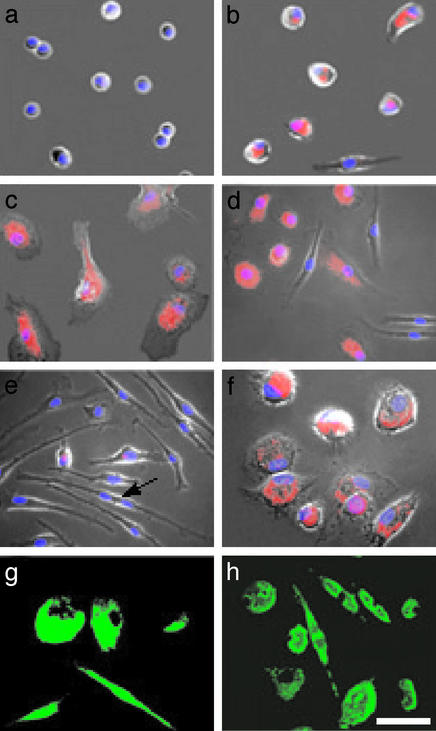
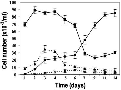
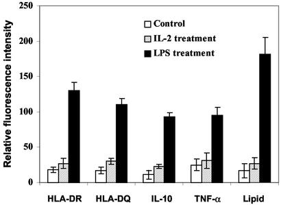
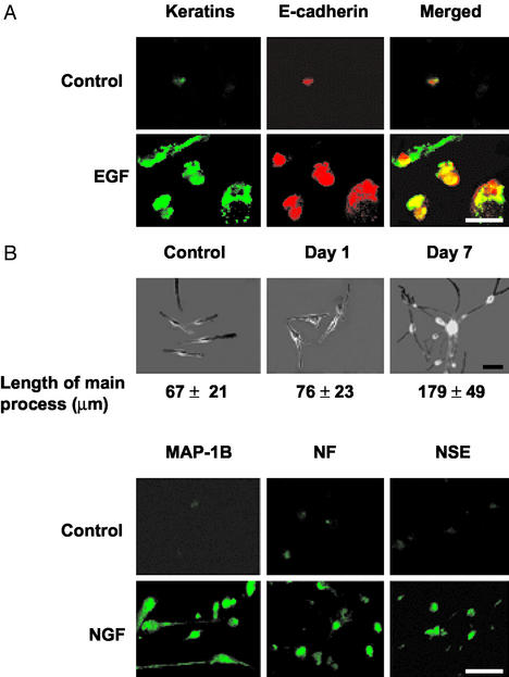
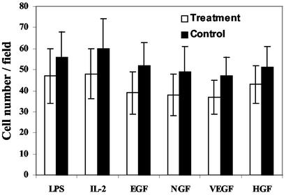
We have identified, cultured, characterized, and propagated adult pluripotent stem cells (PSC) from a subset of human peripheral blood monocytes. These cells, which in appearance resemble fibroblasts, expand in the presence of macrophage colony-stimulating factor and display monocytic and hematopoietic stem cell markers including CD14, CD34, and CD45. We have induced these cells to differentiate into mature macrophages by lipopolysaccharide, T lymphocytes by IL-2, epithelial cells by epidermal growth factor, endothelial cells by vascular endothelial cell growth factor, neuronal cells by nerve growth factor, and liver cells by hepatocyte growth factor. The pluripotent nature of individual PSC was further confirmed by a clonal analysis. The ability to store, expand, and differentiate these PSC from autologous peripheral blood should make them valuable candidates for transplantation therapy.


Figures

Figure 1
Figure 1
Macrophage differentiation of peripheral blood monocytes. (a) Freshly isolated monocytes. (b) Untreated 5-d-old monocyte culture. (c) Five-day PMA-treated culture. (d) Five-day M-CSF-treated culture. (e) Fourteen-day M-CSF-treated culture; the arrow points to a dividing cell. (f) Fourteen-day M-CSF-treated culture incubated for 1 d with LPS. (g) MAC-1 immunostaining of 5-d M-CSF-treated culture. (h) Fluorescence of phagocytized beads in 5-d M-CSF-treated culture. (a–f) Cells visualized by phase-contrast microscopy merged with fluorescence images of lipids stained with Nile red (red) and nuclei stained with 4′,6-diamidino-2-phenylindole (DAPI, blue). (Scale bar, 40 μm.)
Figure 2
Figure 2
Replication of M-CSF-treated f-MΦ. f-MΦ in untreated (x-x) and M-CSF-treated (●-●) cultures. s-MΦ in untreated (▴-▴) and M-CSF-treated (■-■) cultures. The results are the mean ± SD of cell counts from four individuals.
Figure 3
Figure 3
LPS-induced macrophage differentiation of f-MΦ cultures. Fluorescence intensity (mean ± SD of four experiments) is based on 30–50 cells per determination per individual.
Figure 4
Figure 4
Epithelial and neuronal cell differentiation of f-MΦ. (A) EGF-induced epithelial differentiation was assessed by double immunostaining for keratins (green) and E-cadherin (red). Each field contains four to five cells. The control panel was selected to include a positive cell. (B) NGF-induced neuronal differentiation was assessed by length of the main processes (mean ± SD) of 50 randomly selected cells by using SLIDEBOOK software (Upper) and by immunostaining for neuron-specific antigens (Lower). Each immunostained field contains 10–15 cells with the control panel selected to contain positive cells. (Scale bar, 50 μm.)
Figure 5
Figure 5
Relative cell number in f-MΦ cultures treated with or without differentiation inducers. The results are the mean ± SD of five randomly selected microscopic fields each from four different experiments for each treatment.

Similar articles

Cited by

References

    1. Wagers A J, Christensen J L, Weissman I L. Gene Ther. 2002;9:606–612. - PubMed
    1. Griffith L G, Naughton G. Science. 2002;295:1009–1014. - PubMed
    1. Jiang Y, Jahgirdar B N, Reinhadt R L, Schwartz R E, Keene C D, Ortiz-Gonzales X R, Reyes M, Lenvik T, Blackstad M, Du J, et al. Nature. 2002;418:1–9. - PubMed
    1. Terada N, Hamazaki T, Oka M, Hoki M, Mastalerz D M, Nakano Y, Meyer E M, Morel L, Petersen B E, Scott E W. Nature. 2002;416:542–545. - PubMed
    1. Ying Q-L, Nichols J, Evans E P, Smith A G. Nature. 2002;416:545–548. - PubMed

Publication types

MeSH terms