. 2003 Mar 4;100(5):2284-9.
doi: 10.1073/pnas.0530298100. Epub 2003 Feb 26.

The p53-inducible TSAP6 gene product regulates apoptosis and the cell cycle and interacts with Nix and the Myt1 kinase

Affiliations

The p53-inducible TSAP6 gene product regulates apoptosis and the cell cycle and interacts with Nix and the Myt1 kinase

Brent J Passer et al. Proc Natl Acad Sci U S A. .

Abstract

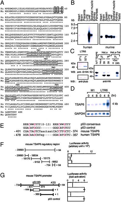
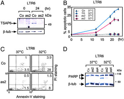
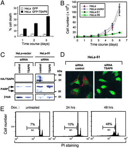
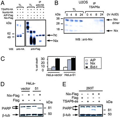
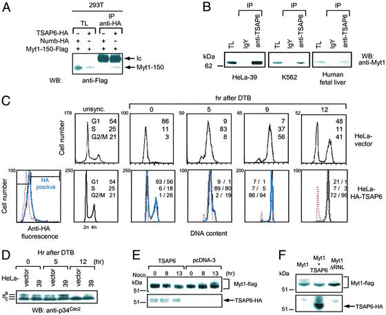
The p53 tumor suppressor protein plays a crucial role in tumorigenesis by controlling cell-cycle progression and apoptosis. We have previously described a transcript designated tumor suppressor activated pathway-6 (TSAP6) that is up-regulated in the p53-inducible cell line, LTR6. Cloning of the murine and human full-length TSAP6 cDNA revealed that it encodes a 488-aa protein with five to six transmembrane domains. This gene is the murine and human homologue of the recently published rat pHyde. Antibodies raised against murine and human TSAP6 recognize a 50- to 55-kDa band induced by p53. Analysis of the TSAP6 promoter identified a functional p53-responsive element. Functional studies demonstrated that TSAP6 antisense cDNA diminished levels of the 50- to 55-kDa protein and decreased significantly the levels of p53-induced apoptosis. Furthermore, TSAP6 small interfering RNA inhibited apoptosis in TSAP6-overexpressing cells. Yeast two-hybrid analysis followed by GST/in vitro-transcribed/translated pull-down assays and in vivo coimmunoprecipitations revealed that TSAP6 associated with Nix, a proapoptotic Bcl-2-related protein and the Myt1 kinase, a negative regulator of the G(2)/M transition. Moreover, TSAP6 enhanced the susceptibility of cells to apoptosis and cooperated with Nix to exacerbate this effect. Cell-cycle studies indicated that TSAP6 could augment Myt1 activity. Overall, these data suggest that TSAP6 may act downstream to p53 to interface apoptosis and cell-cycle progression.


Figures

Figure 1
Figure 1
TSAP6 encodes a putative five- to six-transmembrane protein that is transactivated by p53. (A) Amino acid sequence comparison of human (Hu) and murine (Mu) TSAP6. The five-transmembrane regions (TM1–5) are underlined. The conserved Rossmann-fold motif is boxed in bold. *, amino acid identities between human and mouse TSAP6. (B) Multitissue Northern blots probed with either human (Left) or murine (Right) TSAP6. (C) Anti-murine TSAP6 (S15N) Western blot analysis of extracts from LTR6 cells either before (lane 1) or after (lane 2) temperature shift to 32°C to induce p53 activity. Anti-HA immunoblot detects a doublet of ≈50 and 55 kDa from nonradiolabeled IVT TSAP6-HA, HeLa-39 lysates, and HeLa-TRex cells 24 h after the addition of doxycycline. Anti-TSAP6a immunoprecipitated (IP) the same species (Right, lane 4). (D) Northern blot analysis of TSAP6 expression levels in LTR6 cells compared with its parental p53 null cell line, M1, before and after temperature shift. (E) Sequences of the potential p53 response elements within the mouse and human TSAP6 promoters aligned with the consensus p53-binding site (14) (where R indicates purine, Y indicates pyrimidine, and W indicates A or T). Also shown is the sequence of a consensus p53-binding site (p53 control) used as a positive control in G. (F) The indicated fragments of the mouse TSAP6 promoter were transfected either alone or together with pCMVc50-p53 and tested in luciferase assays. Numbers represent arbitrary units. (G) TSAP6 promoter sequences containing the predicted p53-binding site (p53 BS) are transactivated by p53. Luciferase activity is shown as fold activation. Data represent the mean ± SD (n = 3).
Figure 2
Figure 2
TSAP6 antisense impairs p53-induced apoptosis. LTR6-control (Co) or LTR6-TSAP6 antisense (-as2 and -as4) cells were cultured at 32°C to activate p53. At the indicated time points, cells were harvested and assayed for apoptosis. (A) TSAP6 proteins levels in LTR6-as2 cells are diminished 24 h after p53 activation as detected by immunoblotting with the S15N anti-TSAP6 antibody. β-tub, β-tubulin. (B and C) Apoptosis was measured by flow cytometry by either determining the percentage of nuclei undergoing DNA fragmentation (B) or annexin V staining (C). The percentage of cell death represents the mean of three independent experiments. LTR6 (Co) represents LTR6 cells with or without vector. PI, propidium iodide. (D) Reduced PARP cleavage in LTR6-as2 and -as4 cells after p53 activation. Total lysates were generated and analyzed by immunoblotting with an anti-PARP antibody.
Figure 3
Figure 3
TSAP6 sensitizes cells toward apoptosis. (A) Transient expression of GFP-TSAP6 in HeLa cells promotes apoptosis. Plasmids expressing GFP or GFP-TSAP6 were transfected into HeLa cells. GFP+ cells were scored for apoptosis by monitoring nuclear shrinkage. (B) HeLa and HeLa-vector control cells were analyzed in parallel with HeLa-39 and -51 clones for cell growth. At the indicated time points, cells were harvested and counted. Experiments were performed in triplicate (mean ± SD). (C) HeLa-vector (Left) and HeLa-51 cells (Right) were transfected with either an siRNA control or siRNA TSAP6. Immunoblot analysis with an anti-HA antibody confirmed the efficient depletion of overexpressed TSAP6 (Top Right), which resulted in reduced PARP cleavage (Middle Right). β-tub, β-tubulin. (D) Immunofluorescence analysis using an anti-HA antibody (red) verified the depletion of overexpressed TSAP6 by siRNA. (E) Induced expression of TSAP6 promotes apoptosis. Flow cytometry was performed on propidium iodide (PI)-stained nuclei derived from HeLa-Trex-HA-TSAP6 cells before or at the indicated times after the addition of doxycycline (Dox.).
Figure 4
Figure 4
Interaction and functional analysis of TSAP6 and Nix. (A) HA-TSAP6 and Flag-Nix interact in 293T cells. 293T cells were transiently cotransfected with the indicated expression plasmids. Nix expression levels in total cell lysates (TL) and anti-HA immunoprecipitates (IP) were determined by immunoblotting with anti-Flag antibody. hc, Ig heavy chains; WB, Western blot. (B) Endogenous TSAP6 and Nix interact in U2OS cells under conditions that promote apoptosis. Cell lysates derived from U2OS cells treated with adriamycin were incubated with an anti-TSAP6 antibody coupled to IgY-agarose followed by immunoblotting with an anti-Nix antibody. The Nix dimer is detected as a 38-kDa protein. ActD, actinomycin D. (C) HeLa-51 cells overexpressing Nix are more susceptible to apoptosis. HeLa-vector or HeLa-51 cells were transiently transfected with expression plasmids encoding Flag-tagged proteins containing the AIP negative control, Nix, or COOH-terminal portion of Bid (Bid-t). Flag-positive cells were scored for apoptosis based on nuclear shrinkage. Experiments were performed in triplicate (mean ± SD). (D) Enhanced PARP cleavage in HeLa-51 cells overexpressing Nix as determined by immunoblot analysis with an anti-PARP antibody. (E) TSAP6 antisense impairs Nix-induced cell death. 293T cells were cotransfected with the indicated expression plasmids. Twenty-four hours after transfection, cell lysates were analyzed for PARP cleavage.
Figure 5
Figure 5
TSAP6 interacts with Myt1 and influences cell-cycle progression. (A) Overexpressed HA-TSAP6 immunoprecipitates (IP) Flag-Myt1-150 from extracts of 293T cells. WB, Western blot; Ic, Ig light chain. (B) Endogenous TSAP6 and Myt1 interact in HeLa-39 cells (Left), K562 cells (Center), or human fetal liver tissue (Right). Lysates were incubated with either a control antibody or anti-TSAP6a antibody followed by incubation with agarose anti-IgY and Western blot analysis with anti-Myt1 antibody. TL, total cell lysate. (C) Overproduction of TSAP6 induces a G2/M delay. Polyclonal HeLa-vector and HeLa HA-TSAP6 cells were synchronized at the G1/S border by a DTB procedure and analyzed by flow cytometry after release from the block. Flow-cytometry analysis was performed separately on either gated HA-positive (solid blue line) or HA-negative (dotted red line) subpopulations (Left). (D) TSAP6 maintains p34cdc2 in an inactive state. Cell lysates derived from either synchronized HeLa vector or HeLa-39 cells were immunoblotted by using an anti-p34Cdc2 antibody. (E) TSAP6 overexpression prevents Myt1 hyperphosphorylation during mitotic entry. 293T cells overexpressing pcDNA3 or pcDNA-TSAP6 in combination with Flag-Myt1 were treated with nocodazole (Noco) for the indicated times. Western blot analysis with anti-Flag (Upper) or anti-HA (Lower) antibodies was used to reveal Myt1 and TSAP6 proteins, respectively, in total cell lysates. (F) The effect of TSAP6 on Myt1 phosphorylation is similar to that of the Myt1ΔRNL mutant alone.

Similar articles

Cited by

References

    1. Amson R B, Nemani M, Roperch J-P, Israeli D, Bougueleret L, Le Gall I, Medhioub M, Linares-Cruz G, Lethrosne F, Pasturaud P, et al. Proc Natl Acad Sci USA. 1996;93:3953–3957. - PMC - PubMed
    1. Yonish-Rouach E, Resnitzky D, Lotem J, Sachs L, Kimchi A, Oren M. Nature. 1991;352:345–347. - PubMed
    1. Nemani M, Linares-Cruz G, Bruzzoni-Giovanelli H, Roperch J-P, Tuynder M, Bougueleret L, Cherif D, Medhioub M, Pasturaud P, Alvaro V, et al. Proc Natl Acad Sci USA. 1996;93:9039–9042. - PMC - PubMed
    1. Hu G, Chung Y L, Glover T, Valentine V, Look A T, Fearon E R. Genomics. 1997;46:103–111. - PubMed
    1. Sherrington R, Rogaev E I, Liang Y, Rogaeva E A, Levesque G, Ikeda M, Chi H, Lin C, Li G, Holman K, et al. Nature. 1995;375:754–760. - PubMed

MeSH terms

Substances

Associated data