. 2003 Mar 4;100(5):2397-402.
doi: 10.1073/pnas.0437997100. Epub 2003 Feb 26.

Differentiation, cell fusion, and nuclear fusion during ex vivo repair of epithelium by human adult stem cells from bone marrow stroma

Affiliations

Differentiation, cell fusion, and nuclear fusion during ex vivo repair of epithelium by human adult stem cells from bone marrow stroma

Jeffrey L Spees et al. Proc Natl Acad Sci U S A. .

Abstract

To investigate stem cell differentiation in response to tissue injury, human mesenchymal stem cells (hMSCs) were cocultured with heat-shocked small airway epithelial cells. A subset of the hMSCs rapidly differentiated into epithelium-like cells, and they restored the epithelial monolayer. Immunocytochemistry and microarray analyses demonstrated that the cells expressed many genes characteristic of normal small airway epithelial cells. Some hMSCs differentiated directly after incorporation into the epithelial monolayer but other hMSCs fused with epithelial cells. Surprisingly, cell fusion was a frequent rather than rare event, in that up to 1% of the hMSCs added to the coculture system were recovered as binucleated cells expressing an epithelial surface epitope. Some of the fused cells also underwent nuclear fusion.


Figures

Figure 1
Figure 1
Phase-contrast and UV microscopy of cultures and cocultures. (A) SAECs in SAEC medium. (B) GFP+ hMSCs grown in complete MSC medium (FITC overlay on phase). (C and D) GFP+ hMSCs cultured in serum-free medium for SAECs. (E and F) Coculture experiment with heat-shocked bronchial epithelial cells at 2 wk. The differentiated GFP+ cell has an epithelial morphology and has repaired the monolayer formed by bronchial epithelium. The cell is binucleated (yellow arrow), as is a GFP bronchial cell above it (arrowhead). (GL) Cocultures of GFP+ hMSCs and SAECs after incubation for 12–120 h. (G and H) GFP+ cell between SAECs undergoing morphological changes (arrow). (I and J) Differentiated GFP+ cell has an epithelial morphology, has repaired the monolayer formed by the small airway epithelium, and has a single nucleus (arrow). Adjacent SAEC is binucleated (arrowhead). (K and L) Differentiated GFP+ cell has three nuclei (yellow arrow). (E, F, K, and L) The outermost cytoplasmic edges of the GFP+ cells are artificially enhanced. (Magnification: AD, ×10; E and F, ×40; GJ, ×20; K and L, ×40.)
Figure 2
Figure 2
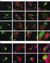
Immunocytochemistry of GFP+ hMSC and SAEC cocultures. (A) Differentiated GFP+ cells express keratins 17, 18, 19, and CC26 (clara cells). (B) Markers of adherens junctions; E-cadherin and β-catenin. First column (left to right) UV with FITC filter. Second column, UV with TRITC filter. Third column, merged images with 4′,6-diamidino-2-phenylindole nuclear staining. Fourth column, enlarged merged image. Arrows, double positive cells. *, Undifferentiated GFP+ hMSCs (note single nuclei). (Magnification: ×40 for first three columns; ×100 for fourth column.)
Figure 3
Figure 3
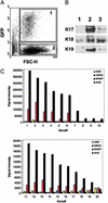
FACS isolation of cells from the cocultures. (A) FACS for GFP and forward scatter of light (FSC-H); GFP+ cells (gate 1), SAECs (gate 2). (B) Immunoblots for keratins 17, 18, and 19 for GFP+ hMSCs before coculture (lane 1), SAECs (lane 2), and GFP+ cells isolated by FACS after coculture with damaged SAECs (lane 3; cells isolated with gate 1 from A). (C) Signal intensities of selected epithelial genes of GFP+ cells from cocultures assayed by microarrays. hMSC, GFP+ hMSCs incubated in complete MSC medium (20% serum). hMSCM, GFP+ hMSCs incubated in SAEC medium (serum-free). EPI/DIFF, GFP+ cells isolated from the cocultures by FACS. Differentiated GFP+ cells (EPI/DIFF) express many of the genes expressed by normal SAECs: lane 1, Stratifin (GenBank accession no. X57348); lane 2, keratin 17 (GenBank accession no. Z19574); lane 3, keratin 6 (GenBank accession no. L42611); lane 4, keratin type II (GenBank accession no. M21389); lane 5, keratin 19 (GenBank accession no. Y00503); lane 6, CAN19 (GenBank accession no. M87068); lane 7, keratin 16 (GenBank accession no. 28439); lane 8, Maspin (GenBank accession no. U04313); lane 9, CD24 (GenBank accession no. L33930); lane 10, Claudin-7 (GenBank accession no. AJ011497); lane 11, cornified envelope precursor (GenBank accession no. AF001691); lane 12, Laminin S B3 chain (GenBank accession no. U17760); lane 13, integrin β 4 (GenBank accession no. X53587); lane 14, E-cadherin (GenBank accession no. Z35402); lane 15, Laminin-related protein (GenBank accession no. L34155); lane 16, lung amelioride sensitive Na-channel protein (GenBank accession no. X76180); lane 17, P-cadherin (GenBank accession no. X63629); lane 18, Laminin γ-2 chain precursor (GenBank accession no. Z15008); lane 19, NES-1 (GenBank accession no. AF055481); lane 20, Mucin 1 (GenBank accession no. X80761).
Figure 4
Figure 4
Time-lapse microscopy of cell fusion in GFP+ hMSC and heat-shocked SAEC cocultures. Three separate fusion events are shown. The same fields were photographed every 20 min by both UV and differential-contrast microscopy. UV photographs are overlain on differential contrast. Selected frames are shown. Note the rapid influx of GFP into target SAECs between the first two frames of each sequence. (E, J, and O) Enlarged images of adjacent frames. White and yellow arrows, GFP+ hMSCs. White and yellow arrowheads, targeted SAECs. Red arrows, multiple nuclei in fused cells. (Magnifications: A–D, F–I, and K–N, ×10; E, J, and O, ×90.)
Figure 5
Figure 5
(A) Sorting of GFP+/CD24+ cells from cocultures (gate 1). (BD) Deconvolution microscopy of single cells that were GFP+/CD24+. Three-dimensional images at 1.0-μm intervals through each cell were analyzed to eliminate overlapping cells. The cells were nuclear-stained with 4′,6-diamidino-2-phenylindole. Note multinucleated cells (arrows), cells with single nuclei, and cells with irregular nuclei (arrowheads). Pseudocoloring is reversed in D to better visualize cell nuclei from C. (Magnification: ×40.)
Figure 6
Figure 6
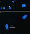
Fluorescent in situ hybridization of GFP+/CD24+ cells isolated from cocultures of male GFP+ hMSCs and female SAECs. Y chromosome, green signal, FITC filter; X chromosome(s), red signal, TRITC filter; 4′,6-diamidino-2-phenylindole nuclear staining, blue, UV filter. (A) Control male normal human bronchial epithelial cells (arrow, Y chromosome; arrowhead, X chromosome). (B) Hybrid cell nucleus derived from fusion of one male GFP+ hMSC nucleus with two female SAEC-derived nuclei (one green signal, five red signals). (C) Hybrid cell nucleus generated from fusion of one male GFP+ hMSC nucleus with one female SAEC nucleus (one green signal, three red signals). (Inset) TRITC filter image from hybrid cell. Upper right, single cell-differentiation (GFP+/CD24+, one green signal, one red signal). (Magnifications: A, ×63; B and C, ×100.)

References

    1. Davidson J M. In: Inflammation: Basic Principles and Clinical Correlates. 2nd Ed. Gallin J I, Goldstein I M, Synderman R, editors. New York: Raven; 1992. p. 809.
    1. Gage F H. Science. 2000;287:1433–1438. - PubMed
    1. Alison M R, Poulsom R, Forbes S J. Liver. 2001;21:367–373. - PubMed
    1. Doherty M J, Ashton B A, Walsh S, Beresford J N, Grant M E, Canfield A E. J Bone Miner Res. 1998;13:828–838. - PubMed
    1. Kuznetsov S A, Mankani M H, Gronthos S, Satomura K, Bianco P, Robey P G. J Cell Biol. 2001;153:1133–1139. - PMC - PubMed

Publication types

MeSH terms