. 2003 Mar;84(3):1756-64.
doi: 10.1016/s0006-3495(03)74983-2.

The lateral diffusion of selectively aggregated peptides in giant unilamellar vesicles

Affiliations

The lateral diffusion of selectively aggregated peptides in giant unilamellar vesicles

Clarence C Lee et al. Biophys J. 2003 Mar.

Erratum in

  • Biophys J. 2003 Aug;85(2):1338

Abstract

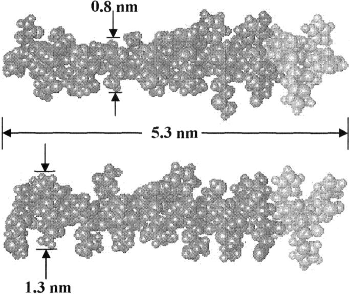
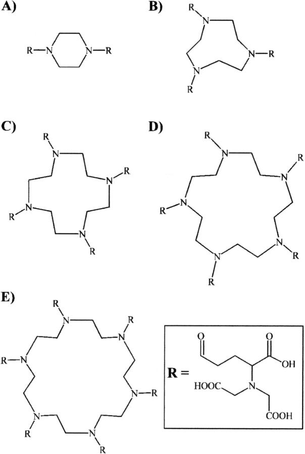
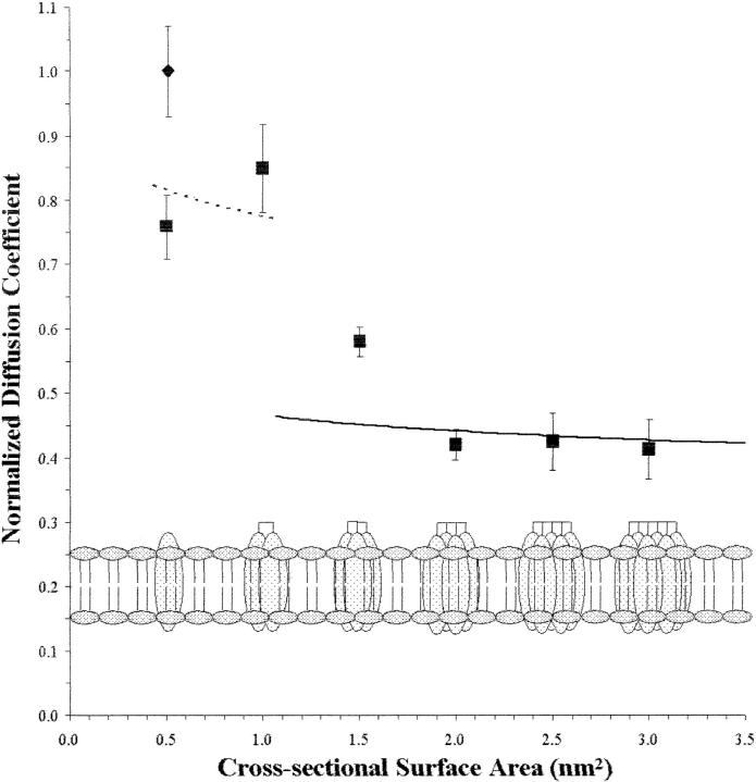
We have systematically investigated the effect of aggregation of a transmembrane peptide on its diffusion in dimyristoylphosphatidylcholine and in palmitoyloleoylphosphatidylcholine model membranes. The hydrophobic segment of the b subunit from E. coli F(1)F(0)-ATP synthase was modified with a histidine tag at the carbonyl terminus and was aggregated selectively by using a series of multivalent, dendritic chelating agents with nitrilotriacetic acid functional groups. Peptide complexes ranging from monomers to hexamers were formed and studied in giant unilamellar vesicles. The rate of diffusion for the transmembrane peptide complexes were found to depend on the size of the complex. The results agree with predictions from the free area model for monomers and dimers, and the hydrodynamic continuum model for tetramers, pentamers, and hexamers. Comparisons with diffusion of lipids confirm that the diffusion of a transmembrane peptide is enhanced by coupling of density fluctuations between the two monolayers.


Figures

FIGURE 1
FIGURE 1
Computer model of Htm-b at two different rotations. The images show Htm-b with the histidine amino acids at the carbonyl terminus (light gray). The simulations show that the length of the peptide is 5.3 nm. The variability of the peptide diameter along the hydrophobic segment of the b subunit is evident. The average cross-sectional area in the hydrophobic region is estimated at 0.5 nm2.
FIGURE 2
FIGURE 2
Images of a mixed monolayer system containing 90:1:9 DPPC/RRX-b/NBD-PC measured through a rhodamine filter set (A–E) and FITC filter set (F) at various surface pressures. Each image is 481 μm × 642 μm. Images of the Langmuir film were recorded through the rhodamine filter set at 10 mN/m (A); 15 mN/m (B); 20 mN/m (C); 25 mN/m (D); and 30 mN/m (E). As surface pressure increases, the contrast between the brighter LE region and the darker lipid LC domains is reduced. Examination of the same film using the FITC filter set at 30 mN/m (F) reveals the continued presence of LC domains, suggesting that the contrast reduction seen from images A–E arises because RRX-b is able to partition into the liquid condensed regions at higher pressures.
FIGURE 3
FIGURE 3
Confocal image showing the incorporation of a derivatized peptide, RRX-b, into POPC GUVs at 20°C. The GUVs were created using the electroformation method. After formation, the vesicles remain attached to the platinum wire, which is located at the bottom left corner of the image. The image size is 69 μm × 69 μm and the large GUV in the center is ∼40 μm in diameter.
FIGURE 4
FIGURE 4
Molecular structures of the dendritic chelating agents (Liu et al., 2002): dimer agent (A); trimer agent (B); tetramer agent (C); pentamer agent (D); and hexamer agent (E). Note that each branch originates at a nitrogen in the ring and terminates with a nitrilotriacetic acid group used to complex with nickel to the histidine tag. The valency of the complexing agent therefore is governed by the number of nitrogens in the ring.
FIGURE 5
FIGURE 5
Normalized diffusion coefficient plotted as a function of the cross-sectional surface area for Htm-b and the peptide aggregates in DMPC bilayers at 35°C. Diffusion coefficients for the transmembrane diffusants (▪) are normalized with the diffusion coefficient for NBD-PE (♦). The schematic representation of the state of aggregation of the transmembrane diffusants in the lipid bilayer is shown for each data point at the bottom of the graph. The error bars represent the mean ± SE at 99% confidence level. Theoretical calculations are shown for the free area theory (solid line) and the Saffman-Delbrück hydrodynamic model (dashed line). The calculations were performed with the following fixed parameters: γa*/a0 = 0.4, β = 0.148, αa = 2.3 × 10⁻³ K−1, Tm = 23.9°C, h = 3.0 nm, μw = 0.00728 Poise, μm = 1.75 Poise.
FIGURE 6
FIGURE 6
Comparison of mean diffusion coefficients for monolayer diffusants (polyamide amphiphiles) (black bar) and bilayer-spanning diffusants (peptide aggregates) (light gray bar) in DMPC at 35°C, with the corresponding values predicted by the triple layer model (cross-hatched bar). The data are averages normalized to the diffusion rate for NBD-PE under the same conditions. (A) corresponds to monomers and dimers reflecting the free area theory and (B) corresponds to the larger molecules or complexes reflecting the continuum theory. In both cases, the predicted diffusion is slower than the observed diffusion, suggesting some interaction between the two monolayers.

Similar articles

Cited by

References

    1. Adam, G., and M. Delbrück. 1968. Reduction of dimensionality in biological diffusion processes. In Structural Chemistry and Molecular Biology. A. Rich and N. Davidson, editors. W. H. Freeman and Company, San Francisco. 198–215.
    1. Angelova, M. I., and D. S. Dimitrov. 1986. Liposome electroformation. Faraday Discuss. Chem. Soc. 81:303–311.
    1. Angelova, M. I., S. Soléau, Ph. Meléard, J. F. Faucon, and P. Bothorel. 1992. Preparation of giant vesicles by external AC fields: kinetics and application. Prog. Colloid Polym. Sci. 89:127–131.
    1. Axelrod, D. 1985. Fluorescence photobleaching techniques and lateral diffusion. In Spectroscopy and the Dynamics of Molecular Biological Systems. P. Bayley and R. Dale, editors. Academic Press, London. 163–176.
    1. Axelrod, D., D. E. Koppel, J. Schlessinger, E. Elson, and W. W. Webb. 1976. Mobility measurement by analysis of fluorescence photobleaching recovery kinetics. Biophys. J. 16:1055–1069. - PMC - PubMed

Publication types

MeSH terms

LinkOut - more resources