

Thermodynamic properties of the kinesin neck-region docking to the catalytic core
- PMID: 12609886
- PMCID: PMC1302753
- DOI: 10.1016/S0006-3495(03)74992-3
Thermodynamic properties of the kinesin neck-region docking to the catalytic core
Abstract
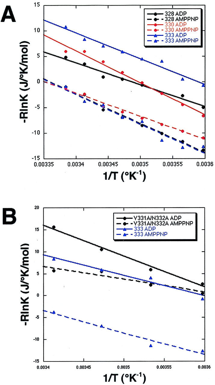
Kinesin motors move on microtubules by a mechanism that involves a large, ATP-triggered conformational change in which a mechanical element called the neck linker docks onto the catalytic core, making contacts with the core throughout its length. Here, we investigate the thermodynamic properties of this conformational change using electron paramagnetic resonance (EPR) spectroscopy. We placed spin probes at several locations on the human kinesin neck linker and recorded EPR spectra in the presence of microtubules and either 5'-adenylylimidodiphosphate (AMPPNP) or ADP at temperatures of 4-30 degrees C. The free-energy change (DeltaG) associated with AMPPNP-induced docking of the neck linker onto the catalytic core is favorable but small, about 3 kJ/mol. In contrast, the favorable enthalpy change (DeltaH) and unfavorable entropy change (TDeltaS) are quite large, about 50 kJ/mol. A mutation in the neck linker, V331A/N332A, results in an unfavorable DeltaG for AMPPNP-induced zipping of the neck linker onto the core and causes motility defects. These results suggest that the kinesin neck linker folds onto the core from a more unstructured state, thereby paying a large entropic cost and gaining a large amount of enthalpy.
Figures


Similar articles
-
EPR spectroscopy shows a microtubule-dependent conformational change in the kinesin switch 1 domain.Biophys J. 2003 May;84(5):3190-6. doi: 10.1016/S0006-3495(03)70043-5. Biophys J. 2003. PMID: 12719248 Free PMC article.
-
Two conformations in the human kinesin power stroke defined by X-ray crystallography and EPR spectroscopy.Nat Struct Biol. 2002 Nov;9(11):844-8. doi: 10.1038/nsb852. Nat Struct Biol. 2002. PMID: 12368902
-
Role of the kinesin neck linker and catalytic core in microtubule-based motility.Curr Biol. 2000 Feb 10;10(3):157-60. doi: 10.1016/s0960-9822(00)00316-x. Curr Biol. 2000. PMID: 10679326
-
Searching for kinesin's mechanical amplifier.Philos Trans R Soc Lond B Biol Sci. 2000 Apr 29;355(1396):449-57. doi: 10.1098/rstb.2000.0586. Philos Trans R Soc Lond B Biol Sci. 2000. PMID: 10836498 Free PMC article. Review.
-
Kinesin: a molecular motor with a spring in its step.Proc Biol Sci. 2002 Nov 22;269(1507):2363-71. doi: 10.1098/rspb.2002.2117. Proc Biol Sci. 2002. PMID: 12495505 Free PMC article. Review.
Cited by
-
Kinesin's second step.Proc Natl Acad Sci U S A. 2004 Mar 9;101(10):3444-9. doi: 10.1073/pnas.0307691101. Epub 2004 Feb 25. Proc Natl Acad Sci U S A. 2004. PMID: 14985504 Free PMC article.
-
Kinesin is an evolutionarily fine-tuned molecular ratchet-and-pawl device of decisively locked direction.Biophys J. 2007 Nov 15;93(10):3363-72. doi: 10.1529/biophysj.107.108233. Epub 2007 Aug 3. Biophys J. 2007. PMID: 17675343 Free PMC article.
-
A seesaw model for intermolecular gating in the kinesin motor protein.Biophys Rev. 2011 Jun;3(2):85-100. doi: 10.1007/s12551-011-0049-4. Epub 2011 Jun 4. Biophys Rev. 2011. PMID: 21765878 Free PMC article.
-
Myosin and Other Energy-Transducing ATPases: Structural Dynamics Studied by Electron Paramagnetic Resonance.Int J Mol Sci. 2020 Jan 20;21(2):672. doi: 10.3390/ijms21020672. Int J Mol Sci. 2020. PMID: 31968570 Free PMC article. Review.
-
Mechanical design of translocating motor proteins.Cell Biochem Biophys. 2009;54(1-3):11-22. doi: 10.1007/s12013-009-9049-4. Epub 2009 May 19. Cell Biochem Biophys. 2009. PMID: 19452133 Free PMC article. Review.
References
-
- Bustamante, C., J. F. Marko, E. D. Siggia, and S. Smith. 1994. Entropic elasticity of lambda-phage DNA. Science 265:1599–1600. - PubMed
-
- Case, R. B., D. W. Pierce, N. Hom-Booher, C. L. Hart, and R. D. Vale. 1997. The directional preference of kinesin motors is specified by an element outside of the motor catalytic domain. Cell 90:959–966. - PubMed
-
- Case, R. B., S. Rice, C. L. Hart, B. Ly, and R. D. Vale. 2000. Role of the kinesin neck linker and catalytic core in microtubule-based motility. Curr. Biol. 10:157–160. - PubMed
-
- Coy, D., M. Wagenbach, and J. Howard. 1999. Kinesin takes one 8-nm step for each ATP it hydrolyzes. J. Biol. Chem. 274:3667–3671. - PubMed
Publication types
MeSH terms
Substances
LinkOut - more resources
Full Text Sources
