

doi: 10.1016/S0006-3495(03)75010-3.
Rietveld refinement on x-ray diffraction patterns of bioapatite in human fetal bones
Affiliations
- PMID: 12609904
- PMCID: PMC1302771
- DOI: 10.1016/S0006-3495(03)75010-3
Item in Clipboard
Rietveld refinement on x-ray diffraction patterns of bioapatite in human fetal bones
Biophys J.
2003 Mar.
Abstract
Bioapatite, the main constituent of mineralized tissue in mammalian bones, is a calcium-phosphate-based mineral that is similar in structure and composition to hydroxyapatite. In this work, the crystallographic structure of bioapatite in human fetuses was investigated by synchrotron radiation x-ray diffraction (XRD) and microdiffraction ( micro -XRD) techniques. Rietveld refinement analyses of XRD and micro -XRD data allow for quantitative probing of the structural modifications of bioapatite as functions of the mineralization process and gestational age.
Figures
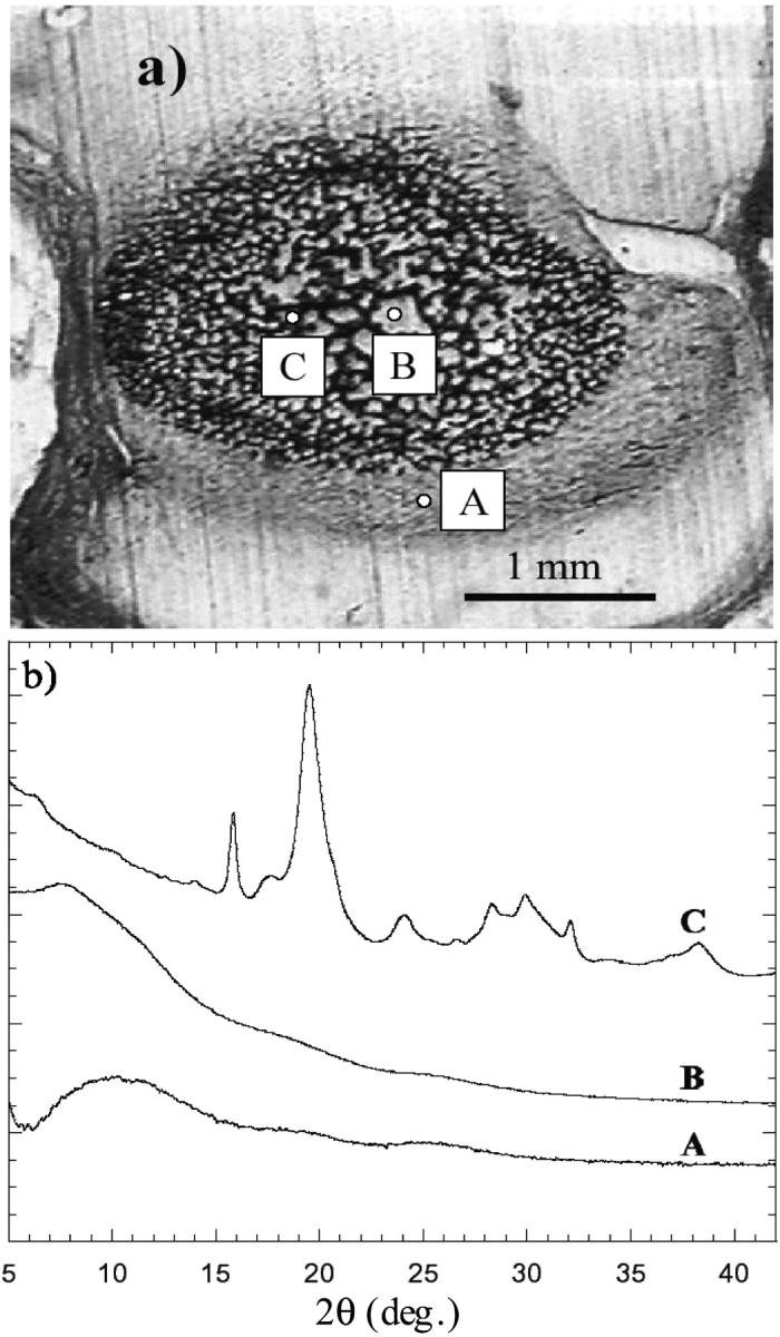
(a) Microscopic image of a thin vertebra slice excised from a 26-week-old fetus. (b) μ-XRD patterns collected from the surrounding region (A), from a clear internal region (B), and from a dark trabecula (C).
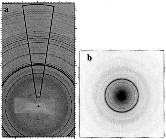
(a) Typical 2-D XRD image collected from synthetic OHA at GILDA; the area used to convert the 2-D image into the equivalent 1-D pattern is shown. (a) Typical 2-D μ-XRD image of bone bioapatite measured at ID13.
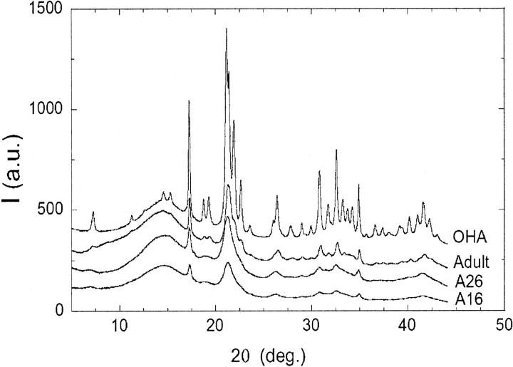
Experimental XRD pattern intensities (I, in arbitrary units, a.u.) relative to synthetic OHA, adult bone sample, and fetal bone samples 26 (A26) and 16 (A16) weeks old. Patterns are vertically shifted for the sake of clarity.
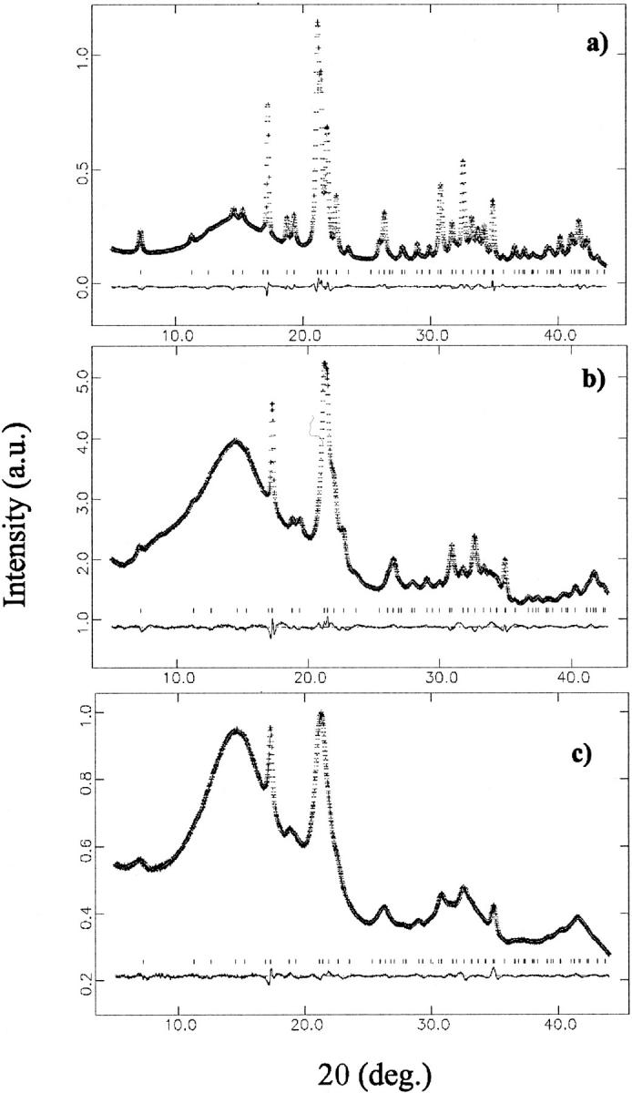
Rietveld refinement for selected samples: (a) synthetic OHA; (b) bioapatite in the adult bone sample; and (c) bioapatite in fetal bone sample (16 weeks old). Each panel reports the observed and refined data, almost indistinguishable (top trace); their difference (bottom trace), shifted for clarity; and the calculated reflection positions (middle points).
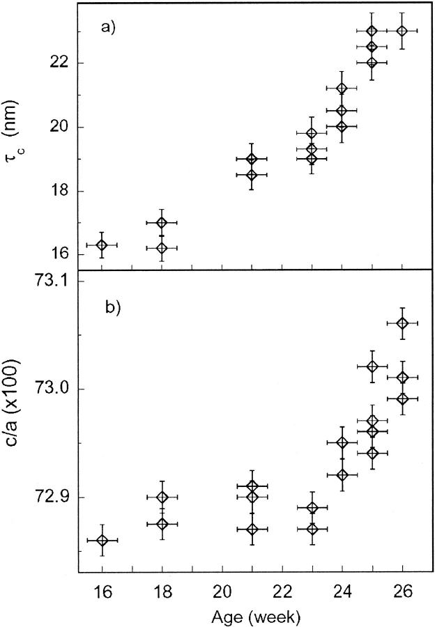
Evolution of the bioapatite crystallite size (a) along the c axis and (b) of the c/a ratio, as a function of gestational age.
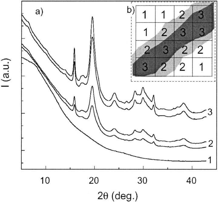
(a) μ-XRD patterns relative to different selected areas of the bone sample. (b) Schematic of the scanning procedure: diffractograms labeled 1 are collected from the intertrabecular region, those labeled 2 are collected from the trabecular surface, and those labeled 3 are collected from the inner region of the trabecula.
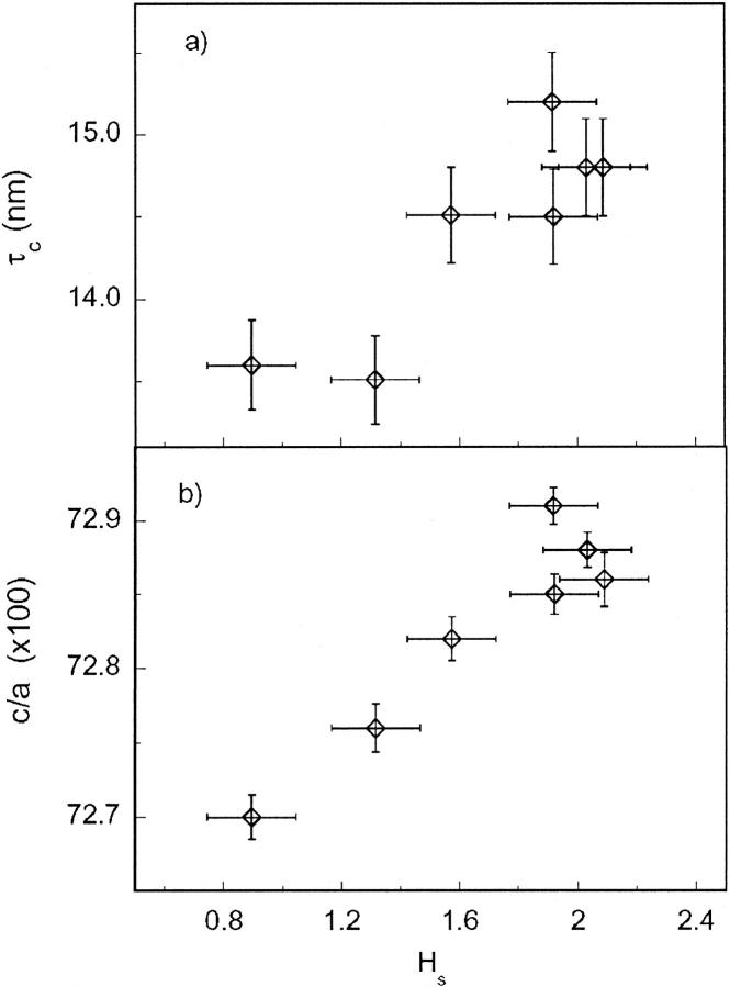
Evolution of bioapatite crystallite size (a) along the c axis and (b) of the c/a ratio, as a function of histogram scale factor (Hs) refined by GSAS (see text for details).
References
-
- Albright, J. A. and H. C. W. Skinner. 1987. Bone: structural organization and remodeling dynamics. In The Scientific Basis of Orthopaedics. J. A. Albright and R. Brand, editors. Appleton and Lange, Norwalk, CT. 161–198.
-
- Bareggi, R., V. Grill, M. A. Sandreucci, G. Baldini, A. De Pol, A. Forabosco, and P. Narducci. 1993. Development pathways of vertebral centra and neural arches in human embryos and fetuses. Anat. Embryol. (Berl.) 187:139–144. - PubMed
-
- Berry, E. E. 1967. The structure and composition of some calcium-deficient apatites. J. Inorg. Nucl. Chem. 29:317–327.
-
- Bigi, A., G. Cojazzi, S. Panzavolta, A. Ripamonti, N. Roveri, M. Romanello, K. Noris Suarez, and L. Moro. 1997. Chemical and structural characterization of the mineral phase from cortical and trabecular bone. J. Inorg. Biochem. 68:45–51. - PubMed
MeSH terms
Substances
LinkOut - more resources
Full Text Sources
