. 2003 Mar;56(3):194-9.
doi: 10.1136/jcp.56.3.194.

Increased Nox2 expression in human cardiomyocytes after acute myocardial infarction

Affiliations

Increased Nox2 expression in human cardiomyocytes after acute myocardial infarction

P A J Krijnen et al. J Clin Pathol. 2003 Mar.

Abstract

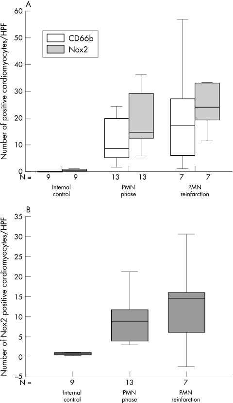
Background/aims: Recent studies indicate the presence of reactive oxygen species (ROS) producing homologues of the enzymatic subunit (Nox2) of phagocytic NADPH oxidase in non-phagocytic cells. Interestingly, in these cells, ROS produced by the Nox2 homologue(s) was shown to play a role in various regulatory processes, including cell death, proliferation, and aging. The purpose of this study was to investigate whether human cardiomyocytes express Nox2.

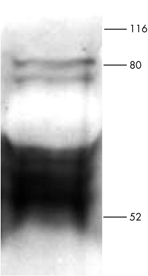
Methods: The expression of Nox2 was studied in human cardiomyocytes using western blot and immunohistochemical analysis. To analyse the putative expression of Nox2 in human heart disease, cardiac samples from patients who had died subsequent to acute myocardial infarction (AMI) were studied.

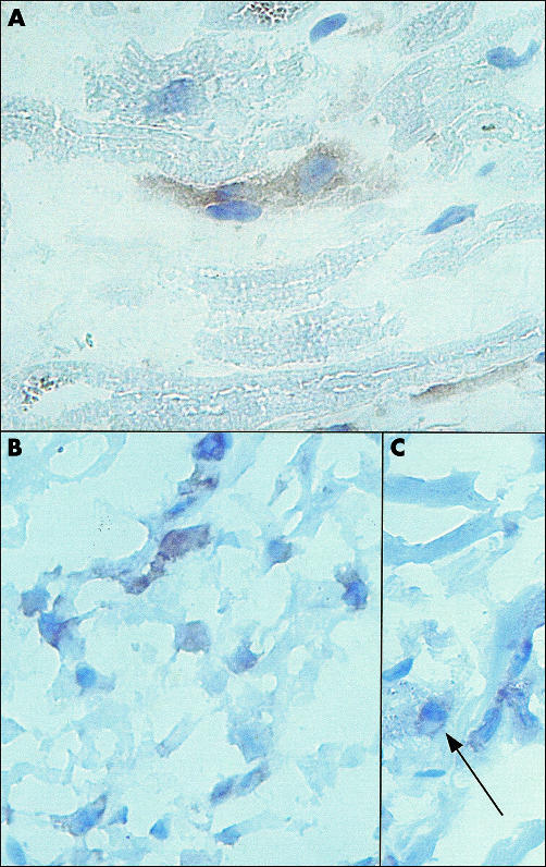
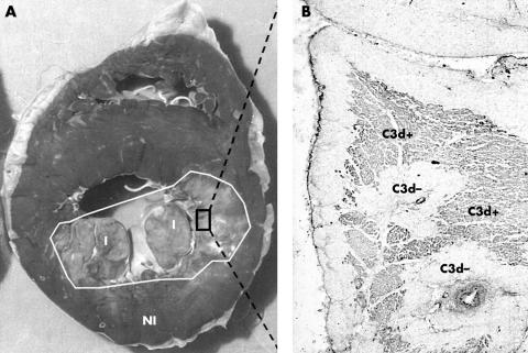
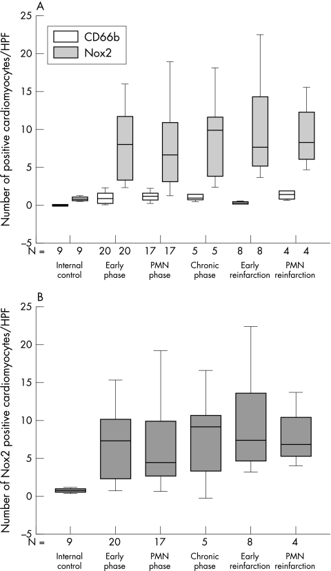
Results: Both in western blot and immunohistochemical studies, Nox2 expression was found in normal human cardiomyocytes. In patients with AMI, a significant increase in Nox2 expression was found both in viable and in jeopardised cardiomyocytes in the infarcted area. In addition, in the "remote from infarction" area, Nox2 expression was present in cardiomyocytes, but was not increased.

Conclusions: Nox2 or its homologue(s) is expressed in normal and jeopardised human cardiomyocytes. This expression is increased in patients with AMI, suggesting a role for this ROS producing Nox2 homologue(s) in the human heart after AMI.


Figures

Figure 1
Figure 1
Western blot analysis of Nox2 expression by isolated human cardiomyocytes. Isolated human cardiomyocytes were subjected to sodium dodecyl sulfate polyacrylamide gel electrophoresis. Proteins were transferred on to nitrocellulose sheets. Nox2 was visualised with monoclonal antibody 48 (1/250 dilution).
Figure 2
Figure 2
Immunohistochemical analysis of human heart tissue. (A) Localisation of Nox2 in the heart of a patient who had died in the absence of heart disease (original magnification, ×630). (B, C) Localisation of Nox2 in a myocardial infarct area in a patient who had died after acute myocardial infarction (original magnification, ×400). Slides were stained with monoclonal antibody 48 (1/150 dilution). (C) The arrow indicates the perinuclear localisation of Nox2.
Figure 3
Figure 3
Example of (A) an infarcted human heart and (B) complement deposition in the infarction area. (A) Cross section of the heart of a patient who had died from acute myocardial infarction. Diminished lactate dehydrogenase (LD) staining is indicative of infarction (I), normal LD content is indicative of unaffected myocardium (NI). (B) Microscopical analysis of complement deposition (complement factor C3d) in the macroscopically determined infarcted myocardium.
Figure 4
Figure 4
Summary of Nox2 expression in relation to the presence of CD66b in complement negative areas within the macroscopical infarction area. Box plots in which the error bars represent minimum and maximum values and in which the boxes represent the lower and upper quartiles. The black lines within the boxes represent the medians. N is the number of patients examined. For each patient the average number of positive cardiomyocytes in each high power field (HPF) was determined from 25 HPFs scored. (A) Box plot of the number of Nox2 (shaded bars) and CD66b (open bars) positive cardiomyocytes in each HPF in the different phases of infarction. (B) Box plot of the number of Nox2 positive cardiomyocytes in each HPF after subtraction of CD66b positive cardiomyocytes. PMN, polymorphonuclear leucocytes.
Figure 5
Figure 5
Statistical analysis of Nox2 and CD66b in complement positive areas within the macroscopical infarction area. Box plots in which the error bars represent minimum and maximum values and in which the boxes represent the lower and upper quartiles. The black lines within the boxes represent the medians. N is the number of patients examined. For each patient the average number of positive cardiomyocytes in each high power field (HPF) was determined from 25 HPFs scored. (A) Box plot of the number of Nox2 (shaded bars) and CD66b (open bars) positive cardiomyocytes in each HPF in the different phases of infarction. (B) Number of Nox2 positive cardiomyocytes in each HPF after subtraction of CD66b positive cardiomyocytes. PMN, polymorphonuclear leucocytes.

References

    1. Duranteau J, Chandel NS, Kulisz A, et al. Intracellular signaling by reactive oxygen species during hypoxia in cardiomyocytes. J Biol Chem 1998;273:11619–24. - PubMed
    1. Rao GN, Berk BC. Active oxygen species stimulate vascular smooth muscle cell growth and proto-oncogene expression. Circ Res 1992;70:593–9. - PubMed
    1. Abe J, Berk BC. Reactive oxygen species as mediators of signal transduction in cardiovascular disease. Trends Cardiovasc Med 2002;8:59–64. - PubMed
    1. Lander HM. An essential role for free radicals and derived species in signal transduction. FASEB J 1997;11:118–24. - PubMed
    1. Katusic ZS. Superoxide anion and endothelial regulation of arterial tone. Free Radic Biol Med 1996;20:443–8. - PubMed

Publication types

MeSH terms