. 2003 Mar;77(6):3516-30.
doi: 10.1128/jvi.77.6.3516-3530.2003.

The stable 2.0-kilobase intron of the herpes simplex virus type 1 latency-associated transcript does not function as an antisense repressor of ICP0 in nonneuronal cells

Affiliations

The stable 2.0-kilobase intron of the herpes simplex virus type 1 latency-associated transcript does not function as an antisense repressor of ICP0 in nonneuronal cells

Edward A Burton et al. J Virol. 2003 Mar.

Abstract

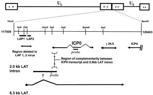
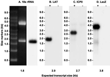
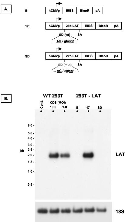
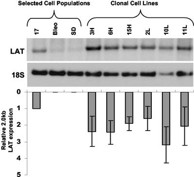
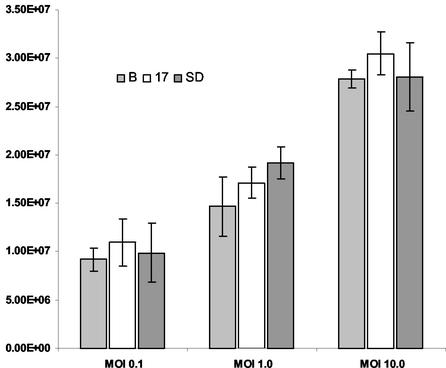
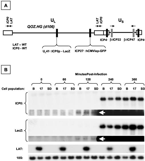
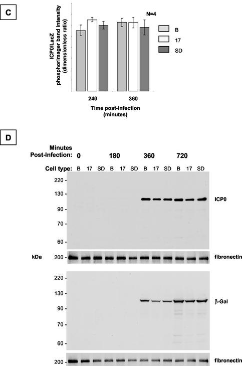
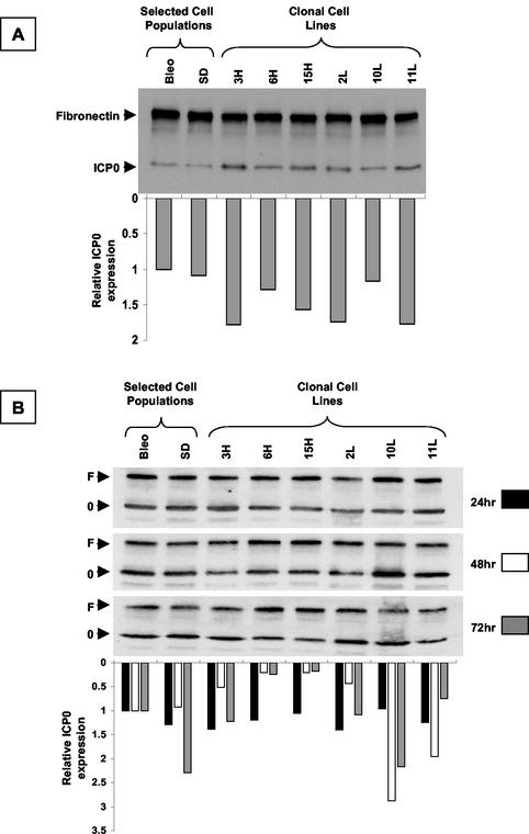
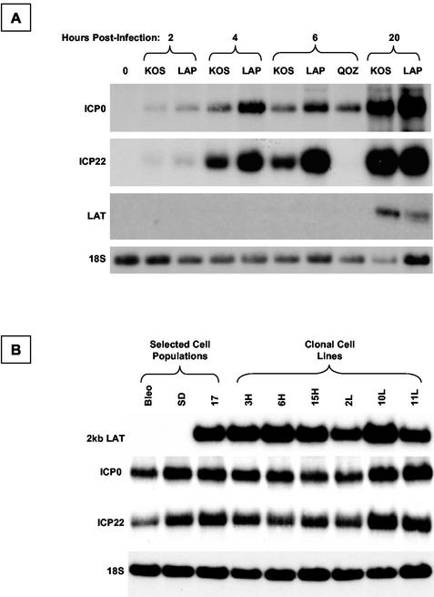
During latency, herpes simplex virus expresses a unique set of latency-associated transcripts (LATs). As the 2.0-kb LAT intron is complementary to, and overlaps, the 3' end of the ICP0 transcript, it has been suggested that the stable LAT intron might function as an antisense repressor of ICP0 expression. We tested this hypothesis in cell culture by dissociating cis- and trans-acting effects of the 2.0-kb LAT, using a series of complementary strategies. Initially, we constructed 293T cell lines that stably express the nuclear 2.0-kb LAT intron to determine whether LAT accumulation in trans affects ICP0 expression. ICP0 mRNA and protein expression profiles were studied (i) following infections with a viral mutant containing wild-type LAT and ICP0 sequences but having deletions of other immediate-early (IE) genes, thus preventing the progression of viral early gene expression, (ii) at early time points after infection with wild-type virus, before viral LAT expression, and (iii) by plasmid transfections. Northern and Western blot analysis showed that trans expression of the 2.0-kb LAT intron does not affect ICP0 mRNA expression, stability, accumulation, splicing, or translation. In addition, suppression of viral replication by overexpression of the 2.0-kb LAT, which has been detected previously in neuronal cell lines, was not found in these nonneuronal cell lines. However, deletion of the latency-active promoter (LAP) region of the virus resulted in overexpression of IE genes, which occurred soon after infection, before viral LAT expression had commenced. This was not complemented by the expression of LAT in trans, suggesting that the LAP deletion affected transcriptional regulation of the IE genes in cis. We conclude that the function of the highly conserved LAT intron is unlikely to involve a direct-acting anti-ICP0 antisense mechanism but that the LAT region could affect ICP0 mRNA expression from the viral genome.


Figures

FIG. 1.
FIG. 1.
Schematic depiction of the HSV-1 genome, with the repeat region flanking the unique long (UL) region of the genome expanded to illustrate the positions of probes used in this study, the positions of LAP1 and LAP2 promoters deleted in the ΔLAP1-2 virus, and the region of antisense overlap between the 2.0-kb LAT intron and the ICP0 transcript.
FIG. 2.
FIG. 2.
Northern blot controls showing that the probes used in this study hybridized specifically with the intended target RNA. (A) 18S RNA oligonucleotide. The RNA sample was from 293T cells. (B) LAT oligonucleotide. RNA was derived from Vero cells infected with HSV-1 strain KOS (+) and uninfected controls (−). (C) ICP0 antisense riboprobe. RNA was from 293T cells infected with QOZ.HG (+) and uninfected controls (−). (D) LacZ antisense riboprobe. RNA samples were as in panel C.
FIG. 3.
FIG. 3.
Construction of cell lines and populations that stably express the 2.0-kb LAT intron. (A) Schematic depiction of transgene expression cassettes introduced into cells by transfection prior to selection. Abbreviations: hCMVp, major IE promoter of human cytomegalovirus; IRES, internal ribosome entry site from encephalomyocarditis virus; BleoR, bleomycin resistance gene. (B) Northern blot hybridization analysis of control 293T cells (cont.), 293T cells 18 h after infection with HSV-1 strain KOS, and cell populations produced with the expression cassettes shown in panel A (293T-LAT). The blot was sequentially hybridized to probes for LAT and 18S rRNA. The 17 cell population expresses the 2.0-kb LAT intron at levels approximating those seen after wild-type infection at an MOI of 10. B, Bleo.
FIG. 4.
FIG. 4.
Northern hybridization analysis of clonal cell lines derived from the 17 cell population. The blot was sequentially hybridized with LAT and 18S rRNA loading control probes. The graph shows the level of LAT expression in each of the clonal cell lines relative to controls and the parent cell population. Error bars indicate standard error of the mean for three measurements.
FIG. 5.
FIG. 5.
RNA in situ hybridization analysis of LAT expression in 293T cell populations. (A) Negative control cells expressing the bleomycin resistance gene only (Bleo); (B) cells from the 17 population expressing the 2-kb LAT intron; (C) cells from the SD population, in which 2.0-kb LAT splicing is prevented by a mutation in the splice donor consensus; (D) positive control 293T cells, 18 h after infection with HSV-1 strain KOS at an MOI of 1. Dark staining in panels B and D shows that the LAT probe specifically hybridizes to the nuclei of infected cells and those expressing the 2.0-kb LAT intron.
FIG. 6.
FIG. 6.
Replication of HSV-1 strain KOS in LAT-expressing cell populations. Multiple samples of each cell population were infected at three different multiplicities. The resulting virus-containing medium was titrated on permissive Vero cells, and the number of PFU per milliliter of medium was determined. HSV KOS replicated equally well in all three cell populations at all three MOI. B, Bleo.
FIG. 7.
FIG. 7.
Dynamic expression profile of ICP0 and LacZ in LAT-expressing cell populations after infection with QOZ.HG. (A) Schematic depiction of the QOZ.HG genome. The virus contains wild-type LAT and ICP0 loci but does not express LAT, as the viral transcriptional program is disrupted by mutations in the ICP4, -22, and -27 loci. (B) Northern blot hybridization analysis of ICP0, LacZ, and LAT expression in LAT-expressing and control cell populations before and at four time points after infection with QOZ.HG. The second and fourth panels show prolonged exposures of the blots depicted in the first and third panels. ICP0 and LacZ mRNAs appear at 120 min postinfection (arrows) in all cell populations and peak at similar levels. (C) Quantitative analysis of the ICP0/LacZ ratio provides a surrogate marker of ICP0 stability (see the text). This is not altered by the presence or absence of the 2.0-kb LAT at any time point. (D) Western blot hybridization analysis of ICP0 and LacZ protein expression in LAT-expressing cell populations and controls before and after infection with QOZ.HG. The upper two and lower two panels represent duplicate blots that were probed for ICP0 or LacZ and then stripped and reprobed for the fibronectin loading control. B, Bleo.
FIG. 7.
FIG. 7.
Dynamic expression profile of ICP0 and LacZ in LAT-expressing cell populations after infection with QOZ.HG. (A) Schematic depiction of the QOZ.HG genome. The virus contains wild-type LAT and ICP0 loci but does not express LAT, as the viral transcriptional program is disrupted by mutations in the ICP4, -22, and -27 loci. (B) Northern blot hybridization analysis of ICP0, LacZ, and LAT expression in LAT-expressing and control cell populations before and at four time points after infection with QOZ.HG. The second and fourth panels show prolonged exposures of the blots depicted in the first and third panels. ICP0 and LacZ mRNAs appear at 120 min postinfection (arrows) in all cell populations and peak at similar levels. (C) Quantitative analysis of the ICP0/LacZ ratio provides a surrogate marker of ICP0 stability (see the text). This is not altered by the presence or absence of the 2.0-kb LAT at any time point. (D) Western blot hybridization analysis of ICP0 and LacZ protein expression in LAT-expressing cell populations and controls before and after infection with QOZ.HG. The upper two and lower two panels represent duplicate blots that were probed for ICP0 or LacZ and then stripped and reprobed for the fibronectin loading control. B, Bleo.
FIG. 8.
FIG. 8.
Western blot hybridization analysis of ICP0 expression in LAT-expressing clonal cell lines. The blots were simultaneously probed with antibodies for ICP0 (0) and fibronectin (F) as a loading control. Quantification of the densitometric ICP0/fibronectin ratio is shown below each lane of the gel. (A) Cells were infected with HSV-1 strain KOS and harvested 5 h postinfection, before viral LAT expression. (B) Cells were transfected with an ICP0 expression plasmid and harvested at the time points shown. There is no systematic difference in ICP0 expression between the different cell lines and controls at any time point following either infection or transfection. As might be expected from a transient-transfection assay, there is more variability at later time points in panel B.
FIG. 9.
FIG. 9.
Analysis of IE gene expression from the HSV-1 LAT-null mutant ΔLAP1-2. (A) Northern blot analysis of viral gene expression from wild-type HSV-1 (strain KOS), ΔLAP1-2, and QOZ.HG at different time points following infection of 293T cells. The expression of ICP0, ICP22, and LAT is shown by sequential hybridization of a Northern blot with different probes; 18S rRNA acts as a loading control. (B) Northern blot hybridization analysis of ICP0 and ICP22 mRNA expression in LAT-expressing cell populations, clonal LAT-expressing cell lines, and controls after infection with ΔLAP1-2 at an MOI of 1. There is no evidence that ICP0 mRNA accumulation is suppressed in the presence of LAT in trans, even though the LAT-null deletion was shown to increase ICP0 mRNA expression (A), presumably by acting in cis.

References

    1. Ahmed, M., M. Lock, C. G. Miller, and N. W. Fraser. 2002. Regions of the herpes simplex virus type 1 latency-associated transcript that protect cells from apoptosis in vitro and protect neuronal cells in vivo. J. Virol. 76:717-729. - PMC - PubMed
    1. Alvira, M. R., W. F. Goins, J. B. Cohen, and J. C. Glorioso. 1999. Genetic studies exposing the splicing events involved in herpes simplex virus type 1 latency-associated transcript production during lytic and latent infection. J. Virol. 73:3866-3876. - PMC - PubMed
    1. Block, T. M., S. Deshmane, J. Masonis, J. Maggioncalda, T. Valyi Nagi, and N. W. Fraser. 1993. An HSV LAT null mutant reactivates slowly from latent infection and makes small plaques on CV-1 monolayers. Virology 192:618-630. - PubMed
    1. Bloom, D. C., J. M. Hill, G. Devi Rao, E. K. Wagner, L. T. Feldman, and J. G. Stevens. 1996. A 348-base-pair region in the latency-associated transcript facilitates herpes simplex virus type 1 reactivation. J. Virol. 70:2449-2459. - PMC - PubMed
    1. Burton, E. A., S. Huang, W. F. Goins, and J. C. Glorioso. 2003. Use of the herpes simplex viral genome to construct gene therapy vectors. Methods Mol. Med. 76:1-31. - PubMed

Publication types