. 2003 Mar;77(6):3669-79.
doi: 10.1128/jvi.77.6.3669-3679.2003.

In vitro selection and characterization of hepatitis C virus serine protease variants resistant to an active-site peptide inhibitor

Affiliations

In vitro selection and characterization of hepatitis C virus serine protease variants resistant to an active-site peptide inhibitor

Caterina Trozzi et al. J Virol. 2003 Mar.

Abstract

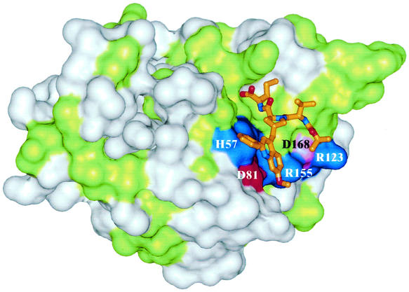
The hepatitis C virus (HCV) serine protease is necessary for viral replication and represents a valid target for developing new therapies for HCV infection. Potent and selective inhibitors of this enzyme have been identified and shown to inhibit HCV replication in tissue culture. The optimization of these inhibitors for clinical development would greatly benefit from in vitro systems for the identification and the study of resistant variants. We report the use HCV subgenomic replicons to isolate and characterize mutants resistant to a protease inhibitor. Taking advantage of the replicons' ability to transduce resistance to neomycin, we selected replicons with decreased sensitivity to the inhibitor by culturing the host cells in the presence of the inhibitor and neomycin. The selected replicons replicated to the same extent as those in parental cells. Sequence analysis followed by transfection of replicons containing isolated mutations revealed that resistance was mediated by amino acid substitutions in the protease. These results were confirmed by in vitro experiments with mutant enzymes and by modeling the inhibitor in the three-dimensional structure of the protease.


Figures

FIG. 1.
FIG. 1.
Structure (A) and mode of inhibition (B) of compound 1. (B) Reactions were performed as described in Materials and Methods with various substrate concentrations in the absence (•) or in the presence of 5 (▪), 10 (♦), and 20 (▾) nM compound 1. The data were fitted to a competitive mechanism. Plotting the slopes of the lines of the double-reciprocal plot yielded a Ki of 6.5 nM.
FIG. 2.
FIG. 2.
Effects of compound 1 on replication and polyprotein processing. (A) Viral (HCV) RNA levels present in cells treated with the indicated concentrations of compound 1 for 48 h were determined by Northern blot hybridization with a plus-strand-specific RNA probe. Hybridization with a probe specific for a cellular mRNA (GAPDH) served as a control. Quantitative data were generated by PhosphorImager analysis as indicated in Materials and Methods. (B) NS3 protein expression in cells treated with the indicated concentrations of compound 1 for 96 h was determined by cell ELISA. (C) Huh-7 (lanes 1 and 2) or HBI10 (lanes 3 to 8) cells were starved for 1 h, pulse-labeled with 35S-labeled amino acids for 15 min at 37°C, and then chased for the time indicated above each lane. Where indicated, starvation, labeling, and chasing were performed in the presence of 50 μM compound (cmp) 1. The labeled proteins were immunoprecipitated with the 55IV anti-NS5A antiserum and analyzed by SDS-10% PAGE. The positions of molecular mass standards (in kilodaltons) and NS5A and uncleaved precursors are indicated.
FIG. 3.
FIG. 3.
Kinetics of polyprotein processing in parental cells and clones resistant to compound 1. Huh-7 (lanes 1 and 2), HBI10 (lanes 3 to 6), R10 (lanes 7 to 10), and R12 (lanes 11 to 14) cells were pulse-labeled with 35S-labeled amino acids for 15 min at 37°C and then chased for the time indicated above each lane. The labeled proteins were immunoprecipitated with the 55IV anti-NS5A antiserum and analyzed by SDS-10% PAGE. The quantitative data below each lane were generated by PhosphorImager analysis as indicated in Materials and Methods and are expressed as percents of the total radioactivity counted in viral proteins. A is the sum of the NS4A-5B, NS4B-5B, and NS5A-5B precursors; B is the sum of the NS4A-5A and NS4B-5A precursors; C is the sum of the p56 and p58 forms of NS5A. The positions of molecular mass standards (in kilodaltons) and NS5A and uncleaved precursors are indicated.
FIG. 4.
FIG. 4.
Structures of compounds 2, 3, and 4.
FIG. 5.
FIG. 5.
Model of compound 1 in the active site of the NS3-4A protease. Conserved residues are shown in green, and nonconserved residues are white. Charged residues are colored according to their charges, with basic residues in blue, acidic residues in red, and aspartate 168 in magenta. Compound 1 is presented as a stick model.

Comment in

References

    1. Barbato, G., D. O. Cicero, M. C. Nardi, C. Steinkuhler, R. Cortese, R. De Francesco, and R. Bazzo. 1999. The solution structure of the N-terminal proteinase domain of the hepatitis C virus (HCV) NS3 protein provides new insights into its activation and catalytic mechanism. J. Mol. Biol. 289:371-384. - PubMed
    1. Bartenschlager, R. 1997. Candidate targets for hepatitis C virus-specific antiviral therapy. Intervirology 40:378-393. - PubMed
    1. Bartenschlager, R. 1999. The NS3/4A proteinase of the hepatitis C virus: unravelling structure and function of an unusual enzyme and a prime target for antiviral therapy. J. Viral Hepat. 6:165-181. - PubMed
    1. Bartenschlager, R., L. Ahlborn-Laake, J. Mous, and H. Jacobsen. 1994. Kinetic and structural analyses of hepatitis C virus polyprotein processing. J. Virol. 68:5045-5055. - PMC - PubMed
    1. Bartenschlager, R., L. Ahlborn-Laake, K. Yasargil, J. Mous, and H. Jacobsen. 1995. Substrate determinants for cleavage in cis and in trans by the hepatitis C virus NS3 proteinase. J. Virol. 69:198-205. - PMC - PubMed

MeSH terms

Substances

LinkOut - more resources