Generation of an infectious clone of VR-2332, a highly virulent North American-type isolate of porcine reproductive and respiratory syndrome virus
- PMID: 12610145
- PMCID: PMC149535
- DOI: 10.1128/jvi.77.6.3702-3711.2003
Generation of an infectious clone of VR-2332, a highly virulent North American-type isolate of porcine reproductive and respiratory syndrome virus
Abstract
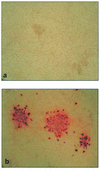
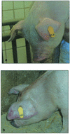
A full-length cDNA clone of the prototypical North American porcine reproductive and respiratory syndrome virus (PRRSV) isolate VR-2332 was assembled in the plasmid vector pOK(12). To rescue infectious virus, capped RNA was transcribed in vitro from the pOK(12) clone and transfected into BHK-21C cells. The supernatant from transfected monolayers were serially passaged on Marc-145 cells and porcine pulmonary alveolar macrophages. Infectious PRRSV was recovered on Marc-145 cells as well as porcine pulmonary macrophages; thus, the cloned virus exhibited the same cell tropism as the parental VR-2332 strain. However, the cloned virus was clearly distinguishable from the parental VR-2332 strain by an engineered marker, a BstZ17I restriction site. The full-length cDNA clone had 11 nucleotide changes, 2 of which affected coding, compared to the parental VR-2332 strain. Additionally, the transcribed RNA had an extra G at the 5' end. To examine whether these changes influenced viral replication, we examined the growth kinetics of the cloned virus in vitro. In Marc-145 cells, the growth kinetics of the cloned virus reflected those of the parental isolate, even though the titers of the cloned virus were consistently slightly lower. In experimentally infected 5.5-week-old pigs, the cloned virus produced blue discoloration of the ears, a classical clinical symptom of PRRSV. Also, the seroconversion kinetics of pigs infected with the cloned virus and VR-2332 were very similar. Hence, virus derived from the full-length cDNA clone appeared to recapitulate the biological properties of the highly virulent parental VR-2332 strain. This is the first report of an infectious cDNA clone based on American-type PRRSV. The availability of this cDNA clone will allow examination of the molecular mechanisms behind PRRSV virulence and attenuation, which might in turn allow the production of second-generation, genetically engineered PRRSV vaccines.
Figures


Similar articles
-
Infectious cDNA clones of porcine reproductive and respiratory syndrome virus and their potential as vaccine vectors.Vet Immunol Immunopathol. 2004 Dec 8;102(3):143-54. doi: 10.1016/j.vetimm.2004.09.019. Vet Immunol Immunopathol. 2004. PMID: 15507301 Free PMC article. Review.
-
Generation of an infectious clone of HuN4-F112, an attenuated live vaccine strain of porcine reproductive and respiratory syndrome virus.Virol J. 2011 Aug 19;8:410. doi: 10.1186/1743-422X-8-410. Virol J. 2011. PMID: 21851649 Free PMC article.
-
A highly pathogenic porcine reproductive and respiratory syndrome virus generated from an infectious cDNA clone retains the in vivo virulence and transmissibility properties of the parental virus.Virology. 2004 Aug 1;325(2):308-19. doi: 10.1016/j.virol.2004.04.046. Virology. 2004. PMID: 15246270 Free PMC article.
-
A full-length cDNA infectious clone of North American type 1 porcine reproductive and respiratory syndrome virus: expression of green fluorescent protein in the Nsp2 region.J Virol. 2006 Dec;80(23):11447-55. doi: 10.1128/JVI.01032-06. Epub 2006 Sep 13. J Virol. 2006. PMID: 16971421 Free PMC article.
-
In vivo growth of porcine reproductive and respiratory syndrome virus engineered nsp2 deletion mutants.Virus Res. 2010 Dec;154(1-2):77-85. doi: 10.1016/j.virusres.2010.07.024. Epub 2010 Jul 29. Virus Res. 2010. PMID: 20673840 Free PMC article. Review.
Cited by
-
Infectious cDNA clones of porcine reproductive and respiratory syndrome virus and their potential as vaccine vectors.Vet Immunol Immunopathol. 2004 Dec 8;102(3):143-54. doi: 10.1016/j.vetimm.2004.09.019. Vet Immunol Immunopathol. 2004. PMID: 15507301 Free PMC article. Review.
-
Development of a chimeric strain of porcine reproductive and respiratory syndrome virus with an infectious clone and a Korean dominant field strain.J Microbiol. 2014 Apr;52(4):345-9. doi: 10.1007/s12275-014-4074-4. Epub 2014 Mar 29. J Microbiol. 2014. PMID: 24682997 Free PMC article.
-
Use of a PRRSV infectious clone to evaluate in vitro quasispecies evolution.Adv Exp Med Biol. 2006;581:435-8. doi: 10.1007/978-0-387-33012-9_78. Adv Exp Med Biol. 2006. PMID: 17037574 Free PMC article. No abstract available.
-
Reverse genetic manipulation of the overlapping coding regions for structural proteins of the type II porcine reproductive and respiratory syndrome virus.Virology. 2009 Jan 5;383(1):22-31. doi: 10.1016/j.virol.2008.09.013. Epub 2008 Nov 5. Virology. 2009. PMID: 18977502 Free PMC article.
-
Molecular assessment of the role of envelope-associated structural proteins in cross neutralization among different PRRS viruses.Virus Genes. 2008 Dec;37(3):380-91. doi: 10.1007/s11262-008-0278-1. Epub 2008 Sep 3. Virus Genes. 2008. PMID: 18770017
References
-
- Albina, E. 1997. Epidemiology of porcine reproductive and respiratory syndrome (PRRS): an overview. Vet. Microbiol. 55:309-316. - PubMed
-
- Allende, R., T. L. Lewis, Z. Lu, D. L. Rock, G. F. Kutish, A. Ali, A. R. Doster, and F. A. Osorio. 1999. North American and European porcine reproductive and respiratory syndrome viruses differ in non-structural protein coding regions. J. Gen. Virol. 80:307-315. - PubMed
-
- Allende, R., G. F. Kutish, W. laegréid, Z. Lu, T. L. Lewis, D. L. Rock, J. Friesen, J. A. Galeota, A. R. Doster, and F. A. Osorio. 2000. Mutations in the genome of porcine reproductive and respiratory syndrome virus responsible for the attenuation phenotype. Arch. Virol. 145:1149-1161. - PMC - PubMed
-
- Benfield, D. A., E. Nelson, J. E. Collings, L. Harris, S. M. Goyal, D. Robison, W. T. Christianson, R. B. Morrison, and D. Gorcyca. 1992. Characterization of swine infertility and respiratory syndrome (SIRS) virus (isolate ATCC VR-2332). J. Vet. Diagn. Investig. 4:127-133. - PubMed
Publication types
MeSH terms
Substances
Associated data
- Actions
LinkOut - more resources
Full Text Sources
Other Literature Sources

