. 2003 Mar;77(6):3816-23.
doi: 10.1128/jvi.77.6.3816-3823.2003.

Neurovirulence in mice of H5N1 influenza virus genotypes isolated from Hong Kong poultry in 2001

Affiliations

Neurovirulence in mice of H5N1 influenza virus genotypes isolated from Hong Kong poultry in 2001

Aleksandr S Lipatov et al. J Virol. 2003 Mar.

Abstract

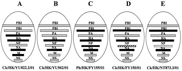
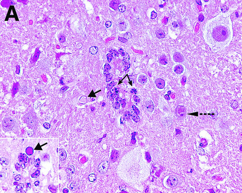
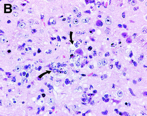
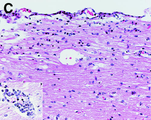
We studied the pathogenicity of five different genotypes (A to E) of highly pathogenic avian H5N1 viruses, which contained HA genes similar to those of the H5N1 virus A/goose/Guangdong/1/96 and five different combinations of "internal" genes, in a mouse model. Highly pathogenic, neurotropic variants of genotypes A, C, D, and E were isolated from the brain after a single intranasal passage in mice. Genotype B virus was isolated from lungs only. The mouse brain variants had amino acid changes in all gene products except PB1, NP, and NS1 proteins but no common sets of mutations. We conclude that the original H5N1/01 isolates of genotypes A, C, D, and E were heterogeneous and that highly pathogenic neurotropic variants can be rapidly selected in mice.


Figures

FIG. 1.
FIG. 1.
Genotypes of Hong Kong 2001 H5N1 viruses. Initial genotyping of H5N1/01 viruses on the basis of partial gene sequences (10) was confirmed in the present study by determination of the full-length sequences. The solid bars indicate genes of A/goose/Guangdong/1/96-like lineage, the hatched bars indicate genes of A/duck/HK/Y280/97-like lineage, the shaded bars indicate genes of wild aquatic bird lineage, and the open bars represent genes of unknown lineage.
FIG. 2.
FIG. 2.
Rate of survival of mice infected with different genotypes of H5N1/01 viruses. Mice were observed for 20 days after intranasal inoculation with 0.1 ml of PBS-diluted allantoic fluid containing ca. 106.5 to 106.75 EID50 of infective virus. Symbols: ♦, Ck/HK/YU822.2/01 (A); ▪, Ck/HK/YU562/01 (B); ▴, Ck/HK/FY155/01 (C); ○, Ck/HK/FY150/01 (D); □, Ck/HK/NT873.3/01 (E).
FIG. 3.
FIG. 3.
Titers of original H5N1 isolates in mouse lungs (A) and brains (B). Virus titers were determined in ca. 10% tissue homogenates prepared in PBS with antibiotic-antimycotic solution. Mice were inoculated intranasally with 0.1 ml of PBS-diluted allantoic fluid containing ca. 106.5 to 107.5 EID50 of infective virus. Lungs were obtained from mice sacrificed on day 3 after inoculation; brains were removed from mice showing signs of CNS disorders (hind-leg paralysis, tremor) that died or were sacrificed on days 8 to 14 after inoculation. ✽, Virus of genotype A was isolated from the brain of one mouse only.
FIG. 4.
FIG. 4.
Titers of MB variants of H5N1 virus in the lungs (A) and brains (B) of infected mice. Mice were intranasally inoculated with 0.1 ml of PBS-diluted allantoic fluid containing 101.5 to 102.5 EID50 of infective virus. Virus titers were determined in approximately 10% tissue homogenates prepared in PBS with antibiotic-antimycotic solution. The lungs and brains were removed from mice that were sacrificed at the terminal stage of illness on day 8 after infection.
FIG. 5.
FIG. 5.
Hematoxylin-and-eosin-stained sections of spinal cords and brain stems from mice infected with Ck/HK/YU822.2/01-MB. Brains and spinal cords were collected from mice sacrificed at the terminal stage of illness on days 10 and 11 after intranasal inoculation with 10 EID50 of virus. (A) Karyorrhexis in two degenerating ependymal cells lining the spinal cord's central canal (two split arrows). An intranuclear inclusion is present in a neuron (broken arrow) and in degenerating glial cells (solid single arrows). The inset shows a section from a deeper part of the same tissue. (B) Inflammatory cells and a focus of degenerating neurons undergoing neuronophagia (arrows) in the spinal cord of a mouse with hind-leg paralysis. (C) Inflammatory cells in the meninges and white matter of the spinal cord and (inset) in the perivascular (Virchow-Robin) space of the medulla oblongata. (D) Dorsal root ganglion (arrow) with degenerating neurons, some of which have an intranuclear inclusion. In the adjacent adipose tissue there is a focus of necrosis with inflammatory cells (arrow head). Original magnifications: (A) ×260, (B) ×260, (C) ×130 (inset, ×260), (D) ×130.
FIG. 5.
FIG. 5.
Hematoxylin-and-eosin-stained sections of spinal cords and brain stems from mice infected with Ck/HK/YU822.2/01-MB. Brains and spinal cords were collected from mice sacrificed at the terminal stage of illness on days 10 and 11 after intranasal inoculation with 10 EID50 of virus. (A) Karyorrhexis in two degenerating ependymal cells lining the spinal cord's central canal (two split arrows). An intranuclear inclusion is present in a neuron (broken arrow) and in degenerating glial cells (solid single arrows). The inset shows a section from a deeper part of the same tissue. (B) Inflammatory cells and a focus of degenerating neurons undergoing neuronophagia (arrows) in the spinal cord of a mouse with hind-leg paralysis. (C) Inflammatory cells in the meninges and white matter of the spinal cord and (inset) in the perivascular (Virchow-Robin) space of the medulla oblongata. (D) Dorsal root ganglion (arrow) with degenerating neurons, some of which have an intranuclear inclusion. In the adjacent adipose tissue there is a focus of necrosis with inflammatory cells (arrow head). Original magnifications: (A) ×260, (B) ×260, (C) ×130 (inset, ×260), (D) ×130.

Similar articles

Cited by

References

    1. Brown, E. G., and J. E. Bailly. 1999. Genetic analysis of mouse-adapted influenza A virus identifies roles for the NA, PB1, and PB2 genes in virulence. Virus Res. 61:63-76. - PubMed
    1. Brown, E. G., H. Liu, L. Chang Kit, S. Baird, and M. Nesrallah. 2001. Pattern of mutation in the genome of influenza A virus on adaptation to increased virulence in mouse lung: identification of functional themes. Proc. Natl. Acad. Sci. USA 98:6883-6888. - PMC - PubMed
    1. Cauthen, A. N., D. E. Swayne, S. Schultz-Cherry, M. L. Perdue, and D. L. Suarez. 2000. Continued circulation in China of highly pathogenic avian influenza viruses encoding the hemagglutinin gene associated with the 1997 H5N1 outbreak in poultry and humans. J. Virol. 74:6592-6599. - PMC - PubMed
    1. Claas, E. C., A. D. Osterhaus, R. van Beek, J. C. de Jong, G. F. Rimmelzwaan, D. A. Senne, S. Krauss, K. F. Shortridge, and R. G. Webster. 1998. Human influenza A H5N1 virus related to a highly pathogenic avian influenza virus. Lancet 351:472-477. - PubMed
    1. de Jong, J. C., E. C. Claas, A. D. Osterhaus, R. G. Webster, and W. L. Lim. 1997. A pandemic warning? Nature 389:554. - PMC - PubMed

Publication types

MeSH terms

Associated data