Comparative Study
. 2003 Mar;23(6):1874-84.
doi: 10.1128/MCB.23.6.1874-1884.2003.

Roles for SR proteins and hnRNP A1 in the regulation of c-src exon N1

Affiliations
Comparative Study

Roles for SR proteins and hnRNP A1 in the regulation of c-src exon N1

Nanette Rooke et al. Mol Cell Biol. 2003 Mar.

Abstract

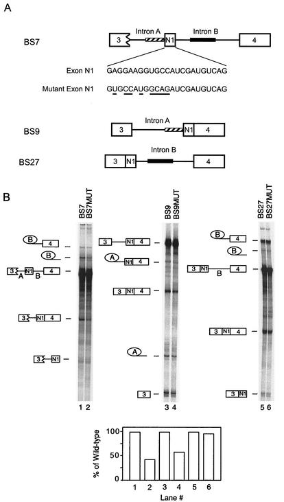
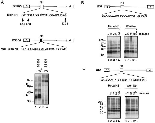
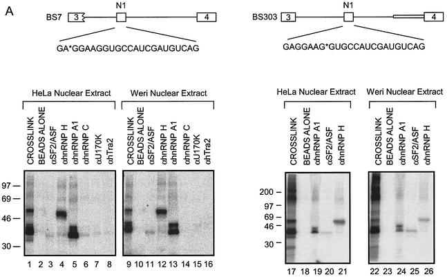
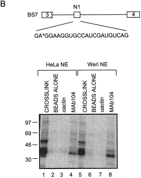
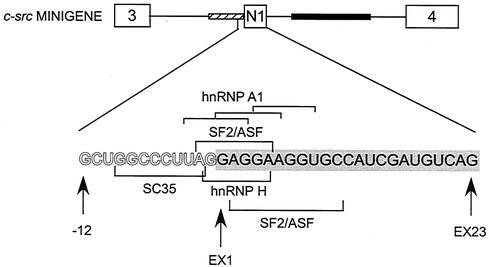
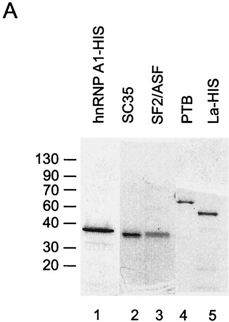
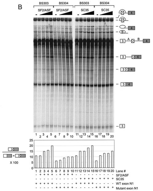
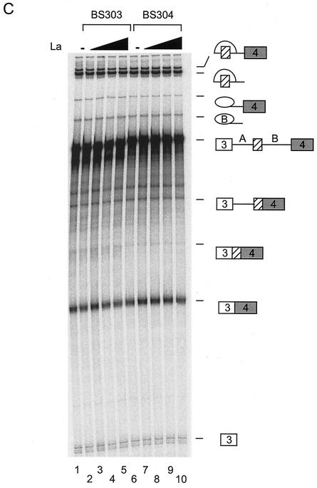
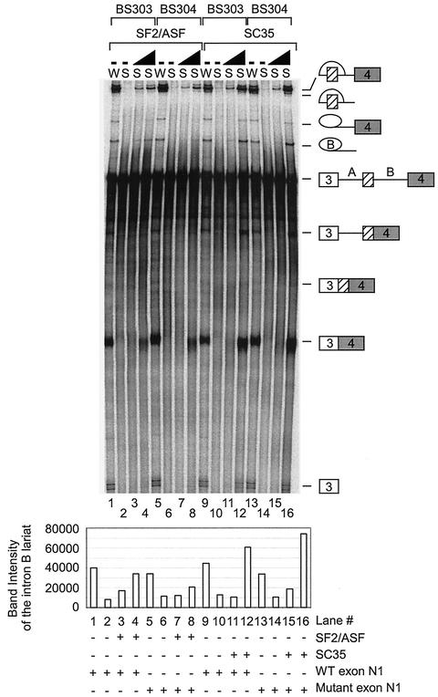
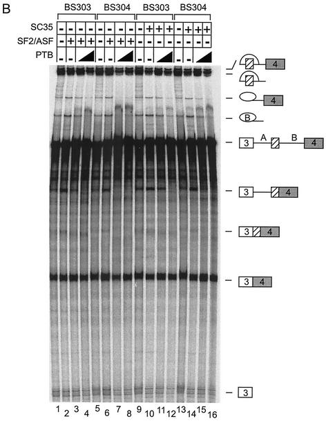
The splicing of the c-src exon N1 is controlled by an intricate combination of positive and negative RNA elements. Most previous work on these sequences focused on intronic elements found upstream and downstream of exon N1. However, it was demonstrated that the 5' half of the N1 exon itself acts as a splicing enhancer in vivo. Here we examine the function of this regulatory element in vitro. We show that a mutation in this sequence decreases splicing of the N1 exon in vitro. Proteins binding to this element were identified as hnRNP A1, hnRNP H, hnRNP F, and SF2/ASF by site-specific cross-linking and immunoprecipitation. The binding of these proteins to the RNA was eliminated by a mutation in the exonic element. The activities of hnRNP A1 and SF2/ASF on N1 splicing were examined by adding purified protein to in vitro splicing reactions. SF2/ASF and another SR protein, SC35, are both able to stimulate splicing of c-src pre-mRNA. However, splicing activation by SF2/ASF is dependent on the N1 exon enhancer element whereas activation by SC35 is not. In contrast to SF2/ASF and in agreement with other systems, hnRNP A1 repressed c-src splicing in vitro. The negative activity of hnRNP A1 on splicing was compared with that of PTB, a protein previously demonstrated to repress splicing in this system. Both proteins repress exon N1 splicing, and both counteract the enhancing activity of the SR proteins. Removal of the PTB binding sites upstream of N1 prevents PTB-mediated repression but does not affect A1-mediated repression. Thus, hnRNP A1 and PTB use different mechanisms to repress c-src splicing. Our results link the activity of these well-known exonic splicing regulators, SF2/ASF and hnRNP A1, to the splicing of an exon primarily controlled by intronic factors.


Figures

FIG. 1.
FIG. 1.
The N1 exonic enhancer element functions in vitro. (A) The pre-mRNAs used in the experiment are diagrammed. The nucleotides in exon N1 and mutant N1 exon are also shown, and the nucleotide changes in the mutant exon are underlined. The wild-type exon contains an inserted ClaI site 3′ of the mutation. This insertion has been previously shown not to affect N1 splicing (1a). The striped bar represents the upstream acceptor sequence, and the black bar represents the downstream enhancer sequence. (B) Splicing of pre-mRNAs with either a wild-type exon or a mutated exon in Weri-1 nuclear extract. Lanes 1 and 2, BS7 and BS7MUT; lanes 3 and 4, BS9 and BS9MUT; lanes 5 and 6, BS27 and BS27MUT. The products and intermediates are shown to the side of the gels. The percentage of RNA converted to mRNA was determined and compared between pairs of substrates, with the value for the mutant shown as a percentage of that for the wild-type.
FIG. 2.
FIG. 2.
Proteins cross-link specifically to N1. (A) UV cross-linking to BS303 and BS304 site-specifically labeled at position EX3 in HeLa (H) and Weri-1 (W) nuclear extracts. The grey bar in intron B in BS303 and BS304 represents adenovirus acceptor sequence. This sequence has previously been shown to increase overall splicing levels but not to affect neural specificity. The nucleotides in the exon are designated EX1 through EX23. The mutated nucleotides are underlined. The asterisk denotes the site of the radioactive phosphate in the exon N1 sequence. Arrows highlight protein bands enriched in Weri-1 extract in lane 2. (B) UV cross-linking to BS7 labeled at position EX3 in HeLa and Weri-1 nuclear extracts. (C) UV cross-linking to BS7 site-specifically labeled at EX8 in HeLa and Weri-1 nuclear extracts.
FIG. 3.
FIG. 3.
SR proteins and hnRNPs bind to exon N1. (A) Immunoprecipitation of proteins cross-linked to positions X3 and X8. Lanes 1, 9, 17, and 22 contain 1/10 of the total cross-linking reaction mixture. Other lanes contain the bound material from immunoprecipitates using the antibodies indicated. (B) A 35-kDa SR protein binds to exon N1. MAb104 was used to immunoprecipitate RS domain proteins that cross-link to position EX3 in BS7.
FIG. 3.
FIG. 3.
SR proteins and hnRNPs bind to exon N1. (A) Immunoprecipitation of proteins cross-linked to positions X3 and X8. Lanes 1, 9, 17, and 22 contain 1/10 of the total cross-linking reaction mixture. Other lanes contain the bound material from immunoprecipitates using the antibodies indicated. (B) A 35-kDa SR protein binds to exon N1. MAb104 was used to immunoprecipitate RS domain proteins that cross-link to position EX3 in BS7.
FIG. 4.
FIG. 4.
Diagram of the 3′ splice site and exon N1. Potential binding sites for hnRNP A1, SF2/ASF, SC35, and hnRNP H in the 3′ splice site of exon N1 and within the exon itself are bracketed. Intron nucleotides at the 3′ splice site are in outline. The nucleotides of exon N1 are filled and encompassed in a grey bar. In the c-src minigene diagram, the upstream acceptor sequence is represented by a striped bar and the downstream enhancer sequence is represented by a solid bar.
FIG. 5.
FIG. 5.
SF2/ASF and SC35 differ in their enhancement of N1. (A) Coomassie staining of purified proteins used in the experiment and separated in an SDS polyacrylamide gel. (B) In vitro splicing reactions in the presence of added SR proteins. Splicing of BS303 (lanes 1 to 5 and 11 to 15) and BS304 (lanes 6 to 10 and 16 to 20) was done in the presence of 100 μg of Weri-1 nuclear extract. Lanes 2 to 5 and 7 to 10 both contain 50, 100, 200, and 400 ng of SF2/ASF, respectively. Lanes 12 to 15 and 17 to 20 both contain 50, 100, 200, and 400 ng of SC35, respectively. Lanes 1, 6, 11, and 16 contain Weri-1 extract without any additional factors. Quantification of the products is shown below the gel. (C) La does not affect splicing of BS303 or BS304. Splicing of BS303 and BS304 was done in the presence of 100 μg of Weri-1 nuclear extract. Lanes 2 to 5 and 7 to 10 both contain 0.200, 0.400, 0.800, and 2 μg of La, respectively. Lanes 1 and 6 contain Weri-1 extract without any additional factors.
FIG. 5.
FIG. 5.
SF2/ASF and SC35 differ in their enhancement of N1. (A) Coomassie staining of purified proteins used in the experiment and separated in an SDS polyacrylamide gel. (B) In vitro splicing reactions in the presence of added SR proteins. Splicing of BS303 (lanes 1 to 5 and 11 to 15) and BS304 (lanes 6 to 10 and 16 to 20) was done in the presence of 100 μg of Weri-1 nuclear extract. Lanes 2 to 5 and 7 to 10 both contain 50, 100, 200, and 400 ng of SF2/ASF, respectively. Lanes 12 to 15 and 17 to 20 both contain 50, 100, 200, and 400 ng of SC35, respectively. Lanes 1, 6, 11, and 16 contain Weri-1 extract without any additional factors. Quantification of the products is shown below the gel. (C) La does not affect splicing of BS303 or BS304. Splicing of BS303 and BS304 was done in the presence of 100 μg of Weri-1 nuclear extract. Lanes 2 to 5 and 7 to 10 both contain 0.200, 0.400, 0.800, and 2 μg of La, respectively. Lanes 1 and 6 contain Weri-1 extract without any additional factors.
FIG. 6.
FIG. 6.
SF2/ASF and SC35 activate splicing of N1 in S100 extract. Splicing of BS303 (lanes 1 to 4 and 9 to 12) and BS304 (lanes 5 to 8 and 13 to 16) was done in the presence of 100 μg of either Weri-1 nuclear extract (lanes 1, 5, 9, and 13) or Weri-1 S100 extract (lanes 2 to 4, 6 to 8, 10 to 12, and 14 to 16). Lanes 3 and 7 contain 400 ng of SF2/ASF; lanes 4 and 8 contain 800 ng of SF2/ASF; lanes 11 and 15 contain 300 ng of SC35; lanes 12 and 16 contain 600 ng of SC35; and lanes 2, 6, 10, and 14 contain Weri S100 extract without any additional factors. Quantification of the intron B lariat is shown below the gel.
FIG. 7.
FIG. 7.
HnRNP A1 can repress splicing of exon N1. (A) Splicing of BS303 (lanes 1 to 4 and 9 to 12) and BS304 (lanes 5 to 8 and 13 to 16) in 100 μg of Weri-1 nuclear extract. Lanes 2 to 4 and 6 to 8 contain 400 ng of SF2/ASF. Lanes 10 to 12 and 14 to 16 contain 300 ng of SC35. Lanes 3 and 4, 7 and 8, 11 and 12, and 15 and 16 contain 100 and 200 ng, respectively, of hnRNP A1. Lanes 1, 5, 9, and 13 contain Weri-1 nuclear extract without additional factors. (B) Splicing of BS303 (lanes 1 to 4 and 9 to 12) and BS304 (lanes 5 to 8 and 13 to 16) in 100 μg of Weri-1 nuclear extract. Lanes 2 to 4 and 6 to 8 contain 400 ng of SF2/ASF. Lanes 10 to 12 and 14 to 16 contain 300 ng of SC35. Lanes 3 and 4, 7 and 8, 11 and 12, and 15 and 16 contain 200, and 1 μg, respectively, of PTB. Lanes 1, 5, 9, and 13 contain Weri-1 nuclear extract without additional factors.
FIG. 7.
FIG. 7.
HnRNP A1 can repress splicing of exon N1. (A) Splicing of BS303 (lanes 1 to 4 and 9 to 12) and BS304 (lanes 5 to 8 and 13 to 16) in 100 μg of Weri-1 nuclear extract. Lanes 2 to 4 and 6 to 8 contain 400 ng of SF2/ASF. Lanes 10 to 12 and 14 to 16 contain 300 ng of SC35. Lanes 3 and 4, 7 and 8, 11 and 12, and 15 and 16 contain 100 and 200 ng, respectively, of hnRNP A1. Lanes 1, 5, 9, and 13 contain Weri-1 nuclear extract without additional factors. (B) Splicing of BS303 (lanes 1 to 4 and 9 to 12) and BS304 (lanes 5 to 8 and 13 to 16) in 100 μg of Weri-1 nuclear extract. Lanes 2 to 4 and 6 to 8 contain 400 ng of SF2/ASF. Lanes 10 to 12 and 14 to 16 contain 300 ng of SC35. Lanes 3 and 4, 7 and 8, 11 and 12, and 15 and 16 contain 200, and 1 μg, respectively, of PTB. Lanes 1, 5, 9, and 13 contain Weri-1 nuclear extract without additional factors.
FIG. 8.
FIG. 8.
hnRNP A1 and PTB use different mechanisms to repress exon N1 splicing. Splicing of BS7 (lanes 1 to 5 and 11 to 15) and BS27 (lanes 6 to 10 and 16 to 20) was done in the presence of 100 μg of Weri-1 nuclear extract. Lanes 2 to 5 and 7 to 10 both contain 50, 100, 200, and 500 ng of PTB, respectively. Lanes 1, 6, 11, and 16 contain Weri-1 nuclear extract without additional factors. Lanes 12 to 15 and 17 to 20 both contain 25, 50, 100 and 200 ng of hnRNP A1, respectively.
FIG. 9.
FIG. 9.
Model for regulation of exon N1. PTB binds to CU repeat sequences in the upstream and downstream introns to repress splicing of N1 in nonneuronal cells. In neuronal cells, PTB is removed to derepress splicing and the enhancer elements are able to activate splicing. Some hnRNP A1 molecules may be replaced with hnRNP A1B molecules in neuronal cells to assist in derepression of N1. SF2/ASF likely enhances splicing at both adjacent splice sites in neuronal cells in cooperation with the proteins binding to the downstream enhancer sequence.

Similar articles

Cited by

References

    1. Black, D. L. 1992. Activation of c-src neuron-specific splicing by an unusual RNA element in vivo and in vitro. Cell 69:795-807. - PubMed
    1. Black, D. L. 1991. Does steric interference between splice sites block the splicing of a short c-src neuron-specific exon in non-neuronal cells? Genes Dev. 5:389-402. - PubMed
    1. Blanchette, M., and B. Chabot. 1999. Modulation of exon skipping by high-affinity hnRNP A1-binding sites and by intron elements that repress splice site utilization. EMBO J. 18:1939-1952. - PMC - PubMed
    1. Blencowe, B. J. 2000. Exonic splicing enhancers: mechanism of action, diversity and role in human genetic diseases. Trends Biochem. Sci. 25:106-110. - PubMed
    1. Burd, C. G., and G. Dreyfuss. 1994. RNA binding specificity of hnRNP A1: significance of hnRNP A1 high-affinity binding sites in pre-mRNA splicing. EMBO J. 13:1197-1204. - PMC - PubMed

Publication types

MeSH terms

Substances