. 2003 Mar;23(6):2055-67.
doi: 10.1128/MCB.23.6.2055-2067.2003.

Principal role of TRAP/mediator and SWI/SNF complexes in Kaposi's sarcoma-associated herpesvirus RTA-mediated lytic reactivation

Affiliations

Principal role of TRAP/mediator and SWI/SNF complexes in Kaposi's sarcoma-associated herpesvirus RTA-mediated lytic reactivation

Yousang Gwack et al. Mol Cell Biol. 2003 Mar.

Abstract

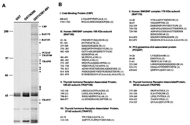
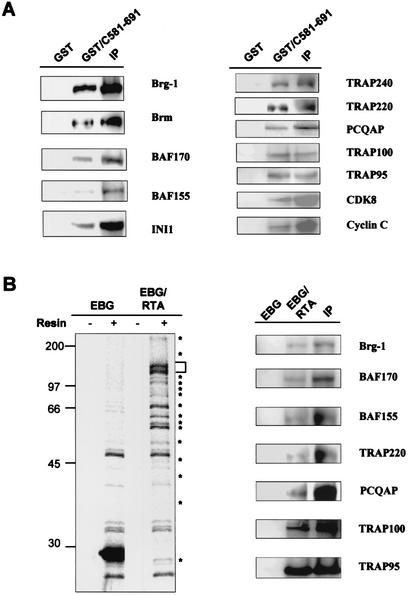
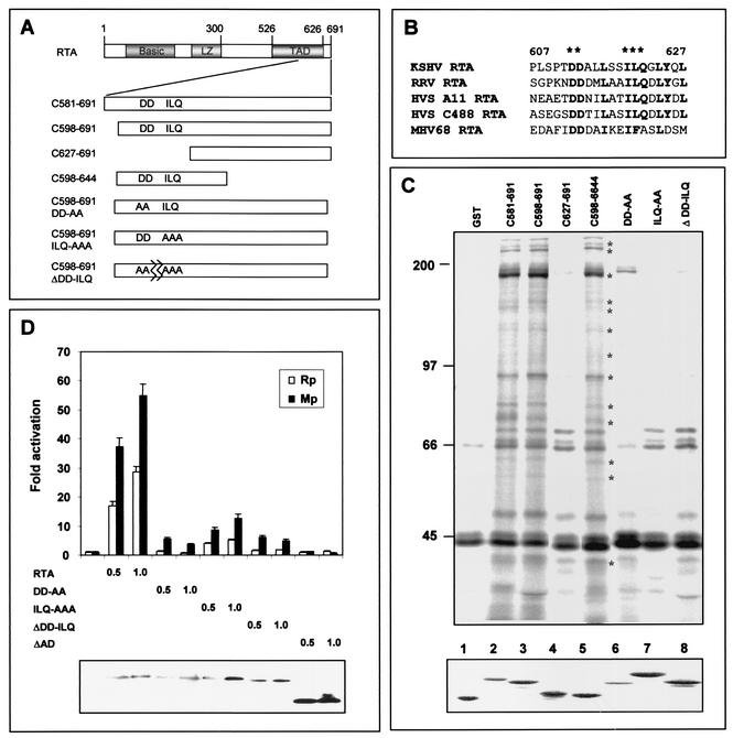
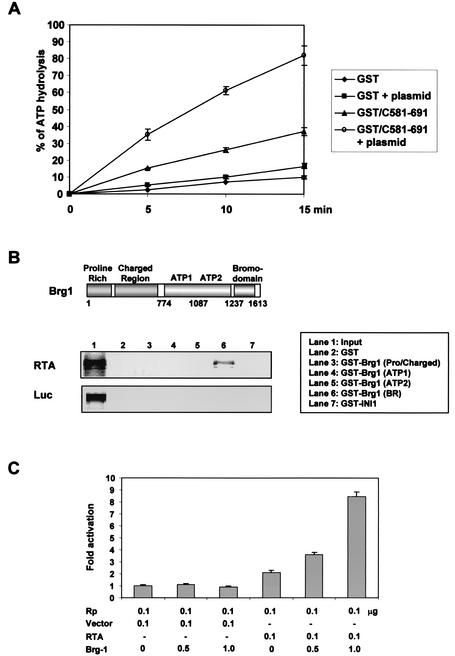
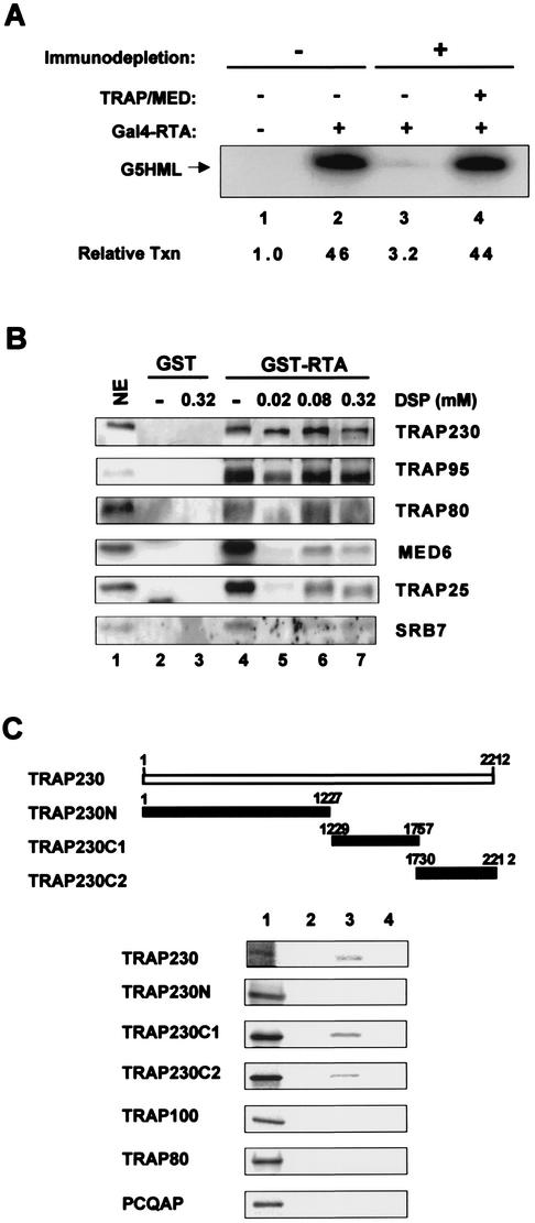
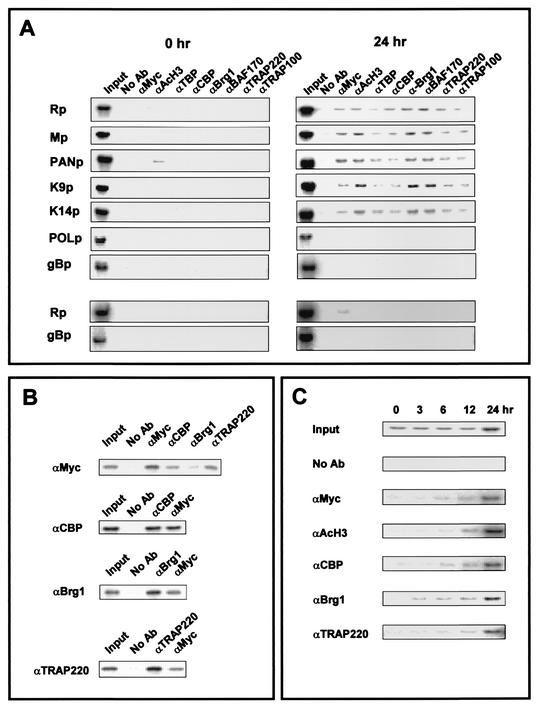
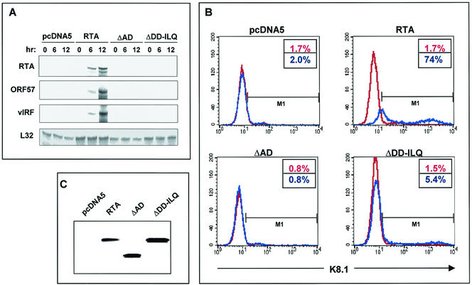
An important step in the herpesvirus life cycle is the switch from latency to lytic reactivation. The RTA transcription activator of Kaposi's sarcoma-associated herpesvirus (KSHV) acts as a molecular switch for lytic reactivation. Here we demonstrate that KSHV RTA recruits CBP, the SWI/SNF chromatin remodeling complex, and the TRAP/Mediator coactivator into viral promoters through interactions with a short acidic sequence in the carboxyl region and that this recruitment is essential for RTA-dependent viral gene expression. The Brg1 subunit of SWI/SNF and the TRAP230 subunit of TRAP/Mediator were shown to interact directly with RTA. Consequently, genetic ablation of these interactions abolished KSHV lytic replication. These results demonstrate that the recruitment of CBP, SWI/SNF, and TRAP/Mediator complexes by RTA is the principal mechanism to direct well-controlled viral gene expression and thereby viral lytic reactivation.


Figures

FIG. 1.
FIG. 1.
Purification and identification of RTA-binding proteins. (A) Identification of RTA-binding proteins. Glutathione-Sepharose beads containing 5 μg of GST, GST-RTA(N1-300), or GST-RTA(C581-691) fusion protein were mixed with lysates of 35S-labeled Raji B cells. RTA-binding proteins were resolved in SDS-PAGE and autoradiographed with a PhosphorImager. Asterisks indicate the proteins that were not identified by mass spectrometry. Sizes are shown in kilodaltons. (B) Mass spectrometry analysis of RTA-binding proteins. The peptide sequence of each protein isolated by the mass spectrometry analysis is presented with its published amino acid sequence number. The number in parentheses indicates the frequency with which a particular peptide sequence was identified from the mass spectrometry analysis.
FIG. 2.
FIG. 2.
Interaction between RTA and cellular transcription cofactors. (A) In vitro interaction between RTA and cellular transcription cofactors. Lysates of Raji B cells were mixed with GST and GST-RTA(C581-691) or used for immunoprecipitation (IP) with the antibodies to cellular transcription cofactors indicated at the right side of the figure. Polypeptides present in GST fusion complexes and immunoprecipitation complexes were separated by SDS-PAGE, followed by immunoblot assay with the same antibodies. (B) Interaction between RTA and cellular transcription cofactors. [35S]methionine/cysteine-labeled 293T cells transfected with the EBG or EBG-RTA vector were used for glutathione-Sepharose affinity chromatography (resin). The bound GST fusion complexes were separated by SDS-PAGE followed by autoradiography (left panel). The bracket indicates the GST-RTA protein, and asterisks indicate the proteins associated with GST-RTA. Lysates were also used for glutathione-Sepharose affinity chromatography or Immunoprecipitation with antibodies to cellular transcription cofactors as indicated at the right side of the figure. Polypeptides present in the GST fusion and immunoprecipitation complexes were separated by SDS-PAGE, followed by immunoblot assay with the same antibodies (right panel). Based on the comparison between GST-RTA pulldown and immunoprecipitation, 12.5% of Brg1, 7.2% of BAF170, 5.4% of BAF55, 4% of TRAP220, 4.1% of PCQAP, 7.3% of TRAP, and 70% of TRAP95 were copurified by GST-RTA protein.
FIG. 3.
FIG. 3.
Identification of region of RTA required for interaction with cellular transcription cofactors and their role in RTA-mediated transcriptional activation. (A) Schematic diagram of RTA mutants. RTA contains an N-terminal basic domain, an internal leucine zipper (LZ) motif, and a C-terminal transcription activation domain (TAD). Each RTA mutant is described in detail in the text. (B) Conserved sequences in the carboxyl transcription activation domain of gamma-2 herpesvirus RTA homologs. Asterisks indicate the conserved amino acid sequences that were mutated. (C) The conserved sequence at the carboxyl activation domain of RTA is necessary for interaction with cellular transcription cofactors. [35S]methionine/cysteine-labeled Raji B cells were mixed with GST, GST-RTA, and GST-RTA mutants. 35S-labeled polypeptides associated with GST fusion proteins were separated by SDS-PAGE, followed by autoradiography. A similar amount of each GST fusion protein was used in this assay (bottom panel). Asterisks indicate the cellular proteins associated with GST-RTA fusion proteins. (D) The conserved sequence at the carboxy-terminal region of RTA is required for efficient transcriptional activation of the RTA (Rp) and ORF57 (Mp) promoters. 293T cells were transfected with an expression vector containing RTA or one of its mutants together with the Rp-luciferase (open rectangle) or Mp-luciferase (solid rectangle) reporter. The RSV-β-galactosidase vector was included as a transfection control. Luciferase activity was measured at 48 h posttransfection, and luciferase values were normalized by β-galactosidase activity. Luciferase activity is represented as the average of three independent experiments. Error bars indicate the standard error. The expression levels of RTA and its mutants are shown (bottom panel).
FIG. 4.
FIG. 4.
ATPase activity in RTA complexes and the role of Brg1 in RTA-mediated transcriptional activation. (A) The presence of ATPase activity in RTA complexes. Raji B-cell lysates were mixed with GST or GST-RTA(C581-691). After extensive washing, GST and GST-RTA(C581-691) complexes were used for ATP hydrolysis in the presence or absence of 20 nM plasmid DNA. The reaction from each time point was separated on polyethyleneimine-cellulose thin-layer chromatography plates, and the ratio of inorganic phosphate to ATP was quantitated by PhosphorImager. (B) Interaction of RTA with the carboxyl region of Brg1. The GST-Brg1 fusion protein contains each domain of Brg1 depicted in the top panel. [35S]methionine-labeled RTA and luciferase proteins from in vitro translation were mixed with GST or GST-Brg1 fusion protein. After extensive washing, 35S-labeled polypeptides associated with GST or GST-Brg1 fusion protein were separated by SDS-PAGE, followed by autoradiography. Lane 1 indicates 10% input amount of RTA or luciferase protein used for the binding assay. (C) The role of Brg1 in RTA-mediated transcriptional activation. SW13 cells were transfected with expression vector and reporter vector as indicated below the figure. Luciferase activity was measured at 48 h posttransfection, and luciferase values were normalized by β-galactosidase activity. Luciferase activity is represented as the average of three independent experiments. Error bars indicate the standard error.
FIG. 5.
FIG. 5.
Interaction of RTA with TRAP/Mediator complex. (A) The essential role of TRAP/Mediator in in vitro RTA-mediated transcription. Each in vitro transcription reaction mixture contained 20 μg of nuclear extract (lanes 1 and 2, untreated; lanes 3 and 4, anti-TRAP25 antibody-depleted nuclear extract [ΔMED]). Gal4-RTA(C581-691) protein was used as an activator, and 50 ng of pG5HML template was added as a template. Relative transcription (Txn) levels, determined by PhosphorImager analysis, are indicated. (B) RTA interacts directly with the TRAP230 subunit when bound to the TRAP/Mediator complex. One-half milliliter of HeLa nuclear extract was incubated with 10 μg of GST or GST-RTA(C581-691) for 7 h at 4°C and washed with phosphate-buffered saline. The bound proteins were exposed to increasing amounts of the crosslinker DSP in dimethylsulfoxide or dimethyl sulfoxide only for 10 min at room temperature. Lane 1, nuclear extract (NE, 5% of starting nuclear extract); lanes 2 and 4, affinity purification of GST and GST-RTA, respectively, before DSP treatment; lanes 3 and 7, 0.32 mM DSP; lane 5, 0.02 mM DSP; lane 6, 0.08 mM DSP. The crosslinking reagent was quenched by addition of Tris (pH 7.5) to a final concentration of 50 mM. After extensive washing with 8 M urea (lanes 3, 5, 6, and 7), polypeptides present in GST and GST-RTA(C581-691) complexes were subjected to the immunoblot assay with antibodies specific to TRAP230, TRAP95, TRAP80, MED6, TRAP25, and SRB7 subunits. (C) Interaction of RTA with TRAP230. [35S]methionine-labeled TRAP230, TRAP230N, TRAP230C1, TRAP230C2, TRAP100, TRAP80, and PCQAP proteins from in vitro translation reactions were mixed with GST (lane 2), GST-RTA(C598-691) (lane 3), or GST-RTA(C598-691ΔDD-ILQ) (lane 4). After extensive washing with lysis buffer, 35S-labeled polypeptides associated with GST fusion proteins were separated by SDS-PAGE, followed by autoradiography. Lane 1 indicates 10% of an input amount of each protein used for the binding assay.
FIG. 6.
FIG. 6.
Recruitment of cellular transcription cofactors to RTA-dependent promoters. (A) Chromatin immunoprecipitation assay of RTA-dependent and RTA-independent promoters. KSHV-infected TRExBCBL1-RTA cells or TRExBCBL1-RTA(ΔDD-ILQ) cells (bottom two panels) were collected without treatment or after 24 h of treatment with 1 μg of doxycycline per ml. Chromatin immunoprecipitation assays were performed with antibodies against the protein indicated at the top. PCR products corresponding to each viral promoter were generated from an aliquot (1/10) of total immunoprecipitated material (Input). (B) ReChip assay. TRExBCBL1-RTA cells were stimulated for 24 h with 1 μg of doxycycline per ml and subjected to the chromatin immunoprecipitation assay as described above. After washing the protein-G-Sepharose beads from the primary immunoprecipitation shown on the left panel, the complexes were eluted by incubation with 10 mM dithiothreitol at 37°C for 30 min and diluted to 40 times the original volume. Eluates were reimmunoprecipitated with the second antibody indicated at the top of figure, followed by PCR amplification. PCR products were generated from an aliquot (1/10) of total immunoprecipitated material (Input). (C) Time course of chromatin immunoprecipitation assay. TRExBCBL1-RTA cells were stimulated with doxycycline for 1, 3, 6, 12, and 24 h and then used for the chromatin immunoprecipitation assay as described above. PCR products were generated from an aliquot (1/10) of total immunoprecipitated material (Input).
FIG. 7.
FIG. 7.
Recruitment of TRAP/Mediator and SWI/SNF complexes is required for RTA-mediated KSHV lytic reactivation. (A) Activation of KSHV RTA, ORF57, and vIRF1 gene expression by RTA and its mutants. After doxycycline treatment for 0, 6, and 12 h, RNA was extracted from KSHV-infected TRExBCBL1-cDNA5, TRExBCBL1-RTA, TRExBCBL1-RTA ΔAD, and TRExBCBL1-RTA(ΔDD-ILQ) cells, and 5 μg of total RNA was subjected to RNase protection assay analysis with 32P-labeled riboprobe templates. The protected RNA fragments were separated by 5% PAGE and visualized with a PhosphorImager. (B) Level of KSHV lytic reactivation. Three days after stimulation with (red) or without (blue) doxycycline, cells [TRExBCBL1-cDNA5, TRExBCBL1-RTA, TRExBCBL1-RTA ΔAD, and TRExBCBL1-RTA(ΔDD-ILQ)] were fixed with paraformaldehyde and reacted with K8.1 rabbit serum, followed by fluorescein isothiocyanate-conjugated anti-rabbit immunoglobulin secondary antibody. Blue numbers and red numbers in the box indicate the percentages of K8.1-positive cells with and without stimulation, respectively. The data were reproduced in three independent experiments. (C) Equivalent amounts of RTA and its mutant proteins. Three days after stimulation with doxycycline, cell lysates were immunoblotted with an anti-Myc antibody to detect the Myc-tagged RTA protein.

References

    1. Agalioti, T., S. Lomvardas, B. Parekh, J. Yie, T. Maniatis, and D. Thanos. 2000. Ordered recruitment of chromatin modifying and general transcription factors to the IFN-beta promoter. Cell 103:667-678. - PubMed
    1. Alexander, L., L. Denekamp, A. Knapp, M. R. Auerbach, B. Damania, and R. C. Desrosiers. 2000. The primary sequence of rhesus monkey rhadinovirus isolate 26-95: sequence similarities to Kaposi's sarcoma-associated herpesvirus and rhesus monkey rhadinovirus isolate 17577. J. Virol. 74:3388-3398. - PMC - PubMed
    1. Armstrong, J. A., J. J. Bieker, and B. M. Emerson. 1998. A SWI/SNF-related chromatin remodeling complex, E-RC1, is required for tissue-specific transcriptional regulation by EKLF in vitro. Cell 95:93-104. - PubMed
    1. Baek, H. J., S. Malik, J. Qin, and R. G. Roeder. 2002. Requirement of TRAP/mediator for both activator-independent and activator-dependent transcription in conjunction with TFIID-associated TAF(II)s Mol. Cell. Biol. 22:2842-2852. - PMC - PubMed
    1. Boshoff, C., T. F. Schulz, M. M. Kennedy, A. K. Graham, C. Fisher, A. Thomas, J. O. McGee, R. A. Weiss, and J. J. O'Leary. 1995. Kaposi's sarcoma-associated herpesvirus infects endothelial and spindle cells. Nat. Med. 1:1274-1278. - PubMed

Publication types

MeSH terms

LinkOut - more resources