Comparative Study
. 2003 Mar;23(6):2135-50.
doi: 10.1128/MCB.23.6.2135-2150.2003.

Electrostatic modulation in steroid receptor recruitment of LXXLL and FXXLF motifs

Affiliations
Comparative Study

Electrostatic modulation in steroid receptor recruitment of LXXLL and FXXLF motifs

Bin He et al. Mol Cell Biol. 2003 Mar.

Abstract

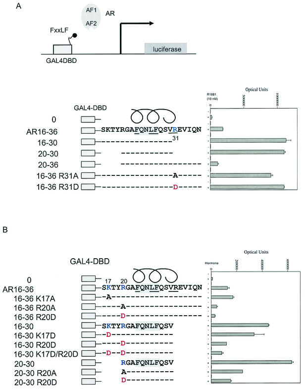
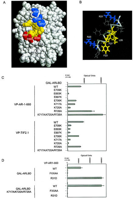
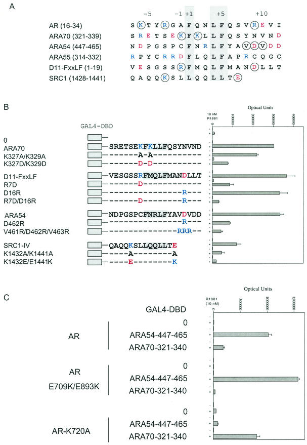
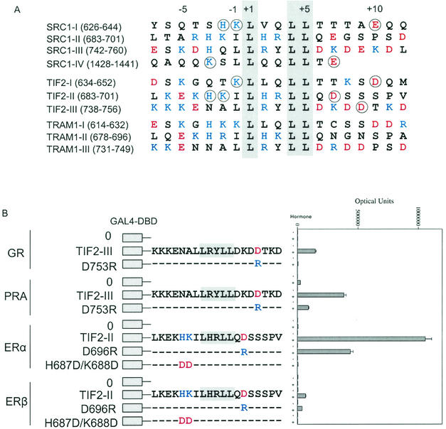
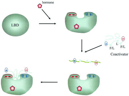
Coactivator recruitment by activation function 2 (AF2) in the steroid receptor ligand binding domain takes place through binding of an LXXLL amphipathic alpha-helical motif at the AF2 hydrophobic surface. The androgen receptor (AR) and certain AR coregulators are distinguished by an FXXLF motif that interacts selectively with the AR AF2 site. Here we show that LXXLL and FXXLF motif interactions with steroid receptors are modulated by oppositely charged residues flanking the motifs and charge clusters bordering AF2 in the ligand binding domain. An increased number of charged residues flanking AF2 in the ligand binding domain complement the two previously characterized charge clamp residues in coactivator recruitment. The data suggest a model whereby coactivator recruitment to the receptor AF2 surface is initiated by complementary charge interactions that reflect a reversal of the acidic activation domain-coactivator interaction model.


Figures

FIG. 1.
FIG. 1.
Effect of charged residues flanking the AR FXXLF in binding AR. (A) Mammalian two-hybrid assays were performed with HepG2 cells by coexpressing GAL-AR peptides containing the GAL4 DNA binding domain (GAL4-DBD) and the indicated AR NH2-terminal FXXLF motif amino acid wild-type or mutant sequence. GAL4-AR-FXXLF peptides (0.05 μg) were cotransfected using Effectine (Qiagen) with pCMVhAR (0.05 μg) coding for full-length human AR and the 5XGAL4Luc3 reporter vector (0.1 μg) in 12-well plates containing 0.2 × 10⁶ HepG2 cells. Transfected cells were incubated for 24 h in the absence and presence of 10 nM R1881 (methyltrienolone), a synthetic androgen. (B) Two-hybrid peptide interaction assays were performed with HepG2 cells as described previously using GAL4-AR peptides with the FXXLF motif region with wild-type or mutant sequence. The GAL-FXXLF peptide vectors with the indicated amino acid residue number and mutations were cotransfected with pCMVhAR to test the role of positively charged residues NH2-terminal to the AR NH2-terminal FXXLF motif.
FIG.2.
FIG.2.
Effect of charge clusters flanking AF2 in the AR LBD on AR-FXXLF and TIF2-LXXLL motif binding. (A) A space-filled model of the DHT-bound AR LBD is based on structural coordinates of rat AR646-901 (48), which has amino acid sequence identical to that of human AR664-919 (37). Positively charged residues K717, K720, and R726 flanking AF2 are shown in blue, negatively charged residues E709, E893, and E897 are shown in red, and AF2 hydrophobic residues are shown in yellow. DHT bound in the core structure would not be evident on the surface. (B) Ideal α-helical structure of the AR NH2-terminal FXXLF motif. The model helix was built using the Biopolymer module of the InsightII molecular modeling system from Accelrys Inc. (www.accelrys.com). (C) Two-hybrid assays were performed with HepG2 cells using GAL-AR-LBD (0.15 μg) coding for the GAL4 DNA binding domain fused to AR LBD containing residues 624 to 919 with wild-type (WT) or the indicated mutant sequences. GAL-AR-LBD was cotransfected with the 5XGAL4Luc3 reporter vector (0.1 μg) and VP-AR1-660 (0.15 μg) containing the VP16 transactivation domain and AR NH2-terminal residues 1 to 660 or VP-TIF2.1 containing the three-LXXLL motif region in TIF2. Assays were performed with HepG2 cells in the absence and presence of 10 nM R1881. (D) Two-hybrid assays were performed using VP-AR1-660 containing wild-type sequence or the 23FQNLF27-to-FQNAA (FXXAA) or R31D mutation. GAL-AR-LBD expressed residues 624 to 919 with wild-type sequence or with a triple mutation, K717A/K720A/R726A, as indicated. HepG2 cells were cotransfected with wild-type and mutant VPAR1-660 together with wild-type or mutant GAL-AR-LBD and the 5XGAL4Luc3 reporter vector. Cells were incubated for 24 h in the absence and presence of 10 nM 1881, and numbers of luciferase optical units were determined.
FIG.3.
FIG.3.
Requirement for charged residues flanking the FXXLF and LXXLL motifs in AR coregulator interaction with AR. (A) FXXLF and LXXLL motifs and flanking sequence of regions that bind AR AF2. Shown are human AR amino acid residues 16 to 34 that contain the FXXLF motif sequence 23FQNLF27, which mediates the N/C interaction (19), and the FXXLF motif regions from AR coregulators ARA70 (residues 321 to 339), ARA54 (residues 447 to 465), and ARA55 (residues 314 to 332) (20). Also shown are the D11-FXXLF peptide (20) and SRC1 carboxyl-terminal LXXLL-IV motif (residues 1428 to 1441) (47). Consensus residues of the binding motifs are shaded, and the relative positions of amino acids are numbered from the start of the core sequence. Residues that were mutated are circled. Basic residues K and R are shown in blue, and acidic residues D and E are shown in red. (B) Two-hybrid peptide interactions with AR. The GAL-ARA70 peptide contained ARA70 residues 321 to 340 with wild-type or mutant sequence as indicated. GAL-D11-FXXLF contained sequence derived from the D11-LXXLL peptide (44) that was changed to an FXXLF motif (20) with wild-type or mutant sequence. Numbering of the D11 peptide was left to right from the beginning of the peptide. The GAL-ARA54 peptide contained residues 447 to 465 with wild-type or mutant sequence. GAL-SRC1-IV contained residues 1428 to 1441 including the fourth and carboxyl-terminal LXXLL motif with WT or mutant sequence. GAL peptide vectors were cotransfected with pCMVhAR coding for full-length human AR and the 5XGAL4Luc3 reporter vector in HepG2 cells as described. Cells were incubated in the absence and presence of 10 nM R1881 for 24 h, and numbers of luciferase optical units were determined. (C) Two-hybrid interaction assays were performed as described above using full-length wild-type AR and AR mutants E709K/E898K or K720A, cotransfected either with empty parent vector (GAL4-DBD-0) or with the GAL4-DBD-peptide fusion proteins. These were cotransfected with GAL-ARA54-447-465 or GAL-ARA70-321-340. Luciferase activity was determined in the absence and presence of 10 nM R1881 as shown.
FIG. 4.
FIG. 4.
Flanking charged residue requirements of p160 coactivator LXXLL motif binding to steroid receptors. (A) Amino acid sequence of the LXXLL motif regions in the human p160 coactivators SRC1 (47), TIF2 (29, 56), and TRAM1 (53). Basic residues K, R, and H are shown in blue, and acidic residues D and E are shown in red. The conserved LXXLL motif is shaded, and the residues that were mutated are circled. The LXXLL motifs are numbered relative to the start of the core motif. (B) Two-hybrid interaction assay of TIF2-LXXLL peptides II and III with full-length steroid receptors. GAL fusion peptide vectors coding for TIF2-LXXLL-II and TIF2-LXXLL-III were cotransfected with the 5XGAL4Luc3 reporter vector and expression vectors for GR (pCMVhGR), PR-A (VP-PR-A), ERα (VP-ERα-LBD coding for residues 312 to 595), and ERβ (VP-ERβ full-length residues 1 to 530). Transfected HepG2 cells were incubated in the absence or presence of 10 nM dexamethasone for GR, 10 nM R5020 for PR-A, and 1 μM 17β-estradiol for ERα and ERβ.
FIG.5.
FIG.5.
Role of charged residues flanking AF2 in ERα LBD in TIF2-LXXLL-II binding. In panel A, the space-filled model of the ERα LBD was based on structural coordinates of ERα amino acids 297 to 554 in the presence of diethylstilbestrol (50). Positively charged residues K362 and R363 are shown in blue, negatively charged residues E380, D538, E542, and D545 are shown in red, and AF2 hydrophobic residues are shown in yellow. Two-hybrid peptide interaction assays were performed by cotransfecting the 5XGAL4Luc3 reporter vector, VP-ERα-LBD with wild-type (WT) or mutant sequence, with either GAL-TIF2-LXXLL-II (B) or GAL-TRAM1-LXXLL-I (C). Cells were incubated in the absence and presence of 1 μM 17β-estradiol (E2), and luciferase activity was determined as described in Materials and Methods.
FIG. 6.
FIG. 6.
Role of charged residues flanking the LXXLL motifs in binding nonsteroid nuclear receptors. Two-hybrid interaction assays were performed using TIF2 and SRC1 LXXLL peptides with TRβ, VDR, and RXRα. GAL-TIF2-LXXLL-I, -II, and -III or GAL-SRC1-LXXLL-I and -IV expression vectors coding for wild-type or mutant sequence were cotransfected with pCMX-VP-F-hTRβ, pVP16-VDR, or VP16-hRXRα. Cells were incubated in the absence and presence of 1 μM triiodothyronine for TRβ, 100 nM 1,25-dihydroxyvitamin D for VDR, and 1 μM 9-cis-retinoic acid for RXRα for 24 h prior to determining luciferase activity. Basic residues K, R, and H are shown in blue, and acidic residues D and E are shown in red.
FIG. 7.
FIG. 7.
Isothermal titration calorimetry measurements of FXXLF and LXXLL peptide binding to the AR LBD. Binding parameters were determined as described in Materials and Methods by titrating ARA54 FXXLF peptide 447-NDPGSPCFNRLFYAVDVDD-465 (A), the AR NH2-terminal FXXLF peptide 20-RGAFQNLFQSV-30 (B), and the TIF2 LXXLL-III peptide 738-KKKENALLRYLLDKDDTK-755 (C) into solutions containing human AR LBD fragment (amino acid residues 663 to 919). Multiple sequential injections (50, 50, and 30, respectively) were performed for each of the peptide-AR LBD interactions. Thermograms are shown in the upper panels, and binding isotherms are shown below.
FIG. 8.
FIG. 8.
Schematic diagram of the charge polarity interaction model for steroid and nuclear hormone receptor recruitment of coactivators and for the AR N/C interaction. Hormone binding induces the formation of the AF2 hydrophobic cleft on the surface of the LBD that is flanked by clusters of oppositely charged residues. The charge patches on either side of AF2 interact with charged residues flanking the FXXLF or LXXLL motif sequences to mediate the androgen-dependent N/C interaction or steroid receptor coactivator recruitment. The less-ordered FXXLF or LXXLL motif is recruited to the AF2 interaction surface by complementary charges on either side of AF2, resulting in stabilization of the α-helix interface at the AF2 hydrophobic cleft.

Similar articles

Cited by

References

    1. Arriza, J. L., C. Weinberger, G. Cerelli, T. M. Glaser, B. L. Handelin, D. E. Housman, and R. M. Evans. 1987. Cloning of human mineralocorticoid receptor complementary DNA: structural and functional kinship with the glucocorticoid receptor. Science 237:268-275. - PubMed
    1. Baker, A. R., D. P. McDonnell, M. Hughes, T. M. Crisp, D. J. Mangelsdorf, M. R. Haussler, J. W. Pike, J. Shine, and B. W. O'Malley. 1988. Cloning and expression of full-length cDNA encoding human vitamin D receptor. Proc. Natl. Acad. Sci. USA 85:3294-3298. - PMC - PubMed
    1. Bledsoe, R. K., V. G. Montana, T. B. Stanley, C. J. Delves, C. J. Apolito, D. D. McKee, T. G. Consler, D. J. Parks, E. L. Stewart, T. M. Willson, M. H. Lambert, J. T. Moore, K. H. Pearce, and H. E. Xu. 2002. Crystal structure of the glucocorticoid receptor ligand binding domain reveals a novel mode of receptor dimerization and coactivator recognition. Cell 110:93-105. - PubMed
    1. Bradshaw, J. M., R. A. Grucza, J. E. Ladbury, and G. Waksman. 1998. Probing the “two-pronged plug two-holed socket” model for the mechanism of binding of the Src SH2 domain to phosphotyrosyl peptides: a thermodynamic study. Biochemistry 37:9083-9090. - PubMed
    1. Brzozowski, A., A. Pike, Z. Dauter, R. Hubbard, T. Bonn, O. Engstrom, L. Ohman, G. Greene, J. Gustafsson, and M. Carlquist. 1997. Molecular basis of agonism and antagonism in the oestrogen receptor. Nature 389:753-758. - PubMed

Publication types

MeSH terms