. 2003 Mar 3;197(5):633-42.
doi: 10.1084/jem.20021756.

Infectious hepatitis C virus pseudo-particles containing functional E1-E2 envelope protein complexes

Affiliations

Infectious hepatitis C virus pseudo-particles containing functional E1-E2 envelope protein complexes

Birke Bartosch et al. J Exp Med. .

Abstract

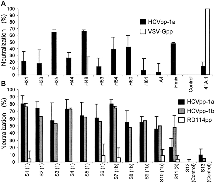
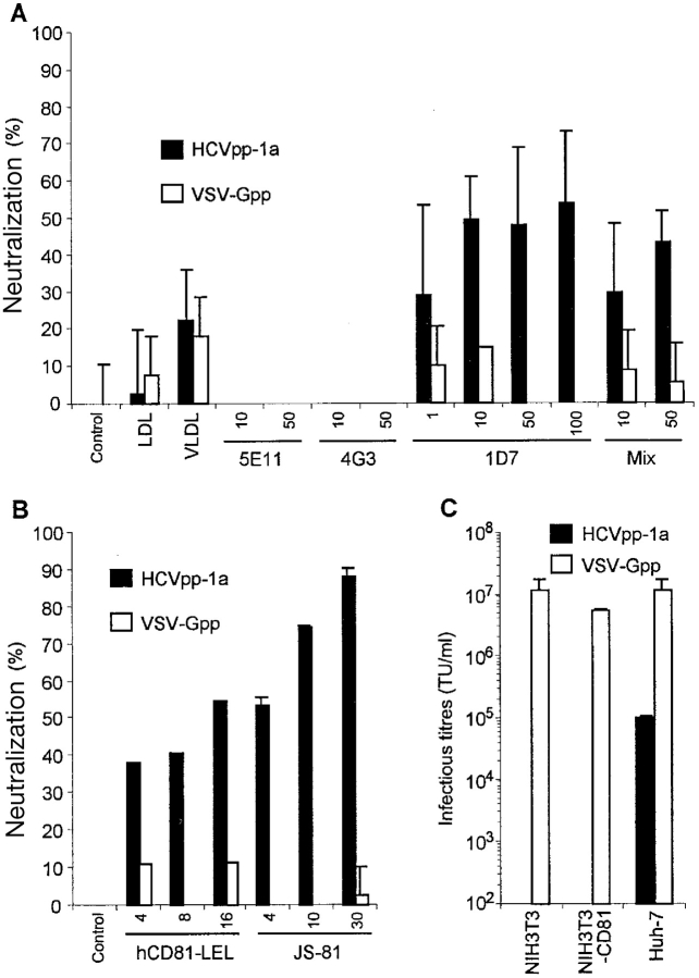
The study of hepatitis C virus (HCV), a major cause of chronic liver disease, has been hampered by the lack of a cell culture system supporting its replication. Here, we have successfully generated infectious pseudo-particles that were assembled by displaying unmodified and functional HCV glycoproteins onto retroviral and lentiviral core particles. The presence of a green fluorescent protein marker gene packaged within these HCV pseudo-particles allowed reliable and fast determination of infectivity mediated by the HCV glycoproteins. Primary hepatocytes as well as hepato-carcinoma cells were found to be the major targets of infection in vitro. High infectivity of the pseudo-particles required both E1 and E2 HCV glycoproteins, and was neutralized by sera from HCV-infected patients and by some anti-E2 monoclonal antibodies. In addition, these pseudo-particles allowed investigation of the role of putative HCV receptors. Although our results tend to confirm their involvement, they provide evidence that neither LDLr nor CD81 is sufficient to mediate HCV cell entry. Altogether, these studies indicate that these pseudo-particles may mimic the early infection steps of parental HCV and will be suitable for the development of much needed new antiviral therapies.


Figures

Figure 1.
Figure 1.
HCV E1E2 and retroviral expression constructs. (A) A cDNA derived from the HCV polyprotein gene was used to express the E1E2 glycoproteins and the COOH terminus of the C protein, which provides the signal peptide for E1 (SP E1). The position of stop codons (asterisk) inserted in the expression constructs to terminate translation of the proteins is shown. The transmembrane domain (TMD) of E1 provides the signal peptide (SP E2) for the E2 glycoprotein (46). NTR, nontranslated region; IRES, internal ribosome entry site. (B) The expression constructs encoding the different components required to assemble infectious pseudo-particles are shown. The shaded boxes represent the viral genes and the marker gene (GFP) transferred to the infected cells. The open boxes show the cis-acting sequences. LTR, long terminal repeat; CMV, cytomegalovirus immediate-early promoter; PBS, primer binding site; Ψ, packaging sequence; PPT, poly-purine track; polyA, polyadenylation site. Vector particles were produced by cotransfection of plasmids harboring the packaging functions, the transfer vector and the viral glycoproteins (GP) into 293T cells. The viral GPs were the HCV E1 and E2 glycoproteins, expressed individually or as a ΔCE1E2 polyprotein (Fig. 1 A), the VSV-G, or the RD114 glycoproteins. The supernatants of transfected cells were collected during transient expression, concentrated by ultracentrifugation, and used for target cell transduction.
Figure 2.
Figure 2.
Assembly of HCV pseudo-particles. (A) Detection of E2 glycoproteins by flow cytometry. E1E2-transfected 293T cells were or were not permeabilized before staining with A4 or H53 monoclonal antibodies against E1 (28) and E2 (black areas; reference 47). Cell-bound antibodies were incubated with FITC-conjugated antibodies before FACS® analysis. The background staining was provided by staining the cells with the conjugated antibodies only (white areas). (B) Immunoblots of lysates of 293T-transfected cells and of pseudo-particles pelleted through 20% sucrose cushions are shown. Expression of E1 and E2 glycoproteins from HCV-1a genotype and of MLV core proteins was revealed in reducing and denaturing conditions with A4 and H52 monoclonal antibodies against E1 (28) and E2 (29) or with an anticapsid (MLV CA) antiserum. VSV-G expressed in control pseudo-particles was detected with the monoclonal antibody P5D4. The positions of the molecular mass markers (kD) are shown. E2 present within the viral pellets migrated slightly slower than the cell-associated forms due to modifications of the associated glycans by Golgi enzymes (not depicted). The presence of VSV-G in viral pellets generated with MLV-G2A assembly-defective core proteins is due to empty vesicles formed by VSV-G itself (48). (C) Immunoblots of lysates of 293T producer cells and of purified pseudo-particles generated with E1 or E2 expressed alone, from two separate expression units, or coexpressed in trans or in cis, from a ΔCE1E2 polyprotein (E1E2).
Figure 3.
Figure 3.
Infectivity of HCV pseudo-particles. (A) The results of experiments performed with different target cell types are displayed as TU per milliliter of supernatant (mean ± SD of up to six experiments) for HCVpp of 1a genotype. The infectivity on Huh-7 cells of HCVpp concentrated 100× by ultracentrifugation is shown. Similar results of infectivity and host-range were obtained for HCVpp of 1b genotype (not depicted). The infectivity of control pseudo-particles generated with VSV-G ranged from 7 × 10⁶ to 2 × 10⁷ TU/ml, depending on the target cell type (not depicted). (B) Infectivity of HCV pseudo-particles generated without E1E2 (a), without retroviral core proteins (b), with MLV-G2A assembly defective core proteins (c), with HIV-1 core proteins (d), with E1 or E2 alone (e or f, respectively), with E1 + E2 expressed in trans from two independents vectors (g), with HCV-1a E1E2 expressed from the same vector in cis (h), or with HCV-1b E1E2 (i). HCVpp were treated with 25 µM AZT (3′-azido-3′-deoxythymidine) before and during infection of target cells (j). Infectious titers (TU/ml) were determined on Huh-7 target cells and are displayed as mean ± SD of up to four experiments.
Figure 4.
Figure 4.
Results of infection on human primary hepatocytes. Infection assays were performed with HCV pseudo-particles generated with core proteins derived from HIV-1 rather than from MLV, which do not permit transduction of nonproliferating target cells (11). (A) Photographs of human adult primary hepatocytes infected by HCVpp of genotype 1a carrying a lacZ marker gene encoding a nuclear-targeted β-galactosidase. The cells were stained 3 d after infection with X-Gal, as described previously (26). Magnification, 150×. The specificity of infection was demonstrated by the absence of transduction when target cells were infected with HIV-1–derived pseudo-particles lacking glycoproteins (pp cores) or by the reduced levels of transduction when target cells were preincubated with 30 µg/ml JS-81 anti-CD81 antibodies before infection (+JS-81). (B) Quantitative analysis of the infectivity of HCVpp on hepatocytes derived from two donors is expressed as a percentage of infectivity determined on Huh-7 cells.
Figure 4.
Figure 4.
Results of infection on human primary hepatocytes. Infection assays were performed with HCV pseudo-particles generated with core proteins derived from HIV-1 rather than from MLV, which do not permit transduction of nonproliferating target cells (11). (A) Photographs of human adult primary hepatocytes infected by HCVpp of genotype 1a carrying a lacZ marker gene encoding a nuclear-targeted β-galactosidase. The cells were stained 3 d after infection with X-Gal, as described previously (26). Magnification, 150×. The specificity of infection was demonstrated by the absence of transduction when target cells were infected with HIV-1–derived pseudo-particles lacking glycoproteins (pp cores) or by the reduced levels of transduction when target cells were preincubated with 30 µg/ml JS-81 anti-CD81 antibodies before infection (+JS-81). (B) Quantitative analysis of the infectivity of HCVpp on hepatocytes derived from two donors is expressed as a percentage of infectivity determined on Huh-7 cells.
Figure 5.
Figure 5.
Neutralization of HCV pseudo-particles. (A) Results of neutralization of HCVpp-1a generated with HCV-1a E1E2 and with MLV core proteins preincubated before infection of Huh-7 cells with 20 µg/ml saturating concentrations of monoclonal antibodies against E1 (A4) or E2 (H31, H33, H35, H44, H48, H53, H54, H60, and H61) glycoproteins of genotype 1a. Hmix: neutralization assays with pooled antibodies. Negative control experiments were performed using no antibodies (Control) or using pseudo-particles generated with VSV-G (VSV-Gpp). Neutralization of the infectivity of these control pseudo-particles was achieved by using the VSV-G neutralizing 41A.1 monoclonal antibody. Results are expressed as the percentages of inhibition of the average infectious titers ± SD relative to incubation in the absence of antibodies. (B) Results of neutralization of HCVpp with HCV patient sera. HCVpp of genotypes 1a or 1b were preincubated for 30 min at room temperature with sera from chronically HCV-infected patients diluted 1:50 before infection of Huh-7 target cells. The genotype of HCV diagnosed in these patients is indicated in brackets. Results are expressed as percentages of inhibition of the average infectious titers ± SD relative to incubation with control sera from healthy individuals. Control experiments were performed using pseudo-particles generated with RD114 glycoproteins (RD114pp), rather than with VSV-G, which exhibits sensitivity to human complement (15). Efficient neutralization of the control pseudo-particles (not depicted) was demonstrated with a hyper-immune goat serum raised against the RD114 SU glycoprotein.
Figure 6.
Figure 6.
Cell entry receptor usage by HCV pseudo-particles. (A) Results of LDL receptor competition assays was performed with 10 µg/ml purified LDLs and VLDLs or using the monoclonal antibodies 5E11, 4G3 (anti-ApoB), or 1D7 (anti-ApoE) preincubated with the pseudo-particles before infection at the indicated concentrations (µg/ml) are shown. Mix: the 5E11, 4G3, and 1B7 antibodies were mixed at the indicated individual concentrations in the neutralization assays. (B) Results of CD81 receptor competition assays are shown. hCD81-LEL GST-fusion polypeptides were preincubated with the pseudo-particles before infection or JS-81 anti-CD81 antibodies were preincubated with the target cells before infection at the indicated concentrations (µg/ml). Results are expressed as percentages of inhibition of the infectious titers obtained on Huh-7 cells relative to titers obtained in the absence of inhibitors. Control experiments were performed using pseudo-particles generated with VSV-G (VSV-Gpp). (C) Comparative results of infection obtained on NIH/3T3, NIH/3T3-hCD81, and Huh-7 cells infected with HCVpp and with VSV-Gpp. Infectious titers (TU/ml) are displayed as mean ± SD of up to three experiments.

Comment in

Similar articles

Cited by

References

    1. Lavanchy, D., R. Purcell, F.B. Hollinger, C. Howard, A. Alberti, M. Kew, G. Dusheiko, M. Alter, E. Ayoola, P. Beutels, et al. 1999. Global surveillance and control of hepatitis C. J. Viral Hepat. 6:35–47. - PubMed
    1. Dieterich, D.T. 2002. Treatment of hepatitis C and anemia in human immunodeficiency virus-infected patients. J. Infect. Dis. 185:S128–137. - PubMed
    1. Lindenbach, B.D., and C.M. Rice. 2001. Flaviviridae: the viruses and their replication. In Fields Virology, 4th ed. D.M. Knipe and P.M. Howley, editors. Lippincott Williams & Wilkins, Philadelphia. 991–1042.
    1. Blight, K.J., A.A. Kolykhalov, and C.M. Rice. 2000. Efficient initiation of HCV RNA replication in cell culture. Science. 290:1972–1974. - PubMed
    1. Lohmann, V., F. Korner, J. Koch, U. Herian, L. Theilmann, and R. Bartenschlager. 1999. Replication of subgenomic hepatitis C virus RNAs in a hepatoma cell line. Science. 285:110–113. - PubMed

Publication types

MeSH terms