

Review
doi: 10.1083/jcb.200210140.
Fluorescence resonance energy transfer (FRET) microscopy imaging of live cell protein localizations
Affiliations
- PMID: 12615908
- PMCID: PMC2173363
- DOI: 10.1083/jcb.200210140
Item in Clipboard
Review
Fluorescence resonance energy transfer (FRET) microscopy imaging of live cell protein localizations
J Cell Biol.
.
Abstract
The current advances in fluorescence microscopy, coupled with the development of new fluorescent probes, make fluorescence resonance energy transfer (FRET) a powerful technique for studying molecular interactions inside living cells with improved spatial (angstrom) and temporal (nanosecond) resolution, distance range, and sensitivity and a broader range of biological applications.
Figures
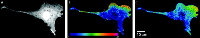
Confocal FRET analysis demonstrates that integrins induce local Rac–effector coupling. NIH-3T3 cells were microinjected with cDNAs encoding the indicated GFP–V12-Rac fusion proteins and then with Alexa–PBD protein (Pozo et al., 2002). Donor (A), uncorrected FRET (B), and corrected FRET (C) images are shown. In the color scale, red represents a high FRET signal and blue represents a low signal.
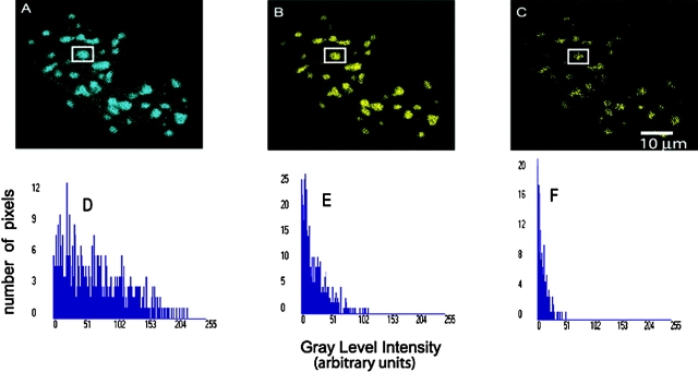
Localization of CFP– and YFP–C/EBPα proteins expressed in live mouse pituitary GHFT1-5 cells studied using Bio-Rad Laboratories MP-FRET microscopy. The donor (A), the uncorrected FRET (B), and processed FRET (C) images and their respective histograms (D, E, and F) representing the signal strength of the selected protein (Elangovan et al., 2003).
References
-
- Adams, S.R., A.T. Harootunian, Y.J. Buechler, S.S. Taylor, and R.Y. Tsien. 1991. Fluorescence ratio imaging of cyclic AMP in single cells. Nature. 349:694–697. - PubMed
-
- Bastiaens, P.I., and A. Squire. 1999. Fluorescence lifetime imaging microscopy: spatial resolution of biochemical processes in the cell. Trends Cell Biol. 9:48–52. - PubMed
-
- Clegg, R.M. 1996. Fluorescence resonance energy transfer. Fluorescence Imaging Spectroscopy and Microscopy. Vol. 137. X.F. Wang and B. Herman, editors. John Wiley & Sons Inc., New York. 179–251.
-
- Cole, N.B., C.L. Smith, N. Sciaky, M. Terasaki, M. Edidin, and J.L. Schwartz. 1996. Diffusional mobility of Golgi proteins in membranes of living cells. Science. 273:797–801. - PubMed
Publication types
MeSH terms
Substances
LinkOut - more resources
Full Text Sources
Other Literature Sources
