. 2003 Mar 3;160(5):709-18.
doi: 10.1083/jcb.200209098.

Regulation of cytochrome c oxidase activity by c-Src in osteoclasts

Affiliations

Regulation of cytochrome c oxidase activity by c-Src in osteoclasts

Tsuyoshi Miyazaki et al. J Cell Biol. .

Abstract

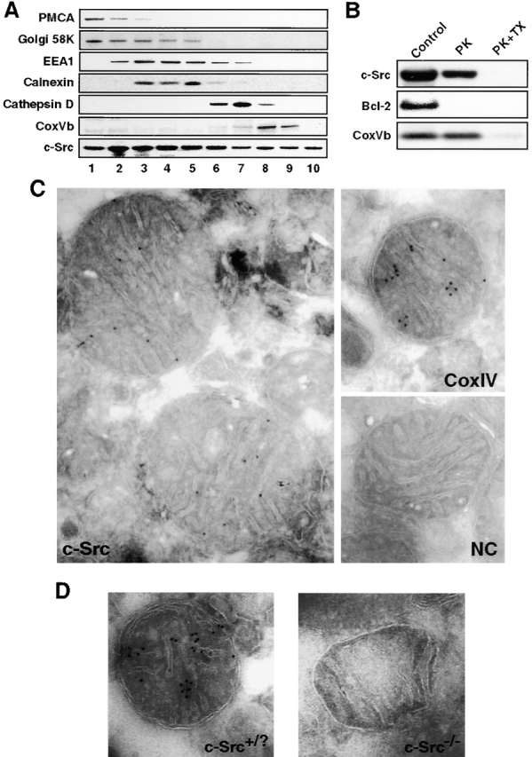
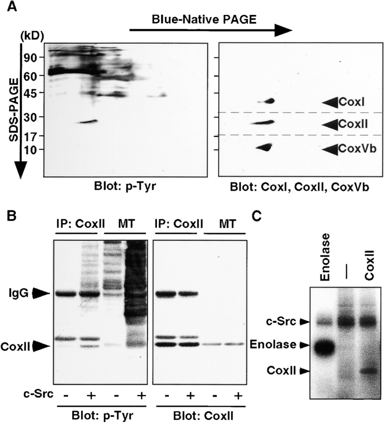
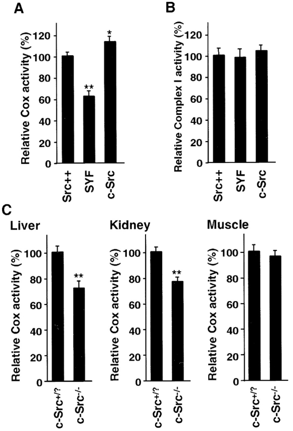
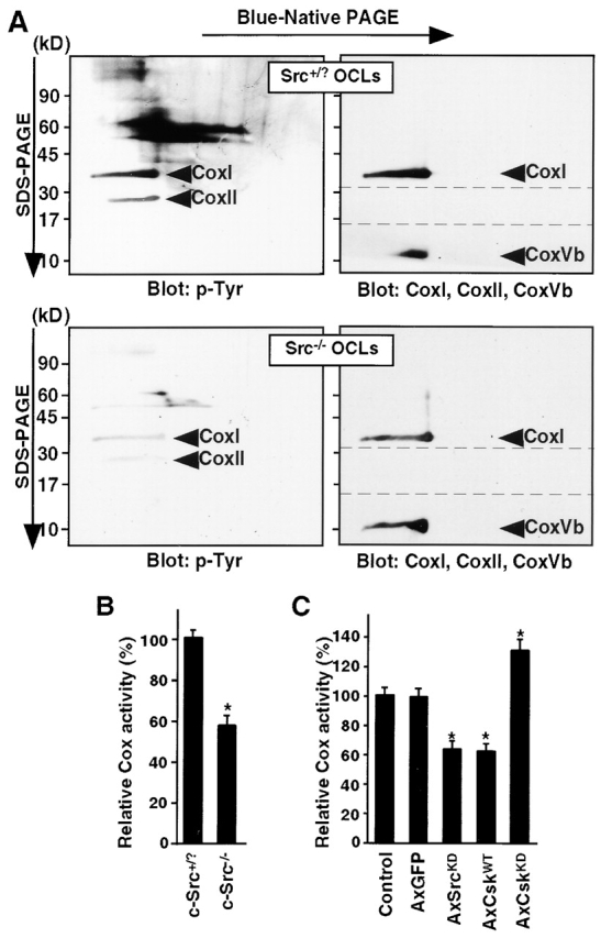
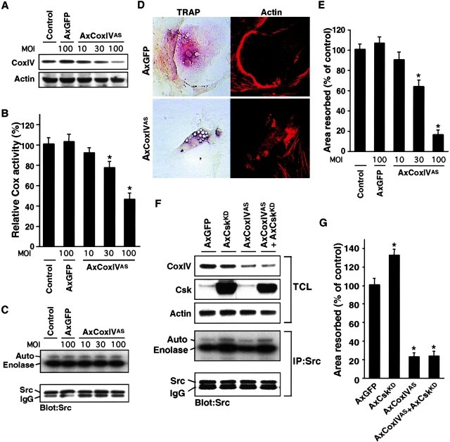
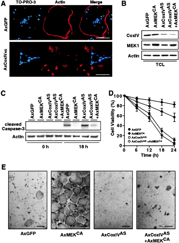
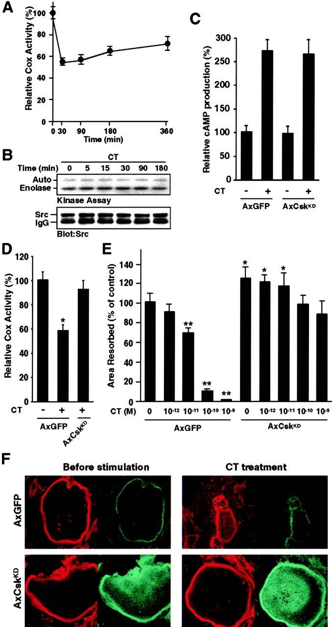
The function of the nonreceptor tyrosine kinase c-Src as a plasma membrane-associated molecular effector of a variety of extracellular stimuli is well known. Here, we show that c-Src is also present within mitochondria, where it phosphorylates cytochrome c oxidase (Cox). Deleting the c-src gene reduces Cox activity, and this inhibitory effect is restored by expressing exogenous c-Src. Furthermore, reducing endogenous Src kinase activity down-regulates Cox activity, whereas activating Src has the opposite effect. Src-induced Cox activity is required for normal function of cells that require high levels of ATP, such as mitochondria-rich osteoclasts. The peptide hormone calcitonin, which inhibits osteoclast function, also down-regulates Cox activity. Increasing Src kinase activity prevented the inhibitory effect of calcitonin on Cox activity and osteoclast function. These results suggest that c-Src plays a previously unrecognized role in maintaining cellular energy stores by activating Cox in mitochondria.


Figures

Figure 1.
Figure 1.
c-Src localization in subcellular fractions and purified mitochondria. (A) Subcellular fractionation of homogenized HEK 293 cells. Cell membranes were fractionated by centrifugation on discontinuous OptiPrep™ gradients, and the resulting fractions were immunoblotted with anti-PMCA, anti-Golgi 58K, anti-EEA1, anti-calnexin, anti-cathepsin D, anti-CoxVb, and anti-Src antibodies. (B) The mitochondrial fraction was isolated as described in Materials and methods and treated with 50 ng/ml proteinase K (PK) in the absence or presence of 0.5% Triton X-100 (TX) at RT for 30 min. The reaction was analyzed by Western blotting using antibodies to c-Src, Bcl-2, and CoxVb. (C) Immunogold labeling of c-Src in isolated mitochondria from HEK 293 cells. As positive control, CoxIV antibody was used for the primary antibody. As negative control (NC), gold-labeled secondary antibody was applied in the absence of c-Src antibody. (D) Immunogold labeling of c-Src in isolated mitochondria from c-Src+/? and c-Src−/− OCLs. c-Src was associated with the inner mitochondrial membrane in c-Src+/? OCLs, whereas no labeling was detected in the mitochondria of c-Src−/− OCLs.
Figure 2.
Figure 2.
Phosphorylation of Cox by c-Src. (A) Immunodetection of phosphotyrosine and Cox subunits in mitochondria from c-Src–overexpressing HEK 293 cells analyzed by two-dimensional electrophoresis. Mitochondrial proteins were separated in the first dimension by nondenaturing Blue-native PAGE and in the second dimension by SDS-PAGE, and were then transferred to nitrocellulose membranes. Membranes were immunoblotted with anti-phosphotyrosine (p-Tyr) antibody, then stripped, cut in three parts at the dotted lines, and reprobed with antibody to Cox subunits. The position of CoxI, CoxII, and CoxVb are indicated on the right. (B) Tyrosine phosphorylation of CoxII in HEK 293 cells with or without transfected c-Src expression vector. Endogenous CoxII protein was immunoprecipitated, and Western blotting was performed with anti-phosphotyrosine antibody. The membrane was then reprobed with anti-CoxII antibody. MT, total mitochondrial fraction. (C) Enolase and CoxII were incubated with c-Src in kinase buffer containing γ[32P]ATP at 30°C for 20 min. Positions of c-Src, enolase, and CoxII are indicated on the left.
Figure 3.
Figure 3.
Cox activity in c-Src–deficient cells. (A) Cox activity in cells expressing endogenous Src (Src++), SYF cells (SYF), and SYF cells transfected with c-Src (c-Src) was measured by determining the increase in OD (450 nm) as described in Materials and methods. The values are means ± SD (n = 8; *, P < 0.05 compared with Src++ cells; **, P < 0.01). (B) Complex I (NADH:ubiquinone oxidoreductase) activity in cells expressing endogenous Src (Src++), SYF cells (SYF), and SYF cells transfected with c-Src (c-Src). No significant difference was found. The values are means ± SD (n = 8). (C) Cox activity of liver, kidney, and muscle in c-Src+/? and c-Src−/− mice. The values are means ± SD (n = 8; **, P < 0.01 compared with c-Src+/? OCLs).
Figure 4.
Figure 4.
c-Src kinase activity modulates Cox activity in osteoclasts. (A) Immunodetection of phosphotyrosine and Cox subunits in mitochondria from c-Src+/? and c-Src−/− OCLs analyzed by two- dimensional electrophoresis. Membranes were immunoblotted with anti-phosphotyrosine (p-Tyr) antibody, then stripped, cut in three parts at the dotted lines, and reprobed with antibody to Cox subunits. Although the CoxII antibody did not recognize mouse CoxII, we can identify CoxII in the 2-D gel by its relative mobility. The position of CoxI, CoxII, and CoxVb are indicated. (B) Cox activity in c-Src+/? and c-Src−/− OCLs. The values are means ± SD (n = 8; *, P < 0.01 compared with uninfected OCLs). (C) Cox activity in OCLs infected with AxGFP, AxSrcKD, AxCskWT, or AxCskKD at an MOI of 100. The values are means ± SD (n = 8; *, P < 0.01 compared with uninfected OCLs).
Figure 5.
Figure 5.
CoxIV antisense prevents bone resorption in a dose-dependent manner. (A) OCLs were infected with AxGFP or AxCoxIVAS at the indicated MOIs. Membranes were immunoblotted with anti-CoxIV antibody, then stripped and reprobed with anti-actin antibody. (B) Cox activity in OCLs infected with AxGFP or AxCoxIVAS at the indicated MOIs. Cox activity of the cells was measured as described in Materials and methods. The values are means ± SD (n = 8; *, P < 0.01 compared with uninfected OCLs). (C) c-Src kinase activity in OCLs infected with AxGFP or AxCoxIVAS at the indicated MOIs was assayed by in vitro kinase assay using enolase as a substrate. (D) Effects of AxCoxIVAS on osteoclast morphology. Co-cultures infected with AxGFP or AxCoxIVAS at an MOI of 100 were plated on serum-coated glass coverslips for 12 h and then costained for TRAP activity (left) and for F-actin using rhodamine phalloidin (right). (E) Dentine-resorbing activity of OCLs expressing CoxIV antisense (AxCoxIVAS). An aliquot of the OCL preparation was transferred to dentine and cultured for an additional 12 h. The resorbed area on the dentine slices was measured as described in Materials and methods. The values are means ± SD (n = 8; *, P < 0.01 compared with uninfected OCLs). (F) OCLs infected with both AxCskKD and AxCoxIVAS expressed Csk and CoxIV at levels similar to those of the cells infected with either AxCskKD or AxCoxIVAS alone. c-Src kinase activity in OCLs coinfected with AxCskKD and AxCoxIVAS was measured by in vitro kinase assay using enolase as a substrate. (G) Dentine-resorbing activity of OCLs coinfected with AxCskKD and AxCoxIVAS. An aliquot of the OCL preparation was transferred onto dentine and cultured for an additional 12 h. The values are means ± SD (n = 8; *, P < 0.01 compared with AxGFP-infected OCLs).
Figure 6.
Figure 6.
Infection with AxCoxIVAS does not affect OCL survival. (A) Co-cultures infected with AxGFP or AxCoxIVAS at an MOI of 100 were plated on serum-coated glass coverslips for 12 h and then stained for F-actin using rhodamine phalloidin (red). To visualize the nuclei of the OCLs, the cells were stained with TO-PRO®-3 (blue). Bars, 50 μm. (B) OCLs coinfected with AxMEKCA and AxCoxIVAS at MOIs of 100 expressed the proteins at levels similar to the levels in the cells infected with either AxMEKCA or AxCOxIVAS alone. (C) Caspase-3 cleavage in OCLs infected with AxMEKCA and/or AxCoxIVAS. The time-dependent change in cleaved caspase-3 in purified OCLs was detected by Western blotting with anti-cleaved caspase-3 antibody. (D) Survival of OCLs infected with AxMEKCA or/and AxCoxIVAS. After purification, infected OCLs were incubated with αMEM/10% FBS for the indicated times. The number of viable cells remaining at the different time points is shown as a percentage of the number of cells at time 0. (E) Typical TRAP staining of OCLs 24 h after purification in the experiment quantified in (D). Bars, 100 μm.
Figure 7.
Figure 7.
The effects of CT treatment on OCLs. (A) Change in Cox activity with time in OCLs treated with CT (10−9 M). Cox activity in OCLs at the different time points is shown as a percentage of that in the cells at time 0. (B) c-Src kinase activity in OCLs treated with CT (10−9 M) was measured by in vitro kinase assay using enolase as a substrate. (C) AxGFP- or AxCskKD-infected OCLs were incubated for 20 min in the presence or absence of CT (10−9 M). cAMP production by the cells was measured as described in Materials and methods (n = 8). There was no significant difference in CT-induced cAMP production by GFP-expressing and CskKD-expressing OCLs. (D) AxGFP- or AxCskKD-infected OCLs were incubated for 30 min in the presence or absence of CT (10−9 M). Cox activity of the cells was measured as described in Materials and methods (n = 8; *, P < 0.01 compared with untreated AxGFP-infected OCLs). (E) Purified OCLs infected with AxGFP or AxCskKD at an MOI of 100 were cultured on dentine for 12 h with increasing concentrations of CT. The values are means ± SD (n = 8; *, P < 0.05 compared with untreated AxGFP-infected OCLs; **, P < 0.01). (F) Double immunofluorescence staining of F-actin (red) and Csk (green) in OCLs infected with AxGFP or AxCskKD at an MOI of 100. Purified GFP- or CskKD-expressing OCLs were treated with or without CT (10−9 M) for 1 h. CskKD expression prevented CT-induced disruption of actin rings.

References

    1. Bard, F., U. Patel, J.B. Levy, W.C. Horne, and R. Baron. 2002. Molecular complexes that contain both c-Cbl and c-Src associate with Golgi membranes. Eur. J. Cell Biol. 81:26–35. - PubMed
    1. Baron, R., J.-H. Ravesloot, L. Neff, M. Chakraborty, D. Chatterjee, A. Lomri, and W. Horne. 1993. Cellular and molecular biology of the osteoclast. Cellular and Molecular Biology of Bone. M. Noda, editor. Academic Press, San Diego. 445–495.
    1. Boyce, B.F., T. Yoneda, C. Lowe, P. Soriano, and G.R. Mundy. 1992. Requirement of pp60c-src expression for osteoclasts to form ruffled borders and resorb bone in mice. J. Clin. Invest. 90:1622–1627. - PMC - PubMed
    1. Brown, M.T., and J.A. Cooper. 1996. Regulation, substrates and functions of src. Biochim. Biophys. Acta. 1287:121–149. - PubMed
    1. Burke, P.A., and R.O. Poyton. 1998. Structure/function of oxygen-regulated isoforms in cytochrome c oxidase. J. Exp. Biol. 201:1163–1175. - PubMed

Publication types

MeSH terms