. 2003 Mar;15(3):612-25.
doi: 10.1105/tpc.008433.

Cell polarity and PIN protein positioning in Arabidopsis require STEROL METHYLTRANSFERASE1 function

Affiliations

Cell polarity and PIN protein positioning in Arabidopsis require STEROL METHYLTRANSFERASE1 function

Viola Willemsen et al. Plant Cell. 2003 Mar.

Abstract

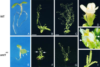
Plants have many polarized cell types, but relatively little is known about the mechanisms that establish polarity. The orc mutant was identified originally by defects in root patterning, and positional cloning revealed that the affected gene encodes STEROL METHYLTRANSFERASE1, which is required for the appropriate synthesis and composition of major membrane sterols. smt1(orc) mutants displayed several conspicuous cell polarity defects. Columella root cap cells revealed perturbed polar positioning of different organelles, and in the smt1(orc) root epidermis, polar initiation of root hairs was more randomized. Polar auxin transport and expression of the auxin reporter DR5-beta-glucuronidase were aberrant in smt1(orc). Patterning defects in smt1(orc) resembled those observed in mutants of the PIN gene family of putative auxin efflux transporters. Consistently, the membrane localization of the PIN1 and PIN3 proteins was disturbed in smt1(orc), whereas polar positioning of the influx carrier AUX1 appeared normal. Our results suggest that balanced sterol composition is a major requirement for cell polarity and auxin efflux in Arabidopsis.


Figures

Figure 1.
Figure 1.
The orc Mutant Alters Plant Morphology. (A) Five-day-old wild-type (WT) seedling grown on half-strength germination medium. (B) Four-week-old wild-type plant. (C) and (D) Six-week-old wild-type plant (C) and wild-type flower (D). (E) Five-day-old orc seedling grown on half-strength germination medium. Arrows indicate lobes of the cotyledons. (F) Three-week-old homozygous orc plant with two primary inflorescences (arrows). (G) Six-week-old homozygous orc plant. (H) orc terminal flower with sepals with frayed edges (arrows). (I) orc terminal flower with fused siliques. Bars in (B) and (C) = 2.5 cm for (B), (C), (F), and (G).
Figure 2.
Figure 2.
The orc Mutation Resides in the C-24 SMT1 Gene. (A) The orc between markers nga 249 and nga 151 (positions in centimorgan). (B) orc location relative to a contig of five BAC clones (T6I14, MSH12, MXE10, MAC12, and MUA22). Numbers above BACs indicate the positions of the markers corresponding to the recombination breakpoints. The number of recombinant seedlings between the marker and the orc locus are shown in parentheses. (C) The full-size TAC that complements the orc mutant. (D) Partial TAC clones used for complementation; only B was complementing. (E) Structure of SMT1. White boxes represent exons. The nucleotide sequence change at the splice site is depicted. (F) The mutation in the splice acceptor site yields two cDNAs: mt cDNA 1 (no splicing), the altered exon depicted with the large black box, and mt cDNA 2 (the next acceptor site used), indicated with a small black box. Horizontal arrows depict the positions of the primers used for reverse transcriptase–mediated PCR: F, forward; R, reverse (see Methods). (G) Alternative splicing leads to an early stop and a truncated protein. Vertical arrows indicate the intron-exon boundaries.
Figure 3.
Figure 3.
Cell Polarity Defects in smt1orc. (A) and (B) In the wild type (WT), amyloplasts are located at the basal ends of columella cells. (C) The nucleus is located at the apical end. (D) Root hairs initiate from the basal end of the polarized trichoblast. (E) and (F) Amyloplast position in smt1orc columella cells. (G) Nuclear localization in smt1orc columella cells. (H) The initiation site of smt1orc root hairs. In (D) and (H), long arrows indicate trichoblast length, and short arrows indicate the root hair initiation site. In (A) to (C) and (E) to (G), plastic sections of 5-day-old seedling roots are shown; whole mounts are shown in (D) and (H) under Nomarski optics. Bars = 25 μm in (A) and (E), 10 μm in (B) for (B), (C), (F), and (G), and 20 μm in (D) and (H).
Figure 4.
Figure 4.
Quantification of Polarity Defects in smt1orc. (A) Positions of nuclei in columella cells of the wild type (WT) and smt1orc. The relative position is the ratio between the distance from the apical cell wall to the apical side of the nucleus and the cell size. In the wild type, the nucleus is positioned toward the apical side (0, apical end; 1, basal end). (B) to (D) Root hair initiation sites on trichoblasts of the wild type and smt1orc are indicated in frequency distributions of average hair initiation per root (30 roots, 5 trichoblasts per root). Apical-basal initiation ratios indicate relative positions of hair initiation. Wild-type root hairs are located toward the basal end (0, basal end [normal end]; 1, apical end). (B) Results in wild-type and smt1orc seedlings germinated and grown for 5 days on half-strength germination medium. (C) Results in wild-type seedlings germinated and grown for 5 days with and without 20 nM 2,4-D. (D) Results in smt1orc seedlings germinated and grown for 5 days with and without 20 nM 2,4-D. (E) and (F) Polar auxin transport in stem sections. Results of polar auxin transport measurements were obtained from two independent experiments each on primary stem segments of 20 plants. (E) Results in stem segments loaded with 3H-IAA in the presence 2 × 10⁻⁶ M of the polar auxin transport inhibitor NPA at the apical side. Radioactivity was detected after transport and cutting in three sections: apical (column A), middle (column B), and basal (column C). (F) As in (E) but without NPA.
Figure 5.
Figure 5.
DR5-GUS Distribution Indicates Altered Auxin Distribution in smt1orc. (A) DR5-GUS distribution in a wild-type (WT) mature embryo. (B) A maximum of DR5-GUS expression localizes to a wild-type columella root cap of a 5-day-old seedling. (C) and (D) DR5-GUS staining after the application of 0.5 and 5 μM 2,4-D, respectively, in 5-day-old wild-type seedlings. (E) Localization of DR5-GUS in a mature smt1orc embryo. (F) Localization of DR5-GUS in a smt1orc root. (G) and (H) DR5-GUS accumulation in smt1orc roots treated with 0.5 and 5 μM 2,4-D, respectively. Bars = 25 μm in (A) for (A) and (E), 50 μm in (B) for (B) and (F), and 75 μm in (C) for (C), (D), (G), and (H).
Figure 6.
Figure 6.
Defective QC Marker Expression and Organization of Columella Root Cap Cells in smt1orc. (A) Cross-section of a wild-type (WT) stem tissue used for polar auxin transport measurements. (B) Regular radial organization of the root tip of a 5-day-old wild-type seedling. (C) Columella-specific marker 35S-B2-GUS in a 5-day-old wild-type seedling. (D) Cross-section of a smt1orc stem tissue used for polar auxin transport measurements. (E) Radial organization of a 5-day-old smt1orc seedling with irregular cell shape. Extra division in the endodermis is indicated with the arrow. (F) 35S-B2::GUS staining in a 5-day-old smt1orc seedling. (G) to (I) Columella-specific green fluorescent protein markers Q1630 (G) and J2341 (H) and the QC-specific promotor trap QC46 (I) in the wild type. (J) to (L) Columella-specific green fluorescent protein markers Q1630 (J) and J2341 (K) and the QC-specific promotor trap QC46 (L) in smt1orc. (A), (B), (D), and (E) show toluidine blue–stained plastic sections; (C), (F), (I), and (L) show Nomarski optic results after GUS staining; and (G), (H), (J), and (K) show confocal laser scanning microscopy images. Bars = 200 μm in (A) for (A) and (D) and 50 μm in (B), (C), and (L) for all other panels.
Figure 7.
Figure 7.
Development of smt1orc Embryos. (A), (E), and (I) Globular-stage embryos of the wild type (A) and two categories of smt1orc (E) and (I). Asterisks indicate QC progenitors. C, columella progenitor; H, hypophyseal cell. (B), (F), and (J) Heart-stage embryos of the wild type (B) and smt1orc (F) and (J). (C) Magnification of (B). (K) Magnification of (F). (D), (H), and (L) Mature embryos of the wild type (D) and smt1orc (H) and (L). Embryos were examined under Nomarski optics. Black arrows indicate periclinal divisions in the basal cells of smt1orc ground tissue. Arrowheads indicate multiple cotyledon primordia in smt1orc embryos. White arrows indicate undivided hypophyseal cells in smt1orc. Bars = 25 μm.
Figure 8.
Figure 8.
Cotyledon Development and Tropic Responses in smt1orc. (A) DR5-GUS expression in a wild-type (WT) mature embryonic cotyledon. (B) Vascular pattern of a cotyledon of a 5-day-old wild-type seedling. (C) Enlargement of (B). (D) Five-day-old seedlings of the wild type. (E) Scheme of the gravitropic response of wild-type seedling roots after 90° rotation and 24 h in the dark. 0 indicates the direction of the gravity vector after rotation. (F) Multiple DR5-GUS maxima in a smt1orc mature embryonic cotyledon (black arrows). (G) Lobed cotyledon of a 5-day-old smt1orc seedling (arrowheads). (H) Enlargement of (G) showing the discontinuous vascular system and the formation of “vascular islands” (white arrows). (I) Altered tropism in 5-day-old smt1orc seedlings. (J) Gravitropic response of smt1orc seedling roots. (B), (C), (F), and (G) show dark-field images of cotyledons of 5-day-old seedlings cleared with chloral hydrate. Bars = 100 μm in (B) and (C) for (B), (C), (G), and (H), 25 μm in (A) for (A) and (F), and 2 mm in (D) for (D) and (I).
Figure 9.
Figure 9.
Localization of HA::AUX1 and Delocalized Auxin Efflux Carriers in smt1orc and smt1orc gn Double Mutants. (A) and (B) Immunolocalization of HA::AUX1 at the apical end in protophloem cells of the wild type (A) and smt1orc (B) (arrow indicates an oblique cell wall). (C) to (E) Immunolocalization of PIN1 in the vascular bundle. Expression at the basal end in the central cells in the wild type (C) and delocalized expression in vascular cells (D) and (E) (arrows indicate central, properly axialized smt1orc cells). (F) to (H) Immunolocalization of PIN3 in columella cells nearly uniform in the wild type (F) and delocalized in smt1orc (G) and (H) (arrows). (I) to (K) smt1orc gnT391 double-mutant analysis. (I) smt1orc seedlings, with multiple cotyledons indicated by the arrow. (J) gnT391 seedling. (K) Additive phenotypes of smt1orc gnT391 double mutants showing slightly smaller seedlings and multiple cotyledons (arrows). Bars = 10 μm in (A), (C), and (F) for (A) to (H) and 1 mm in (I) and (J) for (I) to (K).

References

    1. Altmann, T. (1998). Recent advances in brassinosteroid molecular genetics. Curr. Opin. Plant Biol. 1, 378–383. - PubMed
    1. Athenstaedt, K., Zweytick, D., Jandrositz, A., Kohlwin, S.D., and Daum, G. (1999). Identification and characterization of major lipid particle proteins of the yeast Saccharomyces cerevisiae. J. Bacteriol. 181, 6441–6448. - PMC - PubMed
    1. Bagnat, M., and Simons, K. (2002). Cell surface polarization during yeast mating. Proc. Natl. Acad. Sci. USA 99, 14183–14188. - PMC - PubMed
    1. Bechtold, N., Ellis, J., and Pelletier, G. (1993). In planta Agrobacterium-mediated gene transfer by infiltration of adult Arabidopsis plants. C. R. Acad. Sci. Paris 316, 1194–1199.
    1. Bligh, E.G., and Dyer, W.J. (1959). A rapid method of total lipid extraction and purification. Can. J. Biochem. Physiol. 37, 91–97. - PubMed

Publication types

MeSH terms