. 2003 Mar;185(6):1768-75.
doi: 10.1128/JB.185.6.1768-1775.2003.

Roles of the enantioselective glutathione S-transferases in cleavage of beta-aryl ether

Affiliations

Roles of the enantioselective glutathione S-transferases in cleavage of beta-aryl ether

Eiji Masai et al. J Bacteriol. 2003 Mar.

Abstract

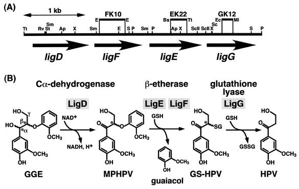
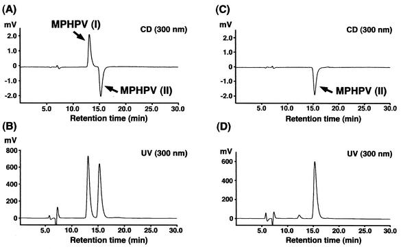
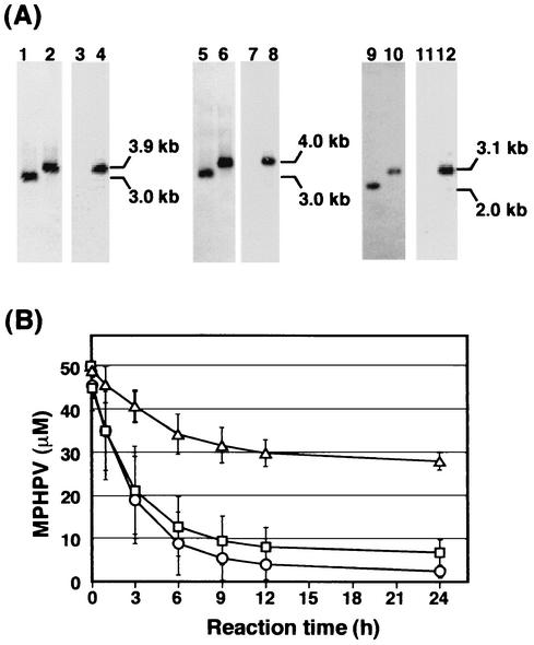
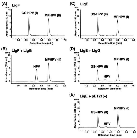
Cleavage of the beta-aryl ether linkage is the most important process in lignin degradation. Here we characterize the three tandemly located glutathione S-transferase (GST) genes, ligF, ligE, and ligG, from low-molecular-weight lignin-degrading Sphingomonas paucimobilis SYK-6, and we describe the actual roles of these genes in the beta-aryl ether cleavage. Based on the identification of the reaction product by electrospray ionization-mass spectrometry, a model compound of beta-aryl ether, alpha-(2-methoxyphenoxy)-beta-hydroxypropiovanillone (MPHPV), was transformed by LigF or LigE to guaiacol and alpha-glutathionyl-beta-hydroxypropiovanillone (GS-HPV). This result suggested that LigF and LigE catalyze the nucleophilic attack of glutathione on the carbon atom at the beta position of MPHPV. High-pressure liquid chromatography-circular dichroism analysis indicated that LigF and LigE each attacked a different enantiomer of the racemic MPHPV preparation. The ligG gene product specifically catalyzed the elimination of glutathione from GS-HPV generated by the action of LigF. This reaction then produces an achiral compound, beta-hydroxypropiovanillone, which is further degraded by this strain. Disruption of the ligF, ligE, and ligG genes in SYK-6 showed that ligF is essential to the degradation of one of the MPHPV enantiomers, and the alternative activities which metabolize the substrates of LigE and LigG are present in this strain.


Figures

FIG. 1.
FIG. 1.
Organization of the ligDFEG gene cluster (A) and deduced functions of the gene products in the β-aryl ether cleavage (B). (A) The ligD, ligF, ligE, and ligG genes are indicated by the thick arrows. Vertical bars above the restriction map indicate the positions of the Kmr gene insertions of ligF (FK10), ligE (EK22), and ligG (GK12) mutants. Abbreviations for restriction enzymes: Ap, ApaI; Bs, BstXI; E, EcoRI; Ec, Eco47III; Ml, MluI; P, PstI; RV, EcoRV; S, SalI; Sc, SacI; ScII, SacII; St, StuI; Sm, SmaI; Tt, Tth111I; and X, XhoI. (B) GGE, guaiacylglycerol-β-guaiacyl ether; GSH, glutathione; GSSG, glutathione disulfide. Asterisks indicate the asymmetric carbons.
FIG. 2.
FIG. 2.
SDS-PAGE analysis of protein fractions. Proteins were separated on an SDS-12% polyacrylamide gel and stained with Coomassie brilliant blue. Lanes: 1, molecular mass markers; 2, crude extract of E. coli BL21(DE3) harboring pET21(+) (10 μg of protein); 3, cell extract of E. coli BL21(DE3) harboring pETF48 (10 μg of protein); 4, HQ fraction (3 μg of protein); 5, PE fraction (1.5 μg of protein). Molecular masses are given on the left.
FIG. 3.
FIG. 3.
Identification of the reaction products from MPHPV catalyzed by LigF. Purified LigF was incubated with the MPHPV preparation in the presence of glutathione. (A and B) HPLC chromatograms at 0 and 10 min of incubation, respectively. Compounds were detected at 280 nm. (C and D) ESI-MS spectra of the reaction mixtures at 0 and 10 min of incubation, respectively. GSH, glutathione; AU, absorbance units.
FIG. 4.
FIG. 4.
Chiral HPLC-CD analysis of LigF enantioselectivity. (A and B) The MPHPV preparation was separated on a chiral column by HPLC and was detected with CD (A) and UV (B) detectors. (C and D) After incubation of the MPHPV preparation with LigF, the remaining MPHPV was analyzed by HPLC and detected with CD (C) and UV (D) detectors.
FIG. 5.
FIG. 5.
Disruption of ligF, ligE, and ligG in S. paucimobilis SYK-6. (A) Southern hybridization analysis of the insertion mutants. Lanes: 1, 3, 5, and 7, total DNA of SYK-6 digested with EcoRV and MluI; 2 and 4, total DNA of the ligF insertion mutant (FK10) digested with EcoRV and MluI; 6 and 8, total DNA of the ligE insertion mutant (EK22) digested with EcoRV and MluI; 9 and 11, total DNA of SYK-6 digested with SalI; and 10 and 12, total DNA of the ligG insertion mutant (GK12) digested with SalI. The 1.3-kb SalI fragment carrying the Kmr gene (lanes 3, 4, 7, 8, 11, and 12), the 1.7-kb XhoI fragment carrying ligF and part of ligE (lanes 1, 2, 5, and 6), and the 0.6-kb SacI-SalI fragment carrying part of ligG (lanes 9 and 10) were used as probes. (B) Degradation of MPHPV by cell extracts of SYK-6 (circles), FK10 (triangles), and EK22 (squares) in the presence of glutathione. The rate of transformation of MPHPV by the cell extract of GK12 was at the same level as that for SYK-6. Error bars indicate standard deviations.
FIG. 6.
FIG. 6.
Detection of the glutathione lyase activity of LigG. (A and C) HPLC chromatograms of the substrates, GS-HPV I and GS-HPV II, generated from the MPHPV preparation by the actions of LigF and LigE enzymes, respectively, in the presence of glutathione. (B) HPLC chromatogram of the reaction product of GS-HPV I incubated for 10 min with the cell extract of E. coli BL21(DE3) harboring pETG9 (10 μg of protein/ml). (D and E) HPLC chromatograms of the reaction products of GS-HPV II incubated for 60 min with the cell extract of E. coli BL21(DE3) harboring pETG9 (500 μg of protein/ml) and with the cell extract of E. coli BL21(DE3) harboring pET21(+) (500 μg of protein/ml), respectively. AU, absorbance units.

References

    1. Adler, E. 1957. Structural elements of lignin. Ind. Eng. Chem. 49:1377-1383.
    1. Adler, E., and E. Eriksoo. 1955. Guaiacylglycerol and its β-guaiacyl ether. Acta Chem. Scand. 9:341-342.
    1. Akiyama, T., K. Magara, Y. Matsumoto, G. Meshitsuka, A. Ishizu, and K. Lundquist. 2000. Proof of the presence of racemic forms of arylglycerol-β-aryl ether structure in lignin: studies on the stereo structure of lignin by ozonation. J. Wood Sci. 46:414-415.
    1. Ausubel, F. M., R. Brent, R. E. Kingston, D. D. Moore, J. G. Seidman, J. A. Smith, and K. Struhl. 1990. Current protocols in molecular biology. John Wiley & Sons, Inc., New York, N.Y.
    1. Board, P. G., M. Coggan, G. Chelvanayagam, S. Easteal, L. S. Jermiin, G. K. Schulte, D. E. Danley, L. R. Hoth, M. C. Griffor, A. V. Kamath, M. H. Rosner, B. A. Chrunyk, D. E. Perregaux, C. A. Gabel, K. F. Geoghegan, and J. Pandit. 2000. Identification, characterization, and crystal structure of the omega class glutathione transferases. J. Biol. Chem. 275:24798-24806. - PubMed

MeSH terms

LinkOut - more resources