. 2003 Mar;111(5):607-16.
doi: 10.1172/JCI17038.

Randall's plaque of patients with nephrolithiasis begins in basement membranes of thin loops of Henle

Affiliations

Randall's plaque of patients with nephrolithiasis begins in basement membranes of thin loops of Henle

Andrew P Evan et al. J Clin Invest. 2003 Mar.

Abstract

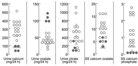
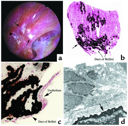
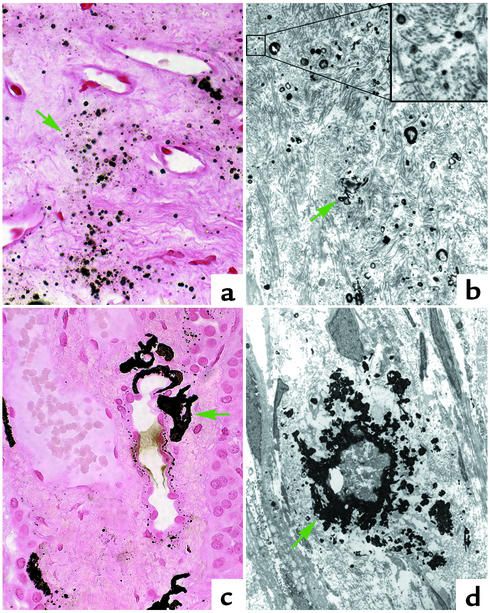
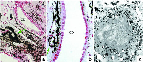
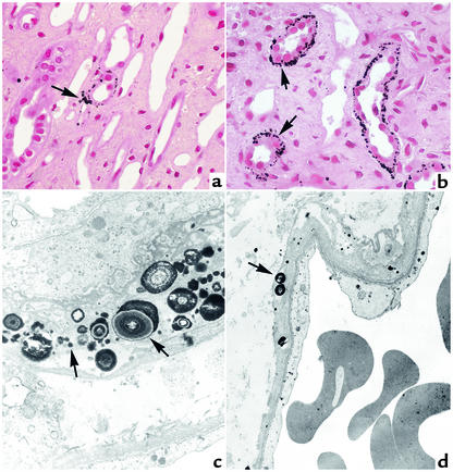
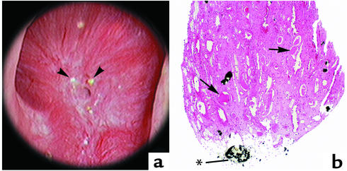
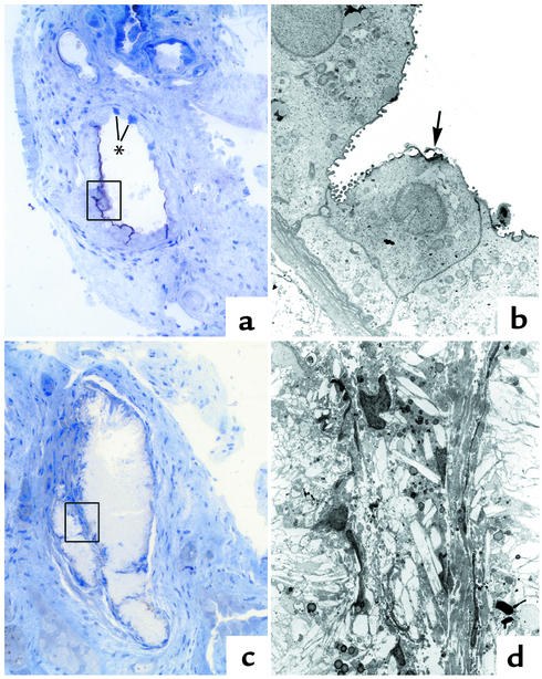
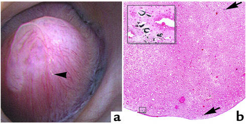
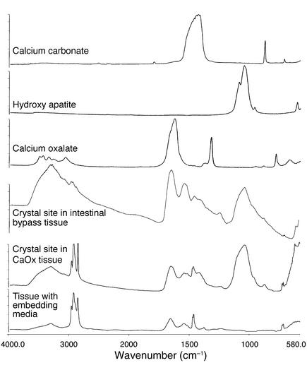
Our purpose here is to test the hypothesis that Randall's plaques, calcium phosphate deposits in kidneys of patients with calcium renal stones, arise in unique anatomical regions of the kidney, their formation conditioned by specific stone-forming pathophysiologies. To test this hypothesis, we performed intraoperative biopsies of plaques in kidneys of idiopathic-calcium-stone formers and patients with stones due to obesity-related bypass procedures and obtained papillary specimens from non-stone formers after nephrectomy. Plaque originates in the basement membranes of the thin loops of Henle and spreads from there through the interstitium to beneath the urothelium. Patients who have undergone bypass surgery do not produce such plaque but instead form intratubular hydroxyapatite crystals in collecting ducts. Non-stone formers also do not form plaque. Plaque is specific to certain kinds of stone-forming patients and is initiated specifically in thin-limb basement membranes by mechanisms that remain to be elucidated.


Figures

Figure 1
Figure 1
Selected urine values. Common-stone formers are represented by open circles, bypass patients by filled circles, and normal subjects by triangles. The values for a comparison of stone formers with bypass patients are as follows: urine calcium, 312 ± 89 versus 81 ± 29; oxalate, 40 ± 13 versus 106 ± 11; citrate, 485 ± 278 versus 144 ± 107; supersaturation with respect to CaOx monohydrate, 11 ± 4 versus 5 ± 2; and supersaturation with respect to calcium phosphate, 1.4 ± 8 versus 0.09 ± .05. P < 0.01 for all values by t test and Kolmogorov-Smirnov nonparametric testing. For normal subjects, values for calcium, oxalate, citrate, supersaturation with respect to CaOx, and supersaturation with respect to calcium phosphate were 113 ± 67, 32 ± 9, 482 ± 193, 3.5 ± 1.3, and 0.42 ± .16, respectively; P = < 0.01 versus stone formers by t test and Kolmogorov-Smirnov for calcium, supersaturation with respect to CaOx, and supersaturation with respect to calcium phosphate; P < 0.0001 versus bypass by t test and Kolmogorov-Smirnov for oxalate; P = 0.022 by t test and 0.0001 by Kolmogorov-Smirnov for supersaturation with respect to calcium phosphate; and P = 0.03 by t test and was not significant by Kolmogorov-Smirnov for citrate. The other comparisons of normal subjects to patient groups were not significant by both t test and Kolmogorov-Smirnov. SS, supersaturation.
Figure 2
Figure 2
Endoscopic and histologic images of Randall’s plaques in CaOx patients. In (a), an example of a papilla from a CaOx-stone former that was video recorded at the time of the mapping is shown. Several sites of Randall’s plaque (arrows) appear as irregular white areas beneath the urothelium in the CaOx patient. In addition, a plaque site was noted that lacked a urothelial layer and is thought to be a site where a stone had been attached to the side of the papilla (arrowhead). In (b), a low-magnification light-microscopic image of a papillary biopsy specimen from a CaOx patient is shown; the sites of calcium deposits (arrows) were stained black by the Yasue metal substitution method for calcium histochemistry. (c) A light micrograph shows large regions of crystal deposits in the interstitial tissue surrounding the ducts of Bellini (arrowhead), proceeding to the urothelium of the papillary tip (arrow), and progressing up the inner medulla. (d) A transmission electron micrograph shows the crystalline material (arrow) comprising Randall’s plaque to be normally covered by a complete layer of urothelium, making the plaque a suburothelial structure. Magnification, ×100 (b); ×600 (c); ×6,000 (d).
Figure 3
Figure 3
Crystalline deposits on interstitial collagen. In (a) and (b), numerous sites of crystal deposition are shown (green arrows) on the interstitial collagen located between the loops of Henle and nearby vascular bundles in the papillary tissue of a CaOx patient. The inset in (b) illustrates the relationship of the initial small sites of crystal formation on individual collagen fibers at a higher magnification. In (c) and (d), a progressive accumulation of crystal deposition (green arrows) is shown in the interstitium near the loops of Henle. Magnification, ×650 (a); ×4,750 (b); ×16,500 (inset in b); ×900 (c); ×3,900 (d).
Figure 4
Figure 4
Accumulation of interstitial crystal deposits. In (a) and (b), extensive accumulation of crystalline deposition (green arrows) is shown around a few loops of Henle and nearby vascular bundles, resulting in the formation of incomplete to complete cuffs (indicated by circles) of crystalline material in the papillary tissue of a CaOx patient. Note the accumulation of crystal material in the basement membrane of nearby collecting duct and the normal appearance of the collecting duct cells. In (c), an electron micrograph shows a dense accumulation of crystalline material around a loop of Henle that appears to be necrotic. Magnification, ×900 (a); ×1,600 (b); ×400 (c). CD, collecting duct.
Figure 5
Figure 5
Initial sites of crystal deposition. This set of illustrations shows the initial sites and size of calcium deposition in the papillary tissue of a CaOx patient as seen by light (a and b) and transmission electron (c and d) microscopy. Sites of crystalline material (arrows) are noted in the basement membranes, near the collagen of the thin loops of Henle (ac), and to a lesser degree in the basement membranes of vasa recta (d). Magnification, ×900 (a); ×1,000 (b); ×15,600 (c); ×5,500 (d).
Figure 6
Figure 6
Endoscopic and histologic images of Randall’s plaques in intestinal bypass patients. In (a), an example of a papilla from an intestinal-bypass stone former that was video recorded at the time of the mapping is shown. Distinct sites of Randall’s plaque material are not found on the papilla of the intestinal-bypass patient; instead, several nodular-appearing structures (arrowheads) were noted near the opening of the ducts of Bellini. In (b), a low magnification light microscopic image of a papillary biopsy specimen from an intestinal-bypass patient is shown. Crystal deposition was only found in the lumens of a few collecting ducts as far down as the ducts of Bellini (*). A large site of crystal material was seen in a duct of Bellini. No other sites of deposits were noted. Note dilated collecting ducts (arrows) with cast material and regions of fibrosis around crystal-deposit–filled collecting ducts. Magnification, ×100 (b).
Figure 7
Figure 7
Pattern of intraluminal crystal deposition in medullary tissue of bypass patients. In (a), early crystal attachment to the apical surface of several collecting duct cells is shown in a 1-μm plastic section. At this stage, these collecting ducts have a mixture of normal and injured (*) cells. In (b), several cells are shown by transmission electron microscopy (see rectangle in a) that possess attached crystals (arrow). In (c), another 1-μm plastic section of a collecting duct shows complete crystallization of the tubular lumen and lining cells. In (d), an electron micrograph of a region of the collecting ducts seen in (c) is shown (see rectangle). No cellular detail remains; only crystalline material is found. Magnification, ×450 (a); ×6,400 (b); ×550 (c); ×4,000 (d).
Figure 8
Figure 8
Endoscopic and crystal deposition in papillary tissue of non–stone formers. In (a), example of a papilla from a non–stone former that was video recorded at the time of the mapping is shown. As in bypass patients, no distinct sites of Randall’s plaque are noted on the papilla of the non–stone formers; instead, a nodular-appearing structure (arrowhead) was seen along the side of the papilla. In (b), a low-magnification light-microscopic image of a large papillary sample of the only non–stone former shows several sites of Yasue-positive material. Crystal deposition was only found surrounding a few loops of Henle (arrows) and several vasa recta (inset). Magnification, ×15 (b); ×119 (inset in b).
Figure 9
Figure 9
μ-FTIR spectra of crystal deposits in CaOx and intestinal-bypass patients. This figure illustrates a series of infrared spectra obtained for a set of standards (calcium carbonate, CaOx, and hydroxyapatite), for a site of calcium deposit (Yasue-positive area) in the tissue of a CaOx and an intestinal-bypass patient, and for the tissue-embedding medium. The infrared spectra of the Yasue-positive area for both the CaOx and intestinal-bypass patients show a spectral band for hydroxyapatite.
Figure 10
Figure 10
X-ray diffraction patterns of crystalline deposits in CaOx and intestinal-bypass patients. The crystal deposits in CaOx-stone formers (a) and intestinal-bypass (b) patients were biological hydroxyapatite. However, the deposits were more poorly crystallized in the CaOx than in the intestinal-bypass patients.

Comment in

References

    1. Randall A. The origin and growth of renal calculi. Ann. Surg. 1937;105:1009–1027. - PMC - PubMed
    1. Coe FL, Parks JH, Moore ES. Familial idiopathic hypercalciuria. N. Engl. J. Med. 1979;300:337–340. - PubMed
    1. Low RK, Stoller ML. Endoscopic mapping of renal papillae for Randall’s plaques in patients with urinary stone disease. J. Urol. 1997;158:2062–2064. - PubMed
    1. Randall R. Papillary pathology as precursor of primary renal calculus. J. Urol. 1940;44:580–589.
    1. Mandel, G.S., and Mandel, N.S. 1996. Crystal-crystal interactions. In Kidney stones: medical and surgical management. F. L. Coe, M. J. Favus, C. Y. C. Pak, J. H. Parks, and G. M. Preminger, editors. Lippincott-Raven Publishers. Philadelphia, Pennsylvania, USA. 115–128.

Publication types

Substances