. 2003 Mar;111(5):707-16.
doi: 10.1172/JCI17423.

Glomerular-specific alterations of VEGF-A expression lead to distinct congenital and acquired renal diseases

Affiliations

Glomerular-specific alterations of VEGF-A expression lead to distinct congenital and acquired renal diseases

Vera Eremina et al. J Clin Invest. 2003 Mar.

Abstract

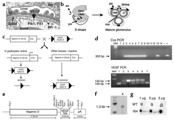
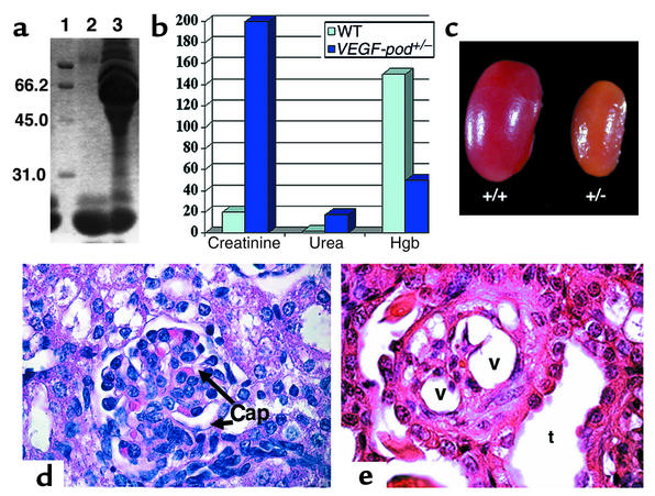
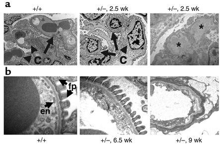
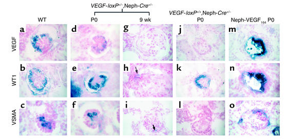
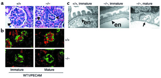
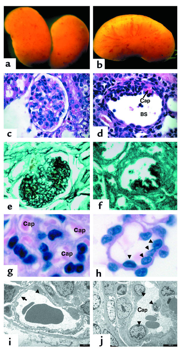
Kidney disease affects over 20 million people in the United States alone. Although the causes of renal failure are diverse, the glomerular filtration barrier is often the target of injury. Dysregulation of VEGF expression within the glomerulus has been demonstrated in a wide range of primary and acquired renal diseases, although the significance of these changes is unknown. In the glomerulus, VEGF-A is highly expressed in podocytes that make up a major portion of the barrier between the blood and urinary spaces. In this paper, we show that glomerular-selective deletion or overexpression of VEGF-A leads to glomerular disease in mice. Podocyte-specific heterozygosity for VEGF-A resulted in renal disease by 2.5 weeks of age, characterized by proteinuria and endotheliosis, the renal lesion seen in preeclampsia. Homozygous deletion of VEGF-A in glomeruli resulted in perinatal lethality. Mutant kidneys failed to develop a filtration barrier due to defects in endothelial cell migration, differentiation, and survival. In contrast, podocyte-specific overexpression of the VEGF-164 isoform led to a striking collapsing glomerulopathy, the lesion seen in HIV-associated nephropathy. Our data demonstrate that tight regulation of VEGF-A signaling is critical for establishment and maintenance of the glomerular filtration barrier and strongly supports a pivotal role for VEGF-A in renal disease.


Figures

Figure 1
Figure 1
Expression and genomic targeting of VEGF-A within the glomerular filtration barrier. (a) Transmission electron micrograph of the glomerular filtration barrier that consists of podocytes (po) and their specialized foot processes (fp), fenestrated endothelium (en), and intervening GBM. VEGF-A is produced in the podocyte; the VEGF receptors Flk1 and Flt1 are expressed in the adjacent endothelial cells. (b) Development of the glomerular filtration barrier. In the S-shape stage, podocyte precursors (po) express VEGF-A. Endothelial cells (en) that express the VEGF receptors migrate into the vascular (Vasc) cleft and differentiate in direct apposition to podocytes. In the mature glomerulus, the fenestrated endothelial capillary loops (cap) remain in intimate contact with the VEGF-expressing podocytes (po). Mesangial cells (me) provide support to the capillary tuft. Urine is formed as blood (bl) is filtered from the capillaries, across the GBM, and through slit diaphragms that connect adjacent podocyte foot processes (fp). (c) Scheme to generate heterozygous and homozygous podocyte-specific VEGF knockout mice. Triangles are 34 bp loxP sites. (d) The Cre recombinase transgene was identified as a 300 bp PCR product. The floxed VEGF allele measures 140 bp by PCR analysis, whereas the wild-type allele measures 100 bp. MW, molecular weight markers. (e) Transgenic construct used to overexpress the 164-isoform of VEGF. pA, poly(A). (f) Presence of the transgenic VEGF-164 gene was identified as a 1.3-kb band (*) by Southern blot analysis. (g) Dot blot analysis of transgene copy number. The transgenic founder mice (164) demonstrated a 30-fold increase in copy number compared with the wild type.
Figure 2
Figure 2
Heterozygous VEGF-loxP+/–,Neph-Cre+/– mice develop nephrotic syndrome and end-stage renal failure by 9 weeks of age. (a) SDS-PAGE analysis was performed using 2 μl of mouse urine. Lane 1 contains molecular weight markers, lane 2 shows urine from a VEGF-loxP–/–,Neph-Cre+/– control aged 9 weeks, and lane 3 shows urine from a 9-week-old sick VEGF-loxP+/–,Neph-Cre+/– animal. The presence of a large amount of albumin measuring 66.2 kDa is identified in the sick mouse and demonstrates damage to the kidney filter. In contrast, low–molecular-weight proteins, which are normally found in mouse urine, are not different. (b) Bar graph showing elevated creatinine (more than ten times higher than normal), elevated urea, and decreased hemoglobin (Hgb) in VEGF-loxP+/–,Neph-Cre+/– mice (VEGF-pod+/–) at 9 weeks of age compared with VEGF-loxP+/–,Neph-Cre–/– and VEGF-loxP–/–,Neph-Cre+/– mice (combined for analysis and considered as wild type). (c) Whole-mount image of a kidney from a sick 9-week-old VEGF-loxP+/–,Neph-Cre+/– (+/–) mouse compared with that of a wild-type littermate (+/+). The affected kidney is pale and shrunken. Magnification: ×60. (d) A wild-type glomerulus. Note the open capillary loops (Cap). ×350. (e) A glomerulus from a heterozygous VEGF-A mouse. All the glomeruli are grossly distorted morphologically. Note the empty cytoplasmic vacuoles (v) that are present in podocytes. No patent capillary loops can be seen. Dilated tubules (t) can be seen and in most places are packed with proteinaceous material, consistent with nephrotic syndrome. Magnification: ×375.
Figure 3
Figure 3
Heterozygous VEGF-loxP+/–,Neph-Cre+/– mice demonstrate endotheliosis and loss of fenestrations. (a) At 2.5 weeks of age, wild-type glomerular capillary loops (c) are open and contain numerous red blood cells. In contrast, podocyte-specific VEGF-A heterozygotes (+/–) demonstrate bloodless glomeruli, and the capillary loops are filled with swollen endothelial cells, demonstrating endotheliosis, the classic renal lesion of preeclampsia. In addition, large subendothelial hyaline deposits (*) can be seen. (b) At 6.5 weeks of age, wild-type filtration barriers (+/+) are characterized by fenestrated endothelial cells (en) and well-formed podocyte foot processes (fp). In the podocyte-specific heterozygotes (+/–), the fenestrations are lost at 6.5 weeks of age, and by 9 weeks of age, the endothelial cells appear necrotic and no podocyte foot processes can be identified.
Figure 4
Figure 4
Digoxigenin-labeled in situ analysis of wild-type and mutant glomeruli. (ac) Capillary loop–stage glomeruli from a newborn VEGF-loxP–/–,Neph-Cre+/– control mouse demonstrate expression of VEGF-A and WT1 in podocytes, while VSMA is expressed in mesangial cells, which are found inside the glomerulus and are required to support the capillary structure. (df) At birth (P0), capillary loop–stage glomeruli from a heterozygous VEGF-loxP+/–,Neph-Cre+/– mouse demonstrate normal levels of expression of WT1 and VSMA, while VEGF expression is consistently reduced at the mRNA level compared with the wild-type controls. (gi) By 9 weeks of age, the heterozygous VEGF mice are clinically unwell. At this time, most glomeruli demonstrate a complete absence of markers of podocyte differentiation (i.e., no VEGF or WT1; both are absent). In h, a single WT1-positive cell can be identified (arrow). (i) VSMA is not usually present in glomeruli at 9 weeks; however, occasional VSMA-positive cells can also be identified and likely represent “activated” mesangial cells (arrow). (jl) In the null VEGF-loxP+/+,Neph-Cre+/– glomeruli at birth, no VEGF is seen in glomeruli as predicted due to podocyte-specific excision of both VEGF alleles. WT1 is present in differentiated podocytes. In contrast, VSMA is absent, demonstrating a defect in migration and/or differentiation of mesangial cells into the glomerulus. (mo) In the nephrin–VEGF-164 mouse, both VEGF and WT1 are expressed in podocytes present within collapsed glomeruli. VEGF is markedly upregulated. In addition, VSMA and mesangial cells are present but appear to surround the collapsed glomerulus in a crescent shape. Magnification: ×350.
Figure 5
Figure 5
VEGF-null glomeruli do not form filtration barriers or fenestrations within endothelial cells. (a) The wild-type (+/+) glomerulus (arrow) has a lacy appearance due to open capillary loops. The VEGF-null glomeruli (–/–) fail to develop fully and lack visible capillary loops. Magnification: ×350. (b) Immunohistochemical staining for WT1 (green), a marker for podocyte cells, and PECAM (red), a marker for endothelial cells, shows a reduced number of endothelial cells in immature (capillary loop–stage) VEGF-null glomeruli. In mature glomeruli, no endothelial cells remain. Magnification: ×300. (c) Transmission EM of the filtration barrier in a wild-type (+/+) glomerulus clearly demonstrates fenestrated endothelium at the late capillary-loop stage, whereas no fenestrations are observed in endothelial cells (en) found in corresponding late capillary loop–stage VEGF-null glomeruli. In mature VEGF-null glomeruli, the basement membrane is seen (arrow), but the endothelial cells are missing. Magnification: ×20,000.
Figure 6
Figure 6
Mice that overexpress the 164 isoform of VEGF-A in their podocytes develop collapsing glomerulopathy. (a and b) Whole-mount images of VEGF-overexpressing kidneys at 5 days. The kidneys demonstrate many surface hemorrhages. (c) A glomerulus stained with H&E from a wild-type littermate. (d) A glomerulus from a transgenic VEGF-overexpressing mouse demonstrates global collapse of the capillary tuft toward the vascular pole of the glomerulus. A single patent capillary loop that appears dilated is identified (Cap). In addition, Bowman’s space (BS) is enlarged. (e) A 5-day-old wild-type glomerulus is stained with silver methenamine that recognizes basement membranes (black). Note the intricate pattern of GBM that lines the capillary loops between endothelial cells and podocytes. (f) In contrast, a transgenic glomerulus demonstrates complete collapse of the capillary network. (g) A high-power view of the capillary loops (Cap) in a wild-type glomerulus. (h) In contrast, the few patent capillary loops identified at 5 days of age in the transgenic mice demonstrate increased diameter and multiple endothelial cell nuclei (arrowheads). (i) A wild-type capillary loop at 5 days of age. Note the fenestrated endothelium (arrow). Although a portion of an endothelial cell body is identified (arrowhead), glomerular endothelial cell nuclei are difficult to find on EM sections. (j) In a transgenic patent capillary loop at 5 days of age, three endothelial cell nuclei are easily identified (arrowheads). Magnification in a and b: ×60; in cf: ×225; in g and h: ×1,000. In i, bar = 2,000 nm; in j, bar = 5,000 nm.

Comment in

References

    1. Mundel P, Reiser J. New aspects of podocyte cell biology. Kidney Blood Press. Res. 1997;20:173–176. - PubMed
    1. Abrahamson DR. Glomerulogenesis in the developing kidney. Semin. Nephrol. 1991;11:375–389. - PubMed
    1. Kriz W, Lemley K. The role of the podocyte in glomerulosclerosis. Curr. Opin. Nephrol. Hypertens. 1999;8:489–497. - PubMed
    1. Kriz W. Evolving role of the podocyte in chronic renal failure. Kidney Blood Press. Res. 1997;20:180–183. - PubMed
    1. Shirato I, et al. The development of focal segmental glomerulosclerosis in masugi nephritis is based on progressive podocyte damage. Virchows Arch. 1996;429:255–273. - PubMed

Publication types

MeSH terms

Substances