. 2003;4(2):R11.
doi: 10.1186/gb-2003-4-2-r11. Epub 2003 Feb 3.

Evolutionary history, structural features and biochemical diversity of the NlpC/P60 superfamily of enzymes

Affiliations

Evolutionary history, structural features and biochemical diversity of the NlpC/P60 superfamily of enzymes

Vivek Anantharaman et al. Genome Biol. 2003.

Abstract

Background: Peptidoglycan is hydrolyzed by a diverse set of enzymes during bacterial growth, development and cell division. The N1pC/P60 proteins define a family of cell-wall peptidases that are widely represented in various bacterial lineages. Currently characterized members are known to hydrolyze D-gamma-glutamyl-meso-diaminopimelate or N-acetylmuramate-L-alanine linkages.

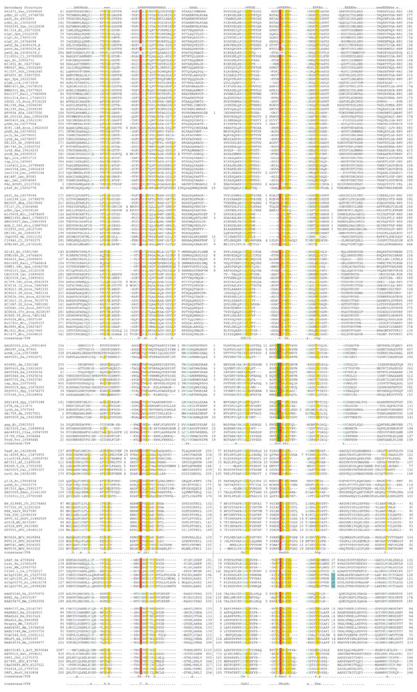
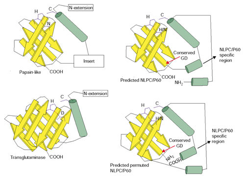
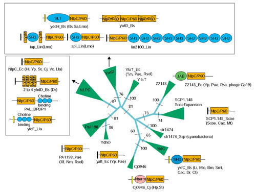
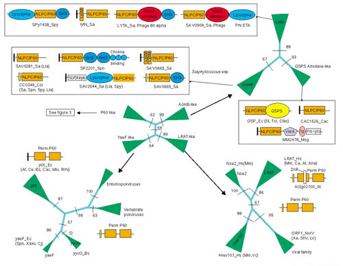
Results: Detailed analysis of the N1pC/P60 peptidases showed that these proteins define a large superfamily encompassing several diverse groups of proteins. In addition to the well characterized P60-like proteins, this superfamily includes the AcmB/LytN and YaeF/YiiX families of bacterial proteins, the amidase domain of bacterial and kinetoplastid glutathionylspermidine synthases (GSPSs), and several proteins from eukaryotes, phages, poxviruses, positive-strand RNA viruses, and certain archaea. The eukaryotic members include lecithin retinol acyltransferase (LRAT), nematode developmental regulator Egl-26, and candidate tumor suppressor H-rev107. These eukaryotic proteins, along with the bacterial YaeF/poxviral G6R family, show a circular permutation of the catalytic domain. We identified three conserved residues, namely a cysteine, a histidine and a polar residue, that are involved in the catalytic activities of this superfamily. Evolutionary analysis of this superfamily shows that it comprises four major families, with diverse domain architectures in each of them.

Conclusions: Several related, but distinct, catalytic activities, such as murein degradation, acyl transfer and amide hydrolysis, have emerged in the N1pC/P60 superfamily. The three conserved catalytic residues of this superfamily are shown to be equivalent to the catalytic triad of the papain-like thiol peptidases. The predicted structural features indicate that the N1pC/P60 enzymes contain a fold similar to the papain-like peptidases, transglutaminases and arylamine acetyltransferases.


Figures

Figure 1
Figure 1
Multiple sequence alignment of the NlpC/P60 superfamily. Multiple sequence alignments of the different families of NlpC/P60 were constructed using T-Coffee [35] after parsing high-scoring pairs from PSI-BLAST search results. The PHD-secondary structure [36] is shown above the alignment with E representing a β strand, and H an α helix. The 85% consensus shown below the alignment was derived using the following amino acid classes: hydrophobic (h, ALICVMYFW, yellow shading), the aliphatic subset of the hydrophobic class are (l, ALIVMC, yellow shading), small (s, ACDGNPSTV, green) and polar (p, CDEHKNQRST, blue). A 'G', 'D', 'C' or 'N' shows the completely conserved amino acid in that group. The numbers colored light blue show the region where a zinc ribbon domain is inserted in the Arabidopsis LRAT homologs. The catalytic residues are highlighted in red, while the conserved cysteine present in LRAT and related proteins is colored red. Specific columns of residues that are peculiar to a particular category of NlpC/P60 (see text) are colored red. The consensus of the individual families and the entire superfamily are shown, and the subgroups of each family are separated by a space. The limits of the domains are indicated by the residue positions, in bold, on each side, or internally in the case of the permuted versions. A dotted line separates the amino and carboxyl termini of the permuted versions. The numbers within the alignment are non-conserved inserts that have not been shown. The sequences are denoted by their gene name followed by the species abbreviation and GeneBank identifier (gi). An alignment covering members of the bacterial P60 family is available in PFAM [78]. The species abbreviations are: Af, Archaeoglobus fulgidus; BPDP1, bacteriophage Dp-1; Ana, Anabaena; Atu, Agrobacterium tumefaciens; Ban, Bacillus anthracis; Bha, Bacillus halodurans; Bs, Bacillus subtilis; Bmel, Brucella melitensis; Cac, Clostridium acetobutylicum; Ccr, Caulobacter crescentus; Cj, Campylobacter jejuni; Ct, Chlamydia trachomatis; Cpn, Chlamydophila pneumoniae; Dr, Deinococcus radiodurans; Ec, Escherichia coli; Hi, Haemophilus influenzae; Hp, Helicobacter pylori; Lla, Lactococcus lactis; Lin, Listeria innocua; Lmo, Listeria monocytogenes; Mle, Mycobacterium leprae; Mlo, Mesorhizobium loti; Mtu, Mycobacterium tuberculosis; Nm, Neisseria meningitidis; Pae, Pseudomonas aeruginosa; Pmu, Pasteurella multocida; Rsol, Ralstonia solanacearum; Rrhi, Rhizobium rhizogenes; St, Salmonella typhimurium; Sme, Sinorhizobium meliloti; Sa, Staphylococcus aureus; Scoe, Streptomyces coelicolor A3; Spn, Streptococcus pneumoniae; Spy, Streptococcus pyogenes; Ssp, Synechocystis sp.; Tm, Thermotoga maritima; Vch, Vibrio cholerae; Xaxo, Xanthomonas axonopodis; Xf, Xylella fastidiosa; Ype, Yersinia pestis; At, Arabidopsis thaliana; Ce, Caenorhabditis elegans; Cfas, Crithidia fasciculata; Hs, Homo sapiens; Mm, Mus musculus; Rn, Rattus norvegicus; Tcr, Trypanosoma cruzi; CV, cowpox virus; FPV, fowlpox virus; MCV, molluscum contagiosum virus; MV, myxoma virus; RFV, rabbit fibroma virus; ShPV, sheeppox virus; SPV, swinepox virus; VV, vaccinia virus; VarV, variola virus; YDV, Yaba-like disease virus; AMV, Amsacta moorei entomopoxvirus; MSV, Melanoplus sanguinipes entomopoxvirus; AiV, Aichi virus; BCV, bovine calicivirus; ChV, Chiba virus; LV, Lordsdale virus; NorV, Norwalk virus; ShV, Southampton virus; Aev, avian encephalomyelitis virus.
Figure 2
Figure 2
Topology diagram of the NlpC/P60 and structurally related proteases. The topology of the papain-like proteases (PDB:1ppn) and transglutaminases (PDB:1fie) are derived from the available crystal structures of these proteins. The predicted topology of the NlpC/P60 family members was derived from secondary structure and sequence conservation profile. Yellow arrows represent β strands and green cylinders represent α helices.
Figure 3
Figure 3
Phylogenetic relationships and domain architectures of NlpC/P60 family proper. The phyletic pattern of each family is shown next to the clade. The RELL bootstrap values for the major branches are shown at their base. The width of a given clade is approximately proportional to number proteins contained within it. N-term is a specific amino-terminal module that is restricted to the Cj0946 clade. The domain abbreviations are: SLT, soluble lytic transglycosidase. The signal peptides are shown as black rectangles, while the transmembrane regions are shown as yellow rectangles. The species abbreviations are given in the legend to Figure 1.
Figure 4
Figure 4
Phylogenetic relationships and domain architectures of NlpC/P60 superfamily. The domain abbreviations are: AM amidases, N-acetylmuramidase; VWA, von Willebrandt factor A; GSPS, glutathionylspermidine synthase; IG, immunoglobulin domain; Fib rpts, fibrinogen-like repeat domains; ZnR, inserted zinc ribbon. The conventions are the same as in Figure 3.

References

    1. Moat AG, Foster JW, Eds . Microbial Physiology. 4th. New York: John Wiley; 2002.
    1. Madigan MM, Parker J, Madigan MT, Martinko JM. Brock Biology of Microorganisms. 10th. Upper Saddle River, NJ: Prentice Hall; 2002.
    1. van Heijenoort J. Formation of the glycan chains in the synthesis of bacterial peptidoglycan. Glycobiology. 2001;11:25R–36R. - PubMed
    1. Healy VL, Lessard IA, Roper DI, Knox JR, Walsh CT. Vancomycin resistance in enterococci: reprogramming of the D-Ala-D-Ala ligases in bacterial peptidoglycan biosynthesis. Chem Biol. 2000;7:R109–R119. - PubMed
    1. Born TL, Blanchard JS. Structure/function studies on enzymes in the diaminopimelate pathway of bacterial cell wall biosynthesis. Curr Opin Chem Biol. 1999;3:607–613. - PubMed

LinkOut - more resources