. 2003 Mar 18;100(6):3404-9.
doi: 10.1073/pnas.0538059100. Epub 2003 Mar 5.

Significance and therapeutic potential of the natriuretic peptides/cGMP/cGMP-dependent protein kinase pathway in vascular regeneration

Affiliations

Significance and therapeutic potential of the natriuretic peptides/cGMP/cGMP-dependent protein kinase pathway in vascular regeneration

Kenichi Yamahara et al. Proc Natl Acad Sci U S A. .

Abstract

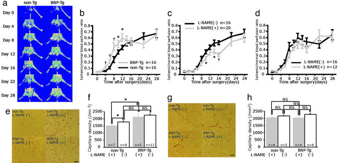
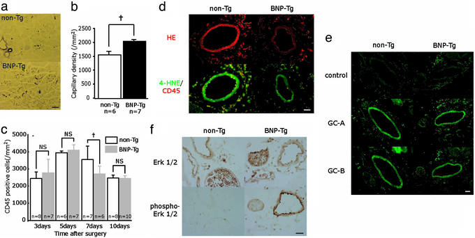
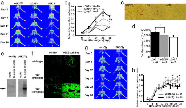
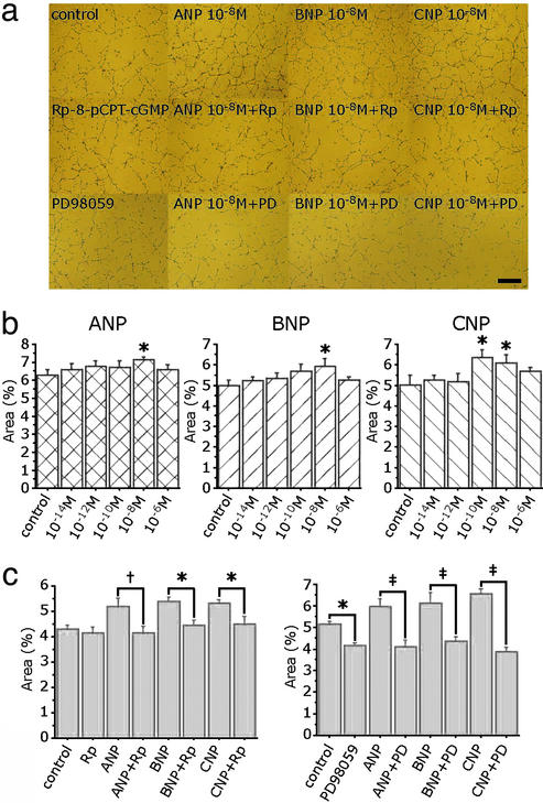
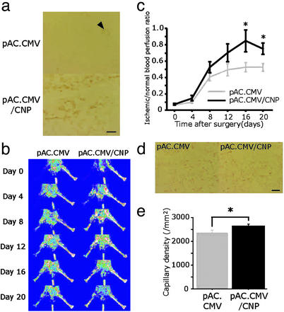
Natriuretic peptides (NPs), which consist of atrial, brain, and C-type natriuretic peptides (ANP, BNP, and CNP, respectively), are characterized as cardiac or vascular hormones that elicit their biological effects by activation of the cGMPcGMP-dependent protein kinase (cGK) pathway. We recently reported that adenoviral gene transfer of CNP into rabbit blood vessels not only suppressed neointimal formation but also accelerated reendothelialization, a required step for endothelium-dependent vasorelaxation and antithrombogenicity. Accordingly, we investigated the therapeutic potential of the NPscGMPcGK pathway for vascular regeneration. In transgenic (Tg) mice that overexpress BNP in response to hindlimb ischemia, neovascularization with appropriate mural cell coating was accelerated without edema or bleeding, and impaired angiogenesis by the suppression of nitric oxide production was effectively rescued. Furthermore, in BNP-Tg mice, inflammatory cell infiltration in ischemic tissue and vascular superoxide production were suppressed compared with control mice. Ischemia-induced angiogenesis was also significantly potentiated in cGK type I Tg mice, but attenuated in cGK type I knockout mice. NPs significantly stimulated capillary network formation of cultured endothelial cells by cGK stimulation and subsequent Erk12 activation. Furthermore, gene transfer of CNP into ischemic muscles effectively accelerated angiogenesis. These findings reveal an action of the NPscGMPcGK pathway to exert multiple vasculoprotective and regenerative actions in the absence of apparent adverse effects, and therefore suggest that NPs as the endogenous cardiovascular hormone can be used as a strategy of therapeutic angiogenesis in patients with tissue ischemia.


Figures

Figure 1
Figure 1
Ischemia-induced angiogenesis was accelerated in BNP-Tg mice, and overexpression of BNP restored delayed angiogenesis induced by NO blockade. (a) Serial LDPI analysis of hindlimb ischemia in BNP-Tg and non-Tg mice. (b) Quantitative analysis of ischemic/normal hindlimb perfusion ratio in BNP-Tg and non-Tg mice. (c and d) Serial LDPI measurements in non-Tg (c) and BNP-Tg (d) mice with and without l-NAME treatment. (e and g) Immunostaining of the ischemic hindlimb tissues with anti-PECAM-1 antibody (bright red) at day 10 (e) and day 28 (g). (f and h) Quantitative analysis of capillary density at day 10 (f) and day 28 (h). *, P < 0.05; †, P < 0.01; ‡, P < 0.001; NS, not significant. (Scale bar, 100 μm.)
Figure 2
Figure 2
Evaluation of ischemic hindlimb tissue of BNP-Tg mice. (a and b) α SMA staining (purple) of the ischemic hindlimb tissues at day 10 (a) and quantitative analysis of capillary density (b). (c) Time course of focal inflammation of ischemic hindlimb obtained from immunostaining with anti-CD45 antibody. (d) Dihydroethidium (HE) staining (Upper; red) and 4-hydroxy-2-nonenal (4-HNE)/CD45 staining (Lower; green fluorescence/red) of the ischemic hindlimb tissue at day 7. (e) Expression of GC-A and GC-B (green fluorescence) in the ischemic hindlimb at day 7. Negative controls for these antibodies are also shown. (f) Immunostaining of the ischemic hindlimb tissues at day 7 with anti-Erk1/2 or phosphor-Erk1/2 antibody (brown). †, P < 0.01. (Scale bars: a, 100 μm; d–f, 25 μm.)
Figure 3
Figure 3
Angiogenesis was blunted in cGKI-knockout mice and accelerated in cGKI-Tg mice. (a and b) Serial LDPI measurements in cGKI-knockout mice. (c and d) PECAM-1 staining (bright red) of the ischemic hindlimb tissues at day 28 in cGKI-Tg mice (c) and quantitative analysis of capillary density (d). (e) The expression of cGKI mRNA in non-Tg and cGKI-Tg mice. (f) Immunostaining of cGKI (green fluorescence) with their negative control in WT, cGKI-knockout, and cGKI-Tg mice. (g and h) Serial LDPI measurements in cGKI-Tg mice. *, P < 0.05; †, P < 0.01. (Scale bars: c, 100 μm; f, 25 μm.)
Figure 4
Figure 4
NPs potentiated capillary network formation of cultured human umbilical vein ECs. (a) Capillary network formation by Matrigel assay in the presence of 10−8 M ANP, BNP, and CNP with or without Rp-8-pCPT-cGMP (Rp: 5 × 10⁻⁶ M), a cGK inhibitor, and PD 98059 (PD: 10−5 M), an Erk1/2 inhibitor. (b) Mean area of tube formation in the presence of various concentrations of ANP, BNP, and CNP. (c and d) Effects of Rp-8-pCPT-cGMP (c) and PD 98059 (d) in network formation induced by NPs. *, P < 0.05; †, P < 0.01; ‡, P < 0.001. (Scale bar, 500 μm.)
Figure 5
Figure 5
Effect of CNP gene transfer in the murine ischemic hindlimb model. (a) CNP immunostaining (brown) after local injection of control vector (pAC.CMV) or pAC.CMV/CNP. (b and c) Serial LDPI measurements in mice receiving pAC.CMV or pAC.CMV/CNP (n = 16 per group). (d and e) PECAM-1 staining (bright red) of the ischemic hindlimb tissues at day 20 in mice injected with pAC.CMV or pAC.CMV/CNP (d), and quantitative analysis of capillary density (e) (n = 10 per group). *, P < 0.05. (Scale bars, 100 μm.)

References

    1. Sugawara A, Nakao K, Morii N, Yamada T, Itoh H, Shiono S, Saito Y, Mukoyama M, Arai H, Nishimura K, et al. J Clin Invest. 1988;81:1962–1970. - PMC - PubMed
    1. Mukoyama M, Nakao K, Hosoda K, Suga S, Saito Y, Ogawa Y, Shirakami G, Jougasaki M, Obata K, Yasue H, et al. J Clin Invest. 1991;87:1402–1412. - PMC - PubMed
    1. Komatsu Y, Itoh H, Suga S, Ogawa Y, Hama N, Kishimoto I, Nakagawa O, Igaki T, Doi K, Yoshimasa T, Nakao K. Circ Res. 1996;78:606–614. - PubMed
    1. Doi K, Ikeda T, Itoh H, Ueyama K, Hosoda K, Ogawa Y, Yamashita J, Chun T H, Inoue M, Masatsugu K, et al. Arterioscler Thromb Vasc Biol. 2001;21:930–936. - PubMed
    1. Ohno N, Itoh H, Ikeda T, Ueyama K, Yamahara K, Doi K, Yamashita J, Inoue M, Masatsugu K, Sawada N, et al. Circulation. 2002;105:1623–1626. - PubMed

MeSH terms

Substances

LinkOut - more resources