

Advances in understanding the regulation of apoptosis and mitosis by peroxisome-proliferator activated receptors in pre-clinical models: relevance for human health and disease
- PMID: 12622871
- PMCID: PMC151270
- DOI: 10.1186/1476-5926-2-3
Advances in understanding the regulation of apoptosis and mitosis by peroxisome-proliferator activated receptors in pre-clinical models: relevance for human health and disease
Abstract
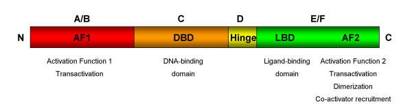
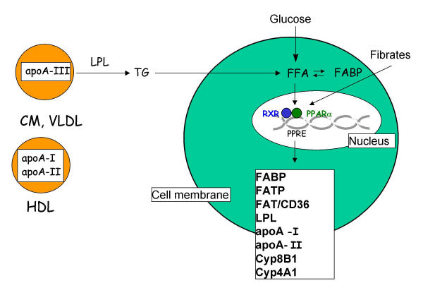
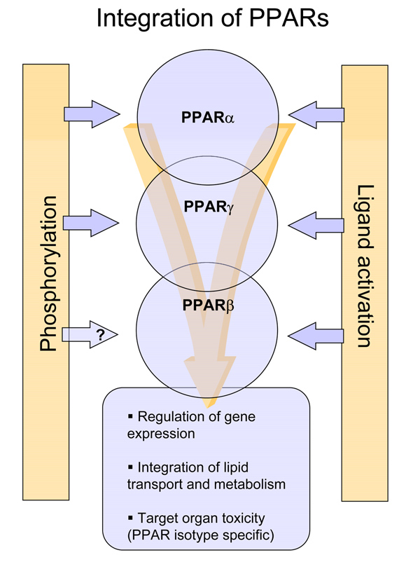
Peroxisome proliferator activated receptors (PPARs) are a family of related receptors implicated in a diverse array of biological processes. There are 3 main isotypes of PPARs known as PPARalpha, PPARbeta and PPARgamma and each is organized into domains associated with a function such as ligand binding, activation and DNA binding. PPARs are activated by ligands, which can be both endogenous such as fatty acids or their derivatives, or synthetic, such as peroxisome proliferators, hypolipidaemic drugs, anti-inflammatory or insulin-sensitizing drugs. Once activated, PPARs bind to DNA and regulate gene transcription. The different isotypes differ in their expression patterns, lending clues on their function. PPARalpha is expressed mainly in liver whereas PPARgamma is expressed in fat and in some macrophages. Activation of PPARalpha in rodent liver is associated with peroxisome proliferation and with suppression of apoptosis and induction of cell proliferation. The mechanism by which activation of PPARalpha regulates apoptosis and proliferation is unclear but is likely to involve target gene transcription. Similarly, PPARgamma is involved in the induction of cell growth arrest occurring during the differentiation process of fibroblasts to adipocytes. However, it has been implicated in the regulation of cell cycle and cell proliferation in colon cancer models. Less in known concerning PPARbeta but it was identified as a downstream target gene for APC/beta-catenin/T cell factor-4 tumor suppressor pathway, which is involved in the regulation of growth promoting genes such as c-myc and cyclin D1. Marked species and tissue differences in the expression of PPARs complicate the extrapolation of pre-clinical data to humans. For example, PPARalpha ligands such as the hypolipidaemic fibrates have been used extensively in the clinic over the past 20 years to treat cardiovascular disease and side effects of clinical fibrate use are rare, despite the observation that these compounds are rodent carcinogens. Similarly, adverse clinical responses have been seen with PPARgamma ligands that were not predicted by pre-clinical models. Here, we consider the response to PPAR ligands seen in pre-clinical models of efficacy and safety in the context of human health and disease.
Figures
References
-
- Smirnov AN. Nuclear receptors: nomenclature, ligands, mechanisms of their effects on gene expression. Biochemistry (Engl Trans Biokhimiya) 2002;67:957–977. - PubMed
-
- Auwerx J, Baulieu E, Beato M, Becker-Andre M, Burbach PH, Camerino G, Chambon P, Cooney A, Dejean A, Dreyer C, Evans RM, Gannon F, Giguere V, Gronemeyer H, Gustafsson JA, Laudet V, Lazar MA, Mangelsdorf DJ, Millbrandt J, Milgrom E, Moore DD, O'Malley B, Parker M, Parker K, Perimann T, Pfahl M, Rosenfeld MG, Samuels H, Schutz G, Sladek FM, Stunnenberg HG, Spedding M, Thummel C, Tsai MJ, Umesono K, Vennstrom B, Wahli W, Weinberg C, Willson TM, Yamamoto K. A unified nomenclature system for the nuclear receptor superfamily. Cell. 1999;97:161–163. - PubMed
-
- Sher T, Yi HF, McBride OW, Gonzalez FJ. cDNA cloning, chromosomal mapping, and functional characterization of the human peroxisome proliferator activated receptor. Biochemistry. 1993;32:5598–5604. - PubMed
LinkOut - more resources
Full Text Sources
Other Literature Sources
Research Materials
Miscellaneous