. 2003 Feb 19:4:1.
doi: 10.1186/1471-2091-4-1. Epub 2003 Feb 19.

Glycosaminoglycans in human retinoblastoma cells: heparan sulfate, a modulator of the pigment epithelium-derived factor-receptor interactions

Affiliations

Glycosaminoglycans in human retinoblastoma cells: heparan sulfate, a modulator of the pigment epithelium-derived factor-receptor interactions

Elena M Alberdi et al. BMC Biochem. .

Abstract

Background: Pigment epithelium-derived factor (PEDF) has binding affinity for cell-surface receptors in retinoblastoma cells and for glycosaminoglycans. We investigated the effects of glycosaminoglycans on PEDF-receptor interactions.

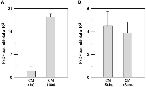
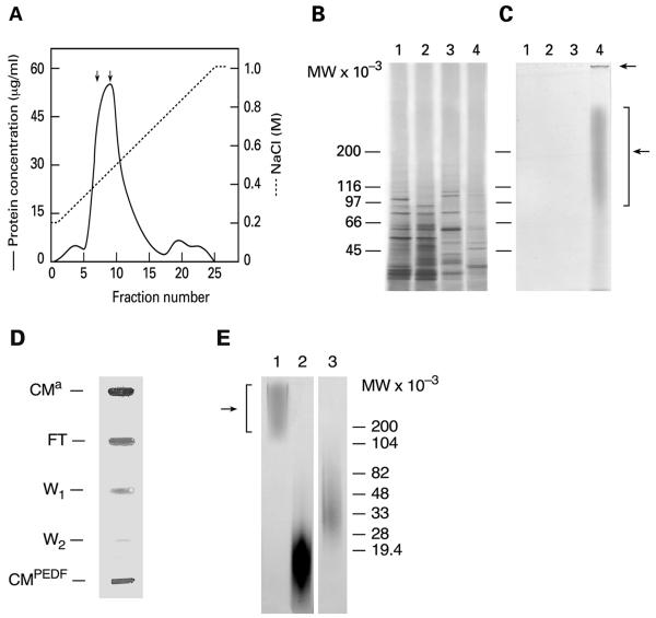
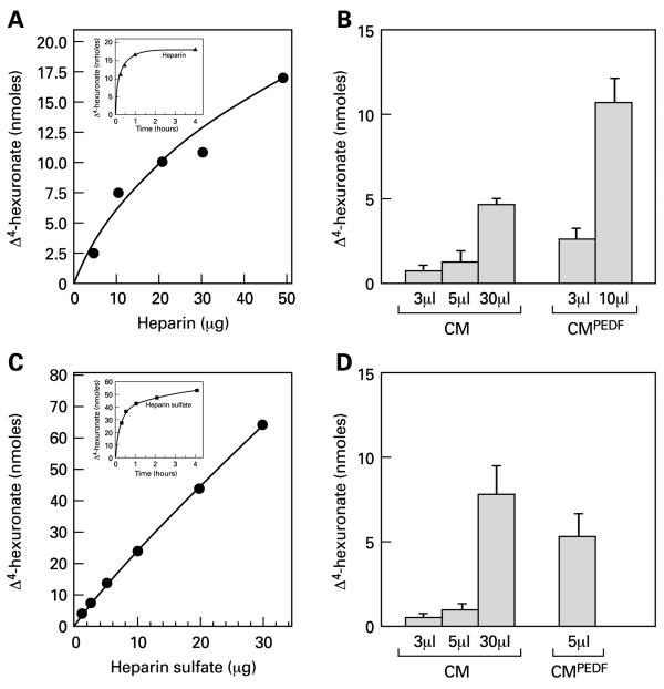
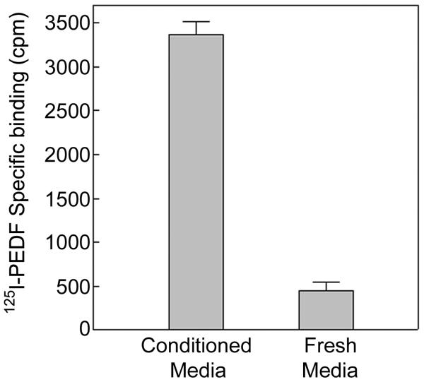
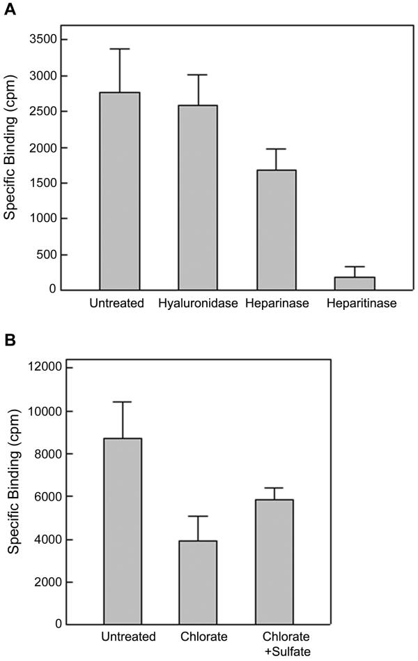
Results: 125I-PEDF formed complexes with protease-resistant components of medium conditioned by human retinoblastoma Y-79 cells. Using specific glycosaminoglycan degrading enzymes in spectrophotometric assays and PEDF-affinity chromatography, we detected heparin and heparan sulfate-like glycosaminoglycans in the Y-79 conditioned media, which had binding affinity for PEDF. The Y-79 conditioned media significantly enhanced the binding of 125I-PEDF to Y-79 cell-surface receptors. However, enzymatic and chemical depletion of sulfated glycosaminoglycans from the Y-79 cell cultures by heparitinase and chlorate treatments decreased the degree of 125I-PEDF binding to cell-surface receptors.

Conclusions: These data indicate that retinoblastoma cells secrete heparin/heparan sulfate with binding affinity for PEDF, which may be important in efficient cell-surface receptor binding.


Figures

Figure 1
Figure 1
Complex formation between PEDF and component(s) in media conditioned by human retinoblastoma Y-79 cells. Binding reactions were performed with 0.75 nM 125I-PEDF and human retinoblastoma Y-79 cell conditioned media (CM) (200 μl) at 4°C for 120 min with gentle rotation. Free and bound PEDF was separated by ultrafiltration through membrane of MWCO = 100 kDa. PEDF bound was calculated by subtracting specific 125I-PEDF binding in fresh media from that in CM and is given as a ratio of the total 125I-PEDF in the reaction. A) Reactions with CM (CM 1X) and 10-fold concentrated CM (CM 10X). B) Reactions with protease-treated CM. CM was concentrated 10-fold and dialyzed against 20 mM Tris pH 8, 10% glycerol. Half of the dialyzate was incubated without and the other with 0.4 μg/ml subtilisin at 37°C for 16 h (-Subt. and +Subt., respectively). The protease reaction was terminated by incubation at 75°C for 25 min to inactivate the protease. All experimental points are given as the average of duplicates. Assays were performed twice.
Figure 2
Figure 2
DEAE-Sephacel and PEDF-affinity column chromatography of media conditioned by retinoblastoma cells. GAGs and polyanions of CM were fractionated by anion-exchange column chromatography (CMa). CM (2.5 mg protein) was applied to a DEAE-Sephacel column and the bound components were eluted with a linear gradient of 0.2–1 M NaCl. Panel A. Protein profile of the DEAE-Sephacel chromatography. Protein (--) and NaCl (-----) concentrations of eluted fractions are indicated. The load, unbound material (FT), and fractions 7 and 9 were resolved by 4–12% polyacrylamide gel electrophoresis under reducing conditions in two duplicate gels. Panels B and C. SDS-PAGE analysis of the DEAE chromatography. Gels stained with Coomassie Brilliant Blue (panel B) and with Toluidine Blue-O (panel C) are shown. Lane 1, load; lane 2, flow-through; lane 3 fraction #7; and lane 4, fraction #9. Panels D and E. PEDF-affinity column chromatography. CMa was subjected to PEDF-affinity column chromatography. Bound GAGs were eluted with 3 M NaCl (CMPEDF). Unbound material is FT and washes W1 and W2., A dot-blot stained with Toluidine Blue-O of fractions (as indicated) is shown in panel D. A 10–20% polyacrylamide gel (SDS-tricine) stained with Toluidine Blue-O is shown in Panel E with: lane 1, CMPEDF; lane 2, 5 μg heparin of average MW 16,000; and lane 3, 5 μg HS. Migration positions of molecular-weight-standards are indicated to the right. Arrows indicate migration positions of GAGs from CM derived samples.
Figure 3
Figure 3
Spectrophotometric assays for heparin and HS-like molecules. Heparin and HS were degraded with heparinase and heparitinase, respectively. Proteins in CM concentrated 10-fold and CMPEDF were digested first with subtilisin. Protease-treated CM and CMPEDF were reacted with heparinase and heparitinase. The appearance of degradation products (Δ4-hexuronate) was measured by absorbance at 235 nm (Extinction coefficient = 5500). Panels A and B show heparinase activity and panels C and D show heparitinase activity. Time course and concentration curves of enzymes using their respective substrates are shown in panels A and C. Panels B and D correspond to reactions with CM and CMPEDF.
Figure 4
Figure 4
Media conditioned by retinoblastoma Y-79 cells enhances the 125 I-PEDF binding to cell-surface receptors. Binding to human retinoblastoma Y-79 cells (1.5 × 10⁵ cells/ml) was performed with 0.8 nM 125I-PEDF (136,000 cpm/point) in their overnight culturing media (conditioned media) or non-conditioned media (fresh media). Free and bound ligand was separated by filtration through glass fiber filters under vacuum. Non-specific binding was binding in the presence of 125-fold molar excess of unlabelled PEDF. Specific binding was calculated by subtracting the non-specific from the total binding. All experimental points are given as the average of triplicates.
Figure 5
Figure 5
Effect of GAG lyases and chlorate on 125 I-PEDF binding to retinoblastoma Y-79 cells. Human retinoblastoma Y-79 cells (5 × 10⁵ cells/ml) were cultured in serum-free media at 37°C and treated with GAG lyases or chlorate. Binding to the treated cells was performed with 2 nM 125I-PEDF. Free and bound PEDF were separated by filtration through glass-fiber filters under vacuum. Non-specific binding was determined from binding reactions in the presence of 50-fold excess of unlabeled ligand. Specific binding was calculated by subtracting non-specific binding from total binding. Panel A. Cells cultured for 16 h were treated with hyaluronidase, heparinase and heparitinase at 37°C for 120 min. All experimental points are given as the average of quadruplicates. Panel B. Cells were cultured with or without 30 mM sodium chlorate and 10 mM sodium sulfate at 37°C for 16 h before the radioligand was added. The binding reactions were at 4°C for 30 min. All experimental points are given as the average of triplicates.

References

    1. Bernfield M, Gotte M, Park PW, Reizes O, Fitzgerald ML, Lincecum J, Zako M. Functions of cell surface heparan sulfate proteoglycans. Annu Rev Biochem. 1999;68:729–777. doi: 10.1146/annurev.biochem.68.1.729. - DOI - PubMed
    1. Park PW, Reizes O, Bernfield M. Cell surface heparan sulfate proteoglycans: selective regulators of ligand-receptor encounters. J Biol Chem. 2000;275:29923–29926. doi: 10.1074/jbc.R000008200. - DOI - PubMed
    1. Steele FR, Chader GJ, Johnson LV, Tombran-Tink J. Pigment epithelium-derived factor: neurotrophic activity and identification as a member of the serine protease inhibitor gene family. Proc Natl Acad Sci U S A. 1993;90:1526–1530. - PMC - PubMed
    1. Cayouette M, Smith SB, Becerra SP, Gravel C. Pigment epithelium-derived factor delays the death of photoreceptors in mouse models of inherited retinal degenerations. Neurobiol Dis. 1999;6:523–532. doi: 10.1006/nbdi.1999.0263. - DOI - PubMed
    1. Cao W, Tombran-Tink J, Chen W, Mrazek D, Elias R, McGinnis JF. Pigment epithelium-derived factor protects cultured retinal neurons against hydrogen peroxide-induced cell death. J Neurosci Res. 1999;57:789–800. doi: 10.1002/(SICI)1097-4547(19990915)57:6<789::AID-JNR4>3.0.CO;2-M. - DOI - PubMed

MeSH terms

LinkOut - more resources