. 2003 May 1;548(Pt 3):881-91.
doi: 10.1113/jphysiol.2003.038778. Epub 2003 Mar 7.

Angiotensin II stimulates hyperplasia but not hypertrophy in immature ovine cardiomyocytes

Affiliations

Angiotensin II stimulates hyperplasia but not hypertrophy in immature ovine cardiomyocytes

N C Sundgren et al. J Physiol. .

Abstract

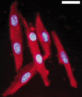
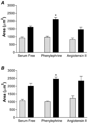
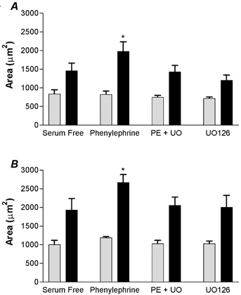
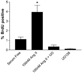
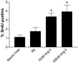
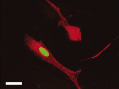
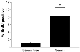
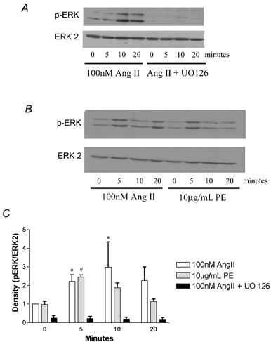
Rat and sheep cardiac myocytes become binucleate as they complete the 'terminal differentiation' process soon after birth and are not able to divide thereafter. Angiotensin II (Ang II) is known to stimulate hypertrophic changes in rodent cardiomyocytes under both in vivo and in vitro conditions via the AT1 receptor and intracellular extracellular regulated kinase (ERK) signalling cascade. We sought to develop culture methods for immature sheep cardiomyocytes in order to test the hypothesis that Ang II is a hypertrophic agent in the immature myocardium of the sheep. We isolated fetal sheep cardiomyocytes and cultured them for 96 h, added Ang II and phenylephrine (PE) for 48 h, and measured footprint area and proliferation (5-bromo-2'-deoxyuridine (BrdU) uptake) separately in mono- vs. binucleate myocytes. We found that neither Ang II nor PE changed the footprint area of mononucleated cells. PE stimulated an increase in footprint area of binucleate cells but Ang II did not. Ang II increased myocyte BrdU uptake compared to serum free conditions, but PE did not affect BrdU uptake. The MAP kinase kinase (MEK) inhibitor UO126 prevented BrdU uptake in Ang II-stimulated cells and prevented cell hypertrophy in PE-stimulated cells. This paper establishes culture methods for immature sheep cardiomyocytes and reports that: (1) Ang II is not a hypertrophic agent; (2) Ang II stimulates hyperplastic growth among mononucleate myocytes; (3) PE is a hypertrophic agent in binucleate myocytes; and (4) the ERK cascade is required for the proliferation effect of Ang II and the hypertrophic effect of PE.


Figures

Figure 1
Figure 1. Dissociated cardiomyocytes from fetal sheep
Cells were fixed after dissociation and viewed under × 400 power (Zeiss Axiophot). Myosin was stained with a secondary antibody conjugated to rhodamine red-X and the nuclei were stained with Hoechst 33342. Mononucleated and binucleated cells appear healthy and intact after dissociation. Bar = 20 μm.
Figure 2
Figure 2. Hypertrophy in culture
Area of LV (A) and RV (B) mononucleated (grey bars) and binucleated (black bars) cardiomyocytes from four separate fetuses. Cells were stimulated with 10 μg ml−1 PE or 100 nm AngII for 48 h. *P < 0.05 compared to serum-free binucleated, n = 4, paired ANOVA.
Figure 3
Figure 3. Hypertrophy in the presence of ERK inhibition
Area of LV (A) and RV (B) mononucleated (grey bars) and binucleated (black bars) cardiomyocytes from four separate fetuses. Inhibition of MEK with UO126 abolishes PE-induced hypertrophy and is not different from serum-free conditions. *P < 0.05 compared to serum-free binucleated myocytes, n = 4, paired ANOVA.
Figure 7
Figure 7. BrdU uptake after 48 h with Ang II in the presence of the MEK inhibitor UO126
UO126 abolishes the increase in BrdU uptake that 100 nm Ang II induces in the absence of the MEK inhibitor. *P < 0.05 compared to serum-free conditions, ANOVA.
Figure 6
Figure 6. BrdU uptake after 48 h in 10 μg ml−1 PE or two different doses of Ang II
*P < 0.05, n = 7; compared to serum-free conditions, ANOVA.
Figure 5
Figure 5. Plated cardiomyocytes after stimulation with 100 nm Ang II in the presence of BrdU for 48 h
Note that one cell has incorporated BrdU into the nucleus (green). Myosin was stained with a secondary antibody conjugated to rhodamine red-X and nuclear BrdU was stained with a secondary antibody conjugated to FITC. Negative controls (without primary antibodies) did not stain for either myosin or BrdU (not shown). Bar = 20 μm.
Figure 4
Figure 4. BrdU uptake after 48 h in serum-free or serum medium conditions
*P < 0.05, student's t test, n = 9.
Figure 8
Figure 8. ERK stimulation
A, shows a typical Western blot (n = 4). Results shown in the left 4 columns were from cells stimulated with 100 nm Ang II in the absence of UO126 for the indicated times. The right 4 columns are from cells pre-treated for 20 min with UO126 before stimulation with Ang II. B, a typical Western blot of Ang II and PE stimulation (n = 4). The left 4 columns are from cells that were stimulated with 100 nm Ang II and the right 4 columns from cells treated with 10 μg ml−1 PE. All membranes were probed for phospho-ERKs, then stripped and probed for ERK 2. C, average band density of Western blots normalized to baseline Ang II stimulation (0 min). Density is plotted in normalized units of p-ERK/ERK 2 levels. At 5 and 10 min, phospho-ERK levels were increased over basal unstimulated levels with Ang II (*P < 0.05, non-parametric ANOVA). At 5 min PE significantly increased phospho-ERK levels over 0 min of PE stimulation (#P < 0.05, non-parametric ANOVA).

References

    1. Anversa P, Kajstura J. Ventricular myocytes are not terminally differentiated in the adult mammalian heart. Circ Res. 1998;83:1–14. - PubMed
    1. Aoki H, Richmond M, Izumo S, Sadoshima J. Specific role of the extracellular signal-regulated kinase pathway in angiotensin II-induced cardiac hypertrophy in vitro. Biochem J. 2000;347:275–284. - PMC - PubMed
    1. Baker KM, Chernin MI, Wixson SK, Aceto JF. Renin-angiotensin system involvement in pressure-overload cardiac hypertrophy in rats. Am J Physiol. 1990;259:H324–332. - PubMed
    1. Barbera A, Giraud GD, Reller MD, Maylie J, Morton MJ, Thornburg KL. Right ventricular systolic pressure load alters myocyte maturation in fetal sheep. Am J Physiol Regul Integr Comp Physiol. 2000;279:R1157–1164. - PubMed
    1. Barlucchi L, Leri A, Dostal DE, Fiordaliso F, Tada H, Hintze TH, Kajstura J, Nadal-Ginard B, Anversa P. Canine ventricular myocytes possess a renin-angiotensin system that is upregulated with heart failure. Circ Res. 2001;88:298–304. - PubMed

Publication types

LinkOut - more resources