Review
. 2003 Apr 15;548(Pt 2):339-52.
doi: 10.1113/jphysiol.2002.037192. Epub 2003 Mar 7.

The human respiratory gate

Collaborators, Affiliations
Review

The human respiratory gate

Dwain L Eckberg. J Physiol. .

Abstract

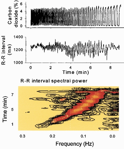
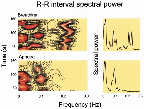
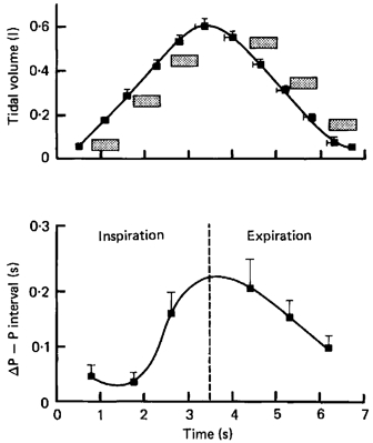
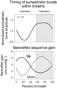
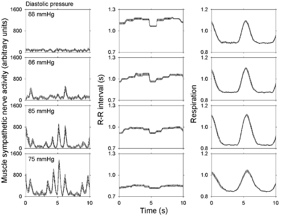
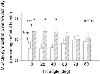
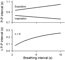
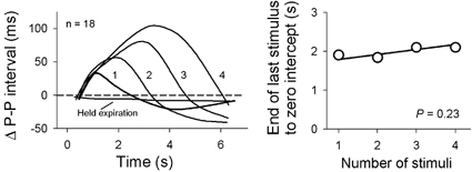
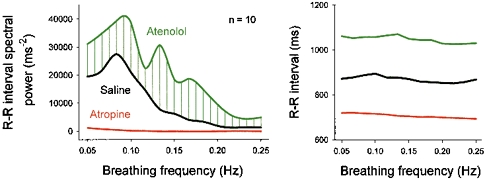
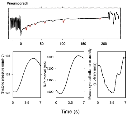
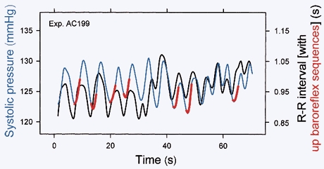
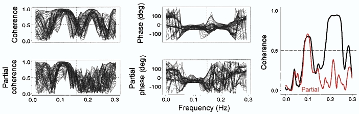
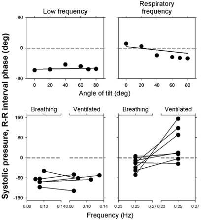
Respiratory activity phasically alters membrane potentials of preganglionic vagal and sympathetic motoneurones and continuously modulates their responsiveness to stimulatory inputs. The most obvious manifestation of this 'respiratory gating' is respiratory sinus arrhythmia, the rhythmic fluctuations of electrocardiographic R-R intervals observed in healthy resting humans. Phasic autonomic motoneurone firing, reflecting the throughput of the system, depends importantly on the intensity of stimulatory inputs, such that when levels of stimulation are low (as with high arterial pressure and sympathetic activity, or low arterial pressure and vagal activity), respiratory fluctuations of sympathetic or vagal firing are also low. The respiratory gate has a finite capacity, and high levels of stimulation override the ability of respiration to gate autonomic responsiveness. Autonomic throughput also depends importantly on other factors, including especially, the frequency of breathing, the rate at which the gate opens and closes. Respiratory sinus arrhythmia is small at rapid, and large at slow breathing rates. The strong correlation between systolic pressure and R-R intervals at respiratory frequencies reflects the influence of respiration on these two measures, rather than arterial baroreflex physiology. A wide range of evidence suggests that respiratory activity gates the timing of autonomic motoneurone firing, but does not influence its tonic level. I propose that the most enduring significance of respiratory gating is its use as a precisely controlled experimental tool to tease out and better understand otherwise inaccessible human autonomic neurophysiological mechanisms.


Figures

Figure 1
Figure 1. Expiratory carbon dioxide concentrations, R–R intervals, and a horizontal section through a sliding fast Fourier transformation of R–R intervals during ramped frequency breathing
Respiratory sinus arrhythmia (middle and bottom panels, black to orange areas) was small at the extremes of breathing frequency. Low frequency spectral power (bottom panel, extreme right) was present throughout the period of ramped breathing. These data indicate that some R–R interval fluctuations closely track breathing.
Figure 2
Figure 2. R–R interval spectral power during frequency-controlled breathing and apnoea
Respiratory-frequency R–R interval spectral power, shown as horizontal sections of a sliding fast Fourier transformation (lower left) or power spectrum (lower right) disappeared during apnoea. Breathing is necessary for the occurrence of respiratory-frequency R–R interval fluctuations; they are absent during apnoea.
Figure 3
Figure 3. Changes of P–P intervals provoked by brief neck suction, applied at different times in the breathing cycle
The stippled boxes show the times and durations of applications of baroreceptor stimulation. Changes of P–P intervals were calculated by subtracting measurements made during breathing from those made at the same times in the breathing cycle after neck suction. Respiratory gating of vagal-cardiac motoneurone responsiveness is continuously variable throughout the breathing cycle. Adapted from Eckberg et al. (1980).
Figure 4
Figure 4. Muscle sympathetic nerve activity and up and down baroreflex slopes, signal-averaged on the beginning of expiration
Respiratory gating of spontaneous upgoing and downgoing baroreflex sequences may be a consequence of respiratory gating of muscle sympathetic neurone firing. Adapted from Rothlisberger et al. (2003).
Figure 5
Figure 5. Signal-averaged mean ±s.e.m. measurements obtained from one supine subject at different arterial pressure levels
The magnitude of respiratory gating depends importantly on level of stimulation of sympathetic-muscle and vagal-cardiac motoneurones. Adapted from Eckberg et al. (1988).
Figure 6
Figure 6. Inspiratory (□) and expiratory () muscle sympathetic nerve activity during supine rest and passive upright tilt
* Significant (P ≤ 0.05) differences between supine and tilt measurements. Insp. = inspiration; Exp. = expiration. Respiratory gating of sympathetic firing declines steadily as the level of sympathetic stimulation increases. Adapted from Cooke et al. (1999).
Figure 7
Figure 7. Schematic representation of the respiratory gate
In this scheme, the level of stimulation of sympathetic or vagal motoneurones is represented as the height of the water to the left of the gates. Fluctuations of neural outflow are greatest at usual levels of stimulation, and are less at low or high levels of stimulation.
Figure 8
Figure 8. Peak and valley P–P intervals at different breathing frequencies
Maximum heart periods become longer, and minimum (Inspiration) heart periods become shorter as breathing rate slows. Adapted from Eckberg (1983).
Figure 9
Figure 9. Average P–P intervals after trains of one, two, three, or four brief neck suction pulses
The right panel shows average times from the peak of P–P interval prolongation to the baseline, calculated with least squares linear regression. The decay of vagal baroreflex inhibition was nearly constant, and its rate was nearly independent of the amount of inhibition. Adapted from Eckberg & Eckberg (1982).
Figure 10
Figure 10. Average R–R interval spectral power and R–R intervals during breathing over a range of breathing frequencies, before (Saline) and after β-adrenergic (Atenolol) or cholinergic (Atropine) blockade
This analysis documents the importance of vagal fluctuations in generating R–R interval fluctuations, and indicates that sympathetic stimulation opposes vagal inhibition at all breathing frequencies. Adapted from Taylor et al. (2001).
Figure 11
Figure 11. Aborted respiratory movements during apnoea, and measurements obtained by signal-averaging on the small pneumograph changes shown as red notches in the upper panel
The subject was not aware of the minute abortive respiratory movements, and the pneumotachograph recording did not register airflow. These responses provide indirect evidence for the importance of respiratory motoneurone activity in modulating autonomic neural firing. Adapted from Badra et al. (2001).
Figure 12
Figure 12. Systolic pressures, R–R intervals and spontaneous baroreflex slopes from one subject during quiet breathing
Systolic pressures and R–R intervals fluctuated together with every breath, and spontaneous baroreflex slopes (in red) occurred less frequently. Therefore, spontaneous baroreflex sequences do not result simply from breathing. Adapted from Rothlisberger et al. (2003).
Figure 13
Figure 13. Sliding systolic pressure and R–R interval coherence and phase from one subject during spontaneous breathing, before and after partialization
Removal of the influence of respiration from systolic pressures and R–R intervals by partialization (dashed line, see text) abolished the systematic coherence and phase differences that were present before partialization. This suggests that parallel respiratory-frequency systolic pressure and R–R interval fluctuations are secondary to breathing, and do not reflect baroreflex physiology. Adapted with permission (Badra et al. 2001).
Figure 14
Figure 14. Low- and respiratory-frequency phase angles derived from cross spectral analysis of systolic pressures and R–R intervals during passive upright tilt (upper panels) and spontaneous breathing or mechanical ventilation (lower panels)
Nearly constant low frequency phase angles are consistent with baroreflex physiology. Conversely, highly variable respiratory-frequency phase angles during tilt and mechanical breathing point toward respiratory influences, and away from baroreflex mechanisms. Adapted from Koh et al. (1998) and Cooke et al. (1999).

References

    1. Angelone A, Coulter NA., Jr Respiratory sinus arrhythmia: a frequency dependent phenomenon. J Appl Physiol. 1964;19:479–482. - PubMed
    1. Anrep GV, Pascual W, Rössler R. Respiratory variations of the heart rate. I-The reflex mechanism of the respiratory arrhythmia. Proc R Soc Lond B Biol Sci. 1936a;119 B:191–217.
    1. Anrep GV, Pascual W, Rössler R. Respiratory variations of the heart rate. II-The central mechanism of the respiratory arrhythmia and the inter-relations between the central and the reflex mechanisms. Proc R Soc Lond B Biol Sci. 1936b;119 B:218–230.
    1. Badra LJ, Cooke WH, Hoag JB, Crossman AA, Kuusela TA, Tahvanainen KUO, Eckberg DL. Respiratory modulation of human autonomic rhythms. Am J Physiol Heart Circ Physiol. 2001;280:H2674–2688. - PubMed
    1. Baselli G, Cerutti S, Badilini F, Biancardi L, Porta A, Pagani M, Lombardi F, Rimoldi O, Furlan R, Malliani A. Model for the assessment of heart period and arterial pressure variability interactions and respiration influences. Med Biol Eng Comput. 1994;32:143–152. - PubMed

Publication types

LinkOut - more resources