Review
. 2003 Mar;67(1):52-65, table of contents.
doi: 10.1128/MMBR.67.1.52-65.2003.

Cytokinesis in bacteria

Affiliations
Review

Cytokinesis in bacteria

Jeffery Errington et al. Microbiol Mol Biol Rev. 2003 Mar.

Abstract

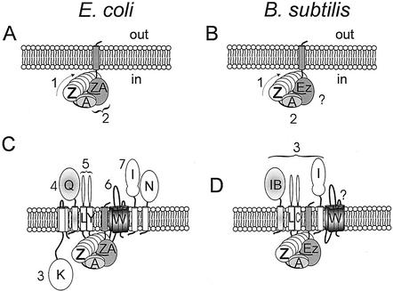
Work on two diverse rod-shaped bacteria, Escherichia coli and Bacillus subtilis, has defined a set of about 10 conserved proteins that are important for cell division in a wide range of eubacteria. These proteins are directed to the division site by the combination of two negative regulatory systems. Nucleoid occlusion is a poorly understood mechanism whereby the nucleoid prevents division in the cylindrical part of the cell, until chromosome segregation has occurred near midcell. The Min proteins prevent division in the nucleoid-free spaces near the cell poles in a manner that is beginning to be understood in cytological and biochemical terms. The hierarchy whereby the essential division proteins assemble at the midcell division site has been worked out for both E. coli and B. subtilis. They can be divided into essentially three classes depending on their position in the hierarchy and, to a certain extent, their subcellular localization. FtsZ is a cytosolic tubulin-like protein that polymerizes into an oligomeric structure that forms the initial ring at midcell. FtsA is another cytosolic protein that is related to actin, but its precise function is unclear. The cytoplasmic proteins are linked to the membrane by putative membrane anchor proteins, such as ZipA of E. coli and possibly EzrA of B. subtilis, which have a single membrane span but a cytoplasmic C-terminal domain. The remaining proteins are either integral membrane proteins or transmembrane proteins with their major domains outside the cell. The functions of most of these proteins are unclear with the exception of at least one penicillin-binding protein, which catalyzes a key step in cell wall synthesis in the division septum.


Figures

FIG. 1.
FIG. 1.
Division site selection in rod-shaped bacteria. (A) Roles of nucleoid occlusion and the Min system. In cells that are about to divide, the correct midcell division site is chosen by the combined action of two negative regulatory systems. The localization of the two nucleoids blocks division in their vicinity, as shown, leaving spaces available for division at the correct midcell site and at the cell poles. The Min system acts to prevent incorrect division at the cell poles. (B) Blocking division at the cell poles. The MinCD inhibitor associates with the cytoplasmic membrane at the cell poles and prevents FtsZ polymerization. The C-terminal domain of MinC (CC) ensures MinC dimerization and interacts with MinD (D), which in turn associates with the membrane. The N-terminal domain of MinC (CN) interacts with FtsZ (Z) and prevents polymerization of FtsZ or interaction of other cell division proteins with the FtsZ ring (see the text). The MinD-MinC stoichiometry, and the presumed action of MinC, preventing the formation of correct FtsZ interactions remains hypothetical. (C and D) Contrasting functioning of the Min system in E. coli (C) and B. subtilis (D). MinD (chevrons [ATP form] and rectangles [ADP form]) is postulated to polymerize in the presence of ATP. In E. coli (A), it forms dynamic filaments emanating from one cell pole. A ring of MinE proteins associates with the ends of the filaments and moves in a poleward manner, by interaction with the MinD filaments. MinE stimulates hydrolysis of ATP by MinD, and this results in the release of monomers, probably following a conformational change (rectangles), which enter the cytoplasm. After exchange of the ADP for ATP, MinD polymers can re-form at the opposite cell pole, possibly requiring a nucleation site at the pole. In B. subtilis (B), MinD forms static filaments restricted to the polar zones as a result of nucleation by DivIVA protein targeted to the cell poles (black triangles). In both cases, MinD is associated with the division inhibitor (MinC; not shown), which blocks Z ring formation. Reproduced reference with permission from the publisher.
FIG. 2.
FIG. 2.
Organization of the cell division apparatus during various steps in the division process. A comparison of the E. coli system (A and C) with that of B. subtilis (B and D) is shown. Numbers beside proteins indicate the hierarchy of assembly; a question mark indicates that this is not yet clear. (A and B) Formation of the Z ring and anchoring of the Z ring to the membrane. FtsZ (Z) interacts with the cytoplasmic cell division protein FtsA (A) via the C-terminal tail of Z, which is flexible. FtsA (at least of B. subtilis [56]) is a dimer. In E. coli, the FtsZ ring is anchored to the membrane, at least partly, by ZipA (ZA), which also binds to the flexible C-terminal FtsZ tail. In most bacteria, functional equivalents of ZipA have not yet been found. In B. subtilis, EzrA protein has a similar topology to ZipA, although it is not essential for division. (C and D) Assembly of the remainder of the cell division machinery. In E. coli (C), the hierarchy for assembly of the remaining proteins is essentially linear, as numbered, except for FtsL (L) and YgbQ (Y). The other proteins are labeled according to their fts suffix. In B. subtilis (D), there is no apparent homologue of FtsN and the FtsK homologue (SpoIIIE) is not required for division. The DivIB (IB), DivIC (C), FtsL (L), and PBP-2B (I) proteins assemble in a concerted manner. The order of assembly of FtsW (W) is not known.

References

    1. Addinall, S. G., E. Bi, and J. Lutkenhaus. 1996. FtsZ ring formation in fts mutants. J. Bacteriol. 178:3877-3884. - PMC - PubMed
    1. Addinall, S. G., C. Cao, and J. Lutkenhaus. 1997. FtsN, a late recruit to the septum in Escherichia coli. Mol. Microbiol. 25:303-309. - PubMed
    1. Addinall, S. G., C. Cao, and J. Lutkenhaus. 1997. Temperature shift experiments with an ftsZ84(Ts) strain reveal rapid dynamics of FtsZ localization and indicate that the Z ring is required throughout septation and cannot reoccupy division sites once constriction has initiated. J. Bacteriol. 179:4277-4284. - PMC - PubMed
    1. Addinall, S. G., and J. Lutkenhaus. 1996. FtsA is localized to the septum in an FtsZ-dependent manner. J. Bacteriol. 178:7167-7172. - PMC - PubMed
    1. Addinall, S. G., and J. Lutkenhaus. 1996. FtsZ-spirals and -arcs determine the shape of the invaginating septa in some mutants of Escherichia coli. Mol. Microbiol. 22:231-237. - PubMed

Publication types

MeSH terms

Substances

LinkOut - more resources