. 2003 Mar 15;31(6):1640-5.
doi: 10.1093/nar/gkg251.

Human mitochondrial DNA is packaged with TFAM

Affiliations

Human mitochondrial DNA is packaged with TFAM

Tanfis Istiaq Alam et al. Nucleic Acids Res. .

Abstract

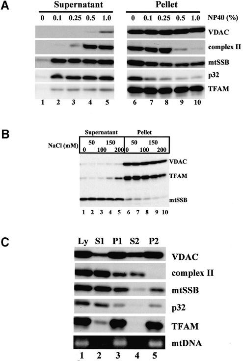
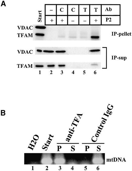
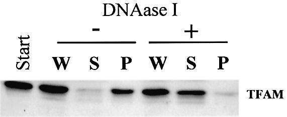
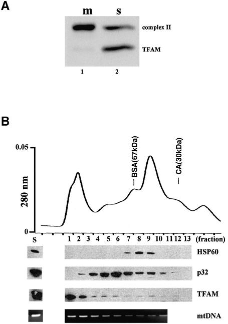
Mitochondrial transcription factor A (TFAM), a member of the high mobility group proteins, is essential for maintenance of mitochondrial DNA (mtDNA). Most TFAM and mtDNA (both of which are normally soluble) was recovered from the particulate fraction of human placental mitochondria when extracted with the non-ionic detergent Nonidet P-40. mtDNA and TFAM were co-immunoprecipitated by anti-TFAM antibodies. TFAM was released into the supernatant by DNase I digestion of mtDNA in the particulate fraction. Thus, TFAM and mtDNA are tightly associated with each other, and it is likely that few TFAM or mtDNA molecules exist in an unbound form in mitochondria. Based on the fact that TFAM is abundant enough to wrap mtDNA entirely, these results suggest that human mtDNA is packaged with TFAM.


Figures

Figure 1
Figure 1
Solubilization of human placental mitochondria. (A) Mito chondria were incubated with various concentrations of NP-40 and then separated into supernatants and pellets. The pellets were re-suspended in the original volume of buffer for easily estimating recovery of proteins. Each protein was detected by western blotting. (B) Mitochondria were incubated with 0.5% NP-40 in the presence of various concentrations of NaCl. Then, the mixtures were separated into supernatants and pellets. (C) Mitochondria were incubated with 0.5% NP-40 (Ly), and separated into the first supernatant (S1) and pellet. The first pellet was re-suspended in the original volume of buffer (P1). The P1 was centrifuged and separated into the second supernatant (S2) and pellet. The second pellet was similarly re- suspended in the original volume of buffer (P2). An mtDNA fragment (np 7319–7603) in each sample was amplified by PCR (lowest row).
Figure 2
Figure 2
Immunoprecipitation by anti-TFAM. P2 samples were incubated with rotation in the presence of antibody-immobilized magnetic beads. The beads were pelleted using a magnet. The resulting supernatants (IP-sup) were removed. After washing three times, proteins were eluted from the beads by heating (IP-pellet). C, control IgG; T, anti-TFAM. (A) VDAC and TFAM were detected by western blotting. In lane 2, unconjugated magnetic beads were used. (B) mtDNA was detected by PCR. P, IP-pellet; S, IP-sup.
Figure 3
Figure 3
DNase I digestion of P2. P2 was digested with DNase I and centrifuged. TFAM was detected by western blotting. W, before centrifugation; S, supernatant; P, pellet.
Figure 4
Figure 4
Immunolabeling of HeLa cells. TFAM in HeLa MRV11 cells was identified using affinity-purified anti-TFAM antibodies and Alexa Fluor 488- conjugated goat anti-rabbit IgG antibodies (a). Mitochondria were stained with Mitotracker Red (b). (c) A merged image. (d) A magnified image of the square outlined region in (c).
Figure 5
Figure 5
Sonication of mitochondria. Mitochondria of U937 cells were sonicated. After centrifugation, soluble (s) and particulate (m) fractions were prepared. The particulate fraction was re-suspended to the original volume. (A) Iron sulfur protein of complex II and TFAM were detected by western blotting. (B) The soluble fraction was separated by size exclusion chromatography. The topmost panel is a profile of UV absorbance. HSP60, p32 and TFAM were detected by western blotting. mtDNA was detected by PCR. BSA, bovine serum albumin; CA, carbonic anhydrase.

References

    1. Fisher R.P. and Clayton,D.A. (1988) Purification and characterization of human mitochondrial transcription factor 1. Mol. Cell. Biol., 8, 3496–3509. - PMC - PubMed
    1. Parisi M.A., Xu,B. and Clayton,D.A. (1993) A human mitochondrial transcription activator can functionally replace a yeast mitochondrial HMG-box protein both in vivo and in vitro. Mol. Cell. Biol., 13, 1951–1961. - PMC - PubMed
    1. Falkenberg M., Gaspari,M., Rantanen,A., Trifunovic,A., Larsson,N.G. and Gustafsson,C.M. (2002) Mitochondrial transcription factors B1 and B2 activate transcription of human mtDNA. Nature Genet., 31, 289–294. - PubMed
    1. McCulloch V., Seidel-Rogol,B.L. and Shadel,G.S. (2002) A human mitochondrial transcription factor is related to RNA adenine methyltransferases and binds S-adenosylmethionine. Mol. Cell. Biol., 22, 1116–1125. - PMC - PubMed
    1. Shadel G.S. and Clayton,D.A. (1997) Mitochondrial DNA maintenance in vertebrates. Annu. Rev. Biochem., 66, 409–435. - PubMed

Publication types

MeSH terms