. 2003 Mar 15;31(6):1725-34.
doi: 10.1093/nar/gkg269.

RPA is an initiation factor for human chromosomal DNA replication

Affiliations

RPA is an initiation factor for human chromosomal DNA replication

Dávid Szüts et al. Nucleic Acids Res. .

Abstract

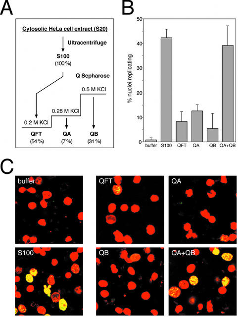
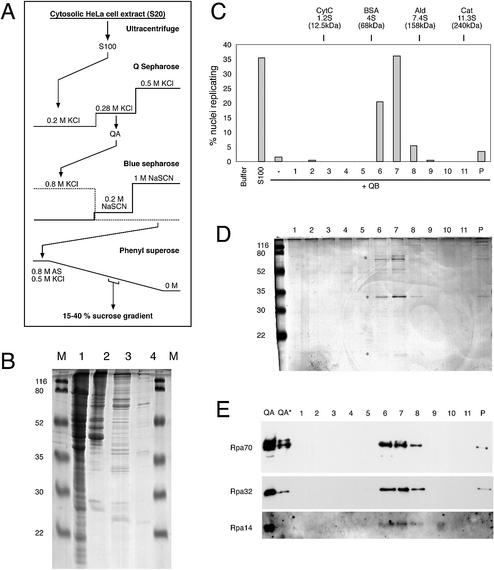
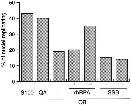
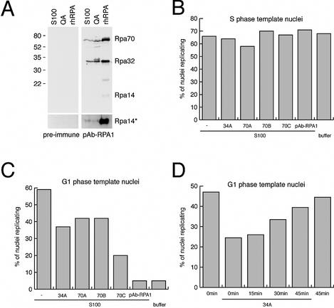
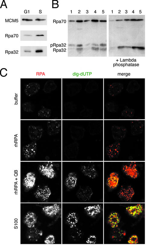
The initiation of chromosomal DNA replication in human cell nuclei is not well understood because of its complexity. To allow investigation of this process on a molecular level, we have recently established a cell-free system that initiates chromosomal DNA replication in an origin-specific manner under cell cycle control in isolated human cell nuclei. We have now used fractionation and reconstitution experiments to functionally identify cellular factors present in a human cell extract that trigger initiation of chromosomal DNA replication in this system. Initial fractionation of a cytosolic extract indicates the presence of at least two independent and non-redundant initiation factors. We have purified one of these factors to homogeneity and identified it as the single-stranded DNA binding protein RPA. The prokaryotic single-stranded DNA binding protein SSB cannot substitute for RPA in the initiation of human chromosomal DNA replication. Antibodies specific for human RPA inhibit the initiation step of human chromosomal DNA replication in vitro. RPA is recruited to DNA replication foci and becomes phosphorylated concomitant with the initiation step in vitro. These data establish a direct functional role for RPA as an essential factor for the initiation of human chromosomal DNA replication.


Figures

Figure 1
Figure 1
Initial fractionation of cytosolic extract. (A) Schematic diagram of fractionation steps. See Materials and Methods for details. The values given in parentheses below each fraction indicate the percentages of total protein recovered in the respective fraction. (B) Quantitative analysis of DNA replication initiated in isolated G1 phase nuclei in vitro. Nuclei from mimosine-arrested HeLa cells were incubated in elongation buffer (buffer), supplemented with 100 µg protein of unfractionated extract (S100), 45 µg of fraction QFT, 15 µg of QA, 35 µg of QB and a combination of 15 µg QA and 35 µg QB as indicated (these masses approximate typical mass percentages of the respective fractions in the S100 extract). Percentages of replicating nuclei were quantitated and mean values and standard deviations of 5–10 independent experiments are shown. The main variance of the percentages of nuclei replicating in the indicated fractions was due to differences between separate preparations of fractions. (C) Representative fields of replicating nuclei. Replication reactions were performed in the presence of the indicated components and nuclei were analysed by confocal fluorescence microscopy. Nuclear DNA is visualised by propidium iodide (red signal) and replicated DNA by fluorescein-conjugated anti-digoxigenin Fab fragments (green signal). Merged images are presented showing sites of replicated nuclear DNA in yellow and non-replicating nuclei in red.
Figure 2
Figure 2
Purification of the initiation factor present in fraction QA. (A) Schematic diagram of purification procedure. See Materials and Methods for details. (B) Protein analysis of key purification steps. Unfractionated S100 extract (100 µg protein, lane 1), fraction QA (20 µg protein, lane 2), Blue Sepharose eluate (10 µg protein, lane 3) and Phenyl Superose eluate (<1 µg, lane 4) were loaded on a 15% polyacrylamide gel. Proteins were visualised by staining with Coomassie brilliant blue. Molecular masses of marker proteins (M) are indicated. (C–E) Final purification and identification of RPA as the initiation activity present in fraction QA by sucrose gradient centrifugation. (C) Quantitative analysis of initiation of DNA replication. Nuclei from mimosine-arrested EJ30 cells were incubated in 35 µg of fraction QB supplemented with a 20 µl volume of the sucrose gradient fractions (1–11) and the pelleted material (P) as indicated. Incubations in elongation buffer (buffer) or 100 µg of unfractionated extract (S100) were used as controls. Percentages of replicating nuclei of a representative experiment are shown. (D) Protein analysis of fractions of the sucrose gradient step. A 20 µl volume of each sucrose gradient fraction (1–11) and the pelleted material (P) were separated on a 15% polyacrylamide gel and stained with silver salts. Molecular masses of marker proteins (M) are indicated. Asterisks denote the position of Rpa70, 32 and 14 subunits. (E) Identification of RPA by western blot analysis. A 20 µl volume of each sucrose gradient fraction (1–11 and P) and 15 and 5 µg (*) of fraction QA were separated on a 15% polyacrylamide gel and analysed by western blotting. The membrane was sequentially probed with a mixture of monoclonal antibodies 70A, 70B and 70C (Rpa70) (36), monoclonal antibody 34A (Rpa32) (36) and polyclonal antibody pAb-RPA1 (visualising Rpa14; see Fig. 4A for a characterisation of this polyclonal antibody).
Figure 3
Figure 3
Recombinant human RPA, but not prokaryotic SSB can substitute for the initiation activity present in fraction QA. Nuclei from mimosine- arrested HeLa cells were incubated in unfractionated extract (S100) and in 35 µg of fraction QB, supplemented with either 20 µg of fraction QA, replication buffer (–), 12.5 ng (+) or 37.5 ng (++) purified recombinant human RPA (rhRPA) or equimolar amounts of E.coli single-stranded DNA binding protein (SSB), respectively. Percentages of replicating nuclei were quantitated and mean values of two or three independent experiments are shown.
Figure 4
Figure 4
Antibodies specific for human RPA inhibit the initiation of chromosomal DNA replication in vitro. (A) Generation of the anti-RPA polyclonal antibody pAb-RPA1 and its specificity on S100 cytosolic extract, fraction QA and rhRPA. The bottom panel shows an overexposed image of the 14 kDa region of the blot. (B) Effect of anti-RPA antibodies on DNA replication in S phase nuclei. Replication initiation reactions using template nuclei from S phase HeLa cells were supplemented with 5 µg of the indicated monoclonal antibodies or 2 µl of pAb-RPA antiserum. Percentages of nuclei replicating were scored and mean values of two independent experiments are shown. (C) Inhibition of initiation in G1 phase template nuclei by the anti-RPA antibodies. Replication initiation reactions using template nuclei from mimosine-arrested late G1 phase HeLa cells were supplemented with the indicated antibodies as detailed in (B). Percentages of nuclei replicating were scored and mean values of two independent experiments are shown. (D) Late addition of antibody 34A no longer inhibits DNA replication. Replication initiation reactions using late G1 phase template nuclei were supplemented with 5 µg of the monoclonal 34A antibody and dig-dUTP at the indicated times. Percentages of nuclei replicating were scored and mean values of two independent experiments are shown.
Figure 5
Figure 5
RPA is recruited to DNA replication foci and becomes phosphorylated in vitro. (A) Comparison of bound RPA in G1 and S phase nuclei in vivo. Nuclear extracts (25 µg protein per lane) from mimosine- arrested late G1 phase and from early S phase cells were analysed by western blot using antibodies specific for MCM5 as a control and polyclonal antibody pAb-RPA1. (B) Nuclear binding and phosphorylation of RPA during initiation of DNA replication in vitro. G1 phase nuclei isolated from in vitro incubations done in replication buffer (lane 1), 100 µg S100 cytosolic extract (lane 2), 30 and 100 ng rhRPA (lanes 3 and 4) and 100 ng rhRPA plus 35 µg of fraction QB (lane 5). The samples shown in the right-hand panel were treated with λ phosphatase before loading onto the same gel. The Rpa70 and Rpa32 subunits were visualised with pAb-RPA1. Phosphorylated Rpa32 is indicated as pRpa32. (C) Confocal microscopy. G1 phase nuclei were incubated in buffer (top row), in 100 ng of rhRPA (second row), in 100 ng rhRPA supplemented with 35 µg QB (third row) and in 100 µg of unfractionated S100 (bottom row). High-resolution micrographs are shown of RPA foci (stained with pAb-RPA1, red), replication foci (dig-UTP, green) and merged images of the same set of nuclei.

Similar articles

Cited by

References

    1. Bell S.P. and Dutta,A. (2002) DNA replication in eukaryotic cells. Annu. Rev. Biochem., 71, 333–374. - PubMed
    1. Blow J.J. and Hodgson,B. (2002) Replication licensing—origin licensing: defining the proliferative state? Trends Cell Biol., 12, 72–78. - PMC - PubMed
    1. Diffley J.F. and Labib,K. (2002) The chromosome replication cycle. J. Cell Sci., 115, 869–872. - PubMed
    1. Lei M. and Tye,B.K. (2001) Initiating DNA synthesis: from recruiting to activating the MCM complex. J. Cell Sci., 114, 1447–1454. - PubMed
    1. Kelly T.J. and Brown,G.W. (2000) Regulation of chromosome replication. Annu. Rev. Biochem., 69, 829–880. - PubMed

Publication types