. 2003 Mar 18;100(6):3095-100.
doi: 10.1073/pnas.0538069100. Epub 2003 Mar 7.

A Saccharomyces cerevisiae mutant strain defective in acetyl-CoA carboxylase arrests at the G2/M phase of the cell cycle

Affiliations

A Saccharomyces cerevisiae mutant strain defective in acetyl-CoA carboxylase arrests at the G2/M phase of the cell cycle

Walid Al-Feel et al. Proc Natl Acad Sci U S A. .

Abstract

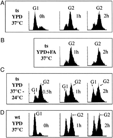
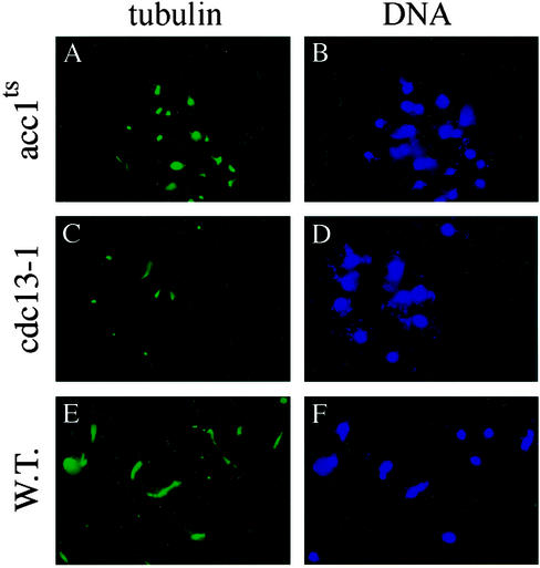
To elucidate the essential functions of acetyl-CoA carboxylase (ACC1FAS3) in Saccharomyces cerevisiae, a temperature-sensitive mutant (acc1(ts)) was constructed. When the acc1(ts) cells were synchronized in G(1) phase with alpha-factor at the permissive temperature of 24 degrees C and then released from the blockade and incubated at the restrictive temperature of 37 degrees C, 95% of the cell population became arrested at the G(2)M phase of the cell cycle despite the presence of fatty acids (C(14)-C(26)) in the medium. These cells developed large undivided nuclei, and the spindles of the arrested mutant cells were short. Shifting the G(2) arrested cells back to the permissive temperature resulted in a reversal of the cell-cycle arrest, with cells initiating mitosis. However, after 3 h of incubation at 37 degrees C, G(2) arrested mutant cells lost viability and displayed a uniquely altered nuclear envelope. Using [1-(14)C]acetate as a precursor for fatty acids synthesis, we identified the phospholipids and sphingolipids derived from acc1(ts) cells and wild-type cells at 24 degrees C and 37 degrees C, respectively. The levels of inositol-ceramides [IPC, MIPC, and M(IP)(2)C] and very long-chain fatty acids C(24) and C(26) declined sharply in the G(2)M arrested cells because of ACC inactivation. Shifting the acc1(ts) cells to 24 degrees C after 2 h of incubation at 37 degrees C resulted in reactivation of the ACC and elevation of the ceramides and very long-chain fatty acid syntheses with normal cell-cycle progression. In contrast, synthesis of wild-type inositol-ceramides, C(24) and C(26), fatty acids were elevated on incubation at 37 degrees C and declined when the cells shifted to the permissive temperature of 24 degrees C.


Figures

Figure 1
Figure 1
Viability of acc1ts yeast at 37°C. Cells harboring the pUN100-acc plasmid (acc1ts) were grown at 24°C in 10 ml of YPD to an optical density of 0.4 at 600 nm, then shifted to 37°C. At the indicated time, a sample was withdrawn, plated onto YPD plates, and incubated at 24°C. The percentages of viable cells were calculated based on the number of colonies formed on the plates relative to the total number of cells as determined by a Coulter counter.
Figure 2
Figure 2
Flow cytometric analyses of DNA contents of acc1ts mutant and wild-type yeast at 37°C. Cells were synchronized in G1 with α-factor at 24°C, as described in Materials and Methods. (A) Cells were released into YPD media and incubated at 37°C. At the indicated times of incubation, samples were withdrawn and fixed with either 70% alcohol or 3.7% formaldehyde and placed, respectively, at −20°C and 4°C. Microscopic and flow cytometric analyses were carried out as described in Materials and Methods. (B) Same as in A plus fatty acids (in all combinations, see text). (C) acc1ts cells were synchronized at the G2 phase by incubation for 2 h at 37°C, as described in A, then shifted to 24°C for 0.5, 1, and 2 h. (D) Wild-type yeast was synchronized with α-factor at 24°C and, after releasing into YPD, was incubated at 37°C. Samples were collected after 1 and 2 h and treated as in A and B. In each figure, the left-most peak represents the G1 population and the right-most peak represents the G2 population, as indicated.
Figure 3
Figure 3
Morphology of acc1ts and wild-type cells. Phase and immunofluorescence microscopy of acc1ts and wild-type cells were carried out as described in Materials and Methods. (A) Phase optics acc1ts cells were arrested at G1 at 24°C by using α-factor and released in YPD and incubated at 37°C for 2 h. (B) After 2 h of incubation at 37°C, the acc1ts cells were shifted to 24°C, and samples were collected after 30 min and examined as in A. (C) The wild-type cells were treated and examined as in A. (A′–C′) The DNA of yeast nuclei of corresponding yeast strains used in A–C were stained with DAPI.
Figure 4
Figure 4
The spindle structures and nuclear morphologies of acc1ts and wild-type cells. Indirect immunofluorescence measurements were obtained with anti-tubulin antibodies of acc1ts, cdc13-1, and wild-type strains after their release into YPD from G1 α-factor arrest at 24°C and incubation at 37°C for 2 h. (A) G2 acc1ts-arrested cells exhibit short spindles. (C) As a control, the G2 cdc13-1-arrested cells also displayed short spindles. (E) Wild-type yeast strain shows normal spindle structure morphology. (B, D, and F) Immunofluorescence microscopy of DAPI-stained nuclei of the corresponding yeast strains used in A, C, and E, respectively.
Figure 5
Figure 5
Transmission electron microscopy of the nuclear envelope of acc1ts cells at a restrictive temperature for 1 h (A) and for 3.5 h (B). The acc1ts cells in B developed an altered nuclear envelope, which is defined by noncharacterized interspace between the outer and inner membrane of the nuclei, similar to the phenotype reported for the mtr7–1 yeast cells (30). The nuclear envelope of the wild-type yeast showed no changes when the cells were treated as in B. I, inner nuclear membrane; O, outer nuclear membrane; N, nucleus.
Figure 6
Figure 6
Incorporation of [14C]acetate into alkaline-stable lipids of acc1ts and wild-type yeast. (A) The acc1ts (ts) and wild-type (wt) yeast cells were arrested at G1 with α-factor at 24°C and then released in YPD and incubated at 37°C for 2 h in the presence of [14C]acetate. In a parallel experiment, comparable (ts and wt) cells were incubated at 37°C for 2 h, then shifted to 24°C and labeled with [14C]acetate for 2 h. The cells were harvested, and the total lipids were extracted and subjected to monomethyl alkaline hydrolysis and analyzed as described in Materials and Methods. The major IPC, MIPC, and M(IP)2C were identified by their Rfvalues and their respective ceramides were labeled with either [3H]inositol or [14C]mannose (27). (B) TLC analysis of [14C]acetate-labeled lipids. The acc1ts, wild-type, and cdc13–1 yeast cells were arrested at G1 with α-factor, then released in YPD and inoculated at 37°C and labeled with [14C]acetate for 2 h as in A. The cells were harvested, and the total lipids were extracted, subjected to monomethyl alkaline hydrolysis, and separated as described in Materials and Methods.
Figure 7
Figure 7
GCMS analysis of IPC, MIPC, and M(IP)2C fatty acids. The IPC, MIPC, and M(IP)2C obtained from wild-type and acc1ts strains labeled with [14C]acetate at 37°C and 24°C as described in Fig. 6 were hydrolyzed, and the very long-chain fatty acids were extracted, purified, and subjected to mass spectroscopic analysis as described in Materials and Methods. The abundance of C24 and C26 was determined by GCMS analysis.

References

    1. Wakil S J, Stoops J K, Joshi V C. Annu Rev Biochem. 1983;52:537–579. - PubMed
    1. Oh C-S, Toke D A, Mandala S, Martin C. J Biol Chem. 1997;272:17376–17384. - PubMed
    1. Toke D, Martin C. J Biol Chem. 1996;271:18413–18422. - PubMed
    1. Silver S, Leplatois P, Josse A, Dupuy P-H, Lanau C, Kaghad M, Dhers C, Rahier A, Taton M, Le Fur G, et al. Mol Cell Biol. 1996;16:2719–2727. - PMC - PubMed
    1. Lester R L, Dickson R C. Adv Lipid Res. 1993;26:253–274. - PubMed

Publication types

LinkOut - more resources