. 2003 Mar 18;100(6):3299-304.
doi: 10.1073/pnas.0434590100. Epub 2003 Mar 7.

Mapping Wnt/beta-catenin signaling during mouse development and in colorectal tumors

Affiliations

Mapping Wnt/beta-catenin signaling during mouse development and in colorectal tumors

Silvia Maretto et al. Proc Natl Acad Sci U S A. .

Abstract

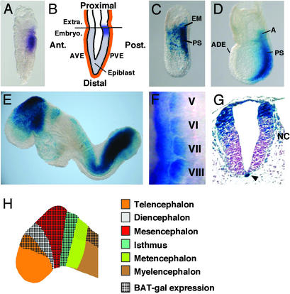
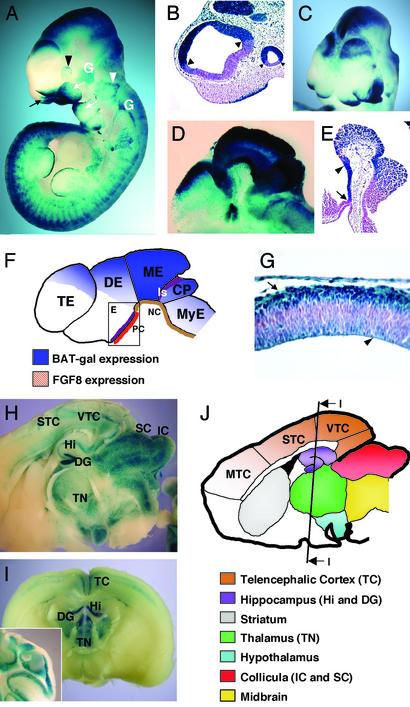
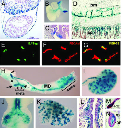
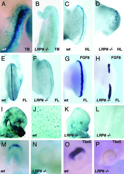
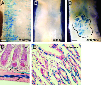
Wntbeta-catenin signaling plays key roles in several developmental and pathological processes. Domains of Wnt expression have been extensively investigated in the mouse, but the tissues receiving the signal remain largely unidentified. To define which cells respond to activated beta-catenin during mammalian development, we generated the beta-catenin-activated transgene driving expression of nuclear beta-galactosidase reporter (BAT-gal) transgenic mice, expressing the lacZ gene under the control of beta-cateninT cell factor responsive elements. Reporter gene activity is found in known organizing centers, such as the midhindbrain border and the limb apical ectodermal ridge. Moreover, BAT-gal expression identifies novel sites of Wnt signaling, like notochord, endothelia, and areas of the adult brain, revealing an unsuspected dynamic pattern of beta-catenin transcriptional activity. Expression of the transgene was analyzed in mutant backgrounds. In lipoprotein receptor-related protein 6-null homozygous mice, which lack a Wnt coreceptor, BAT-gal staining is absent in mutant tissues, indicating that BAT-gal mice are bona fide in vivo indicators of Wntbeta-catenin signaling. Analyses of BAT-gal expression in the adenomatous polyposis coli (multiple intestinal neoplasia+) background revealed betacatenin transcriptional activity in intestinal adenomas but surprisingly not in normal crypt cells. In summary, BAT-gal mice unveil the entire complexity of Wntbeta-catenin signaling in mammals and have broad application potentials for the identification of Wnt-responsive cell populations in development and disease.


Figures

Figure 1
Figure 1
BAT-gal expression during early development. (A) First expression in the posterior epiblast of prestreak stage embryo detected by in situ hybridization with antisense β-galactosidase probe. (B) Diagram indicating the location of lacZ-positive cells (in blue) at the prestreak stage. Ant., anterior; Post., posterior; Extra., extraembryonic region; Embryo., embryonic region; AVE, anterior visceral endoderm; PVE, posterior visceral endoderm. (C and D) Whole-mount X-Gal staining of embryos at early- and late-streak stage, respectively. A, allantois; ADE, anterior definitive endoderm; PS, primitive streak. (E) E8.5 embryo shows staining in the fore-, mid-, and hindbrain, tailbud, newly formed somites, and cardiac region. (F) Lateral view of lacZ in situ hybridization of 9.5-days postcoitum somites V–VIII (anterior is up and dorsal is left). Note the progressive restriction of BAT-gal staining in the dorsal medial portion of the dermomyotome. (G) Cross section through the caudal spinal cord. Arrowhead points to the notochord. Note the migrating neural crest cells (NC). (H) Diagram indicating the expression of BAT-gal in different regions of the brain at E8.5.
Figure 2
Figure 2
Expression of BAT-gal in the developing brain and neural tissue. (A) Lateral view of whole-mount stained E10.5 embryo. Indicated are nasal folds (black arrow), optic vesicle (black arrowhead), otic vesicle (white arrowhead), and first and second branchial arches (white arrows). Note positive staining of cranial nerve ganglia (white, G). (B) Histological section through telencephalic and optic vesicles. Arrowheads point to the sharp borders of BAT-gal expression. (C) Anterior–top view of X-Gal-stained embryo (E9.5). Note the enhanced expression of BAT-gal in the dorsal side of the telencephalic hemispheres. (D) Sagittal section of head from a whole-mount-stained E10.5 embryo. Note expression in fore-, mid-, and hindbrain. (E) Section through ventral diencephalon showing lacZ expression in the region comprised between the infundibulum (arrow) and the mammilary body (arrowhead). (F) Schematic diagram of the β-galactosidase-positive areas of the developing brain at E10.5. PC, prechordal plate; NC, notochord; Is, isthmus; TE, telencephalon; DE, diencephalon; ME, mesencephalon; CP, cerebellar plate; MyE, myelencephalon. The square in the diencephalons is a diagram for E. (G) Section of telencephalic vesicle at E11.5. Staining is restricted in the cortical (arrow) and ventricular (arrowhead) layers of the tissue. (H and I) Expression of the BAT-gal transgene in the adult encephalon. Vibratome sagittal (H) and coronal (I) sections of the organ are shown. Staining is detected in distinct areas of the adult brain (J). Note the unprecedented detection of Wnt signaling in the sensory (STC) and visual (VTC) telencephalic cortex, but not motor cortex (MTC). (I Inset) A section of the adult cerebellum showing staining in the granule cells.
Figure 3
Figure 3
Expression of the BAT-gal transgene during organogenesis. (A) Section of heart and septum transversum of E9.5 embryo. (B) Whole-mount staining of heart and large vessels. Ventral side is to the left. (C) Section of E16.5 embryo showing staining of aortic semilunar valves. (D) lacZ-positive endothelial cells of vessels invading the spinal cord (sc) from the perineural mesenchyme (pm) at E12.5. (E–G) Costaining of blood vessels of the mesenchyme surrounding the spinal cord for the endothelial marker platelet endothelial cell adhesion molecule (PECAM) and β-galactosidase. (E) Expression of the BAT-gal transgene driving nuclear β-galactosidase. (F) PECAM localization identifies endothelial cells. (G) Merge of the previous pictures. (H) Genitourinary organs. Epithelia of mesonephros (meso), mesonephric duct (MD), ureter (u), ureteral buds (UB, arrows), and urogenital sinus (arrowhead) express lacZ, whereas the mesenchyme of metanephros (meta) does not. (I) E13.5, kidney. (J) E11.5, trachea and main bronchi. (K) E13.5, lung. (L) E13.5, section of bronchus showing positive X-Gal reaction in the mesenchyme. (M and N) Histological sections of the skin. Postnatal day 4, hairs in active anagen phase show staining in dermal papilla (DP), matrix (Mx), and precortex (PC).
Figure 4
Figure 4
Alterations of BAT-gal expression in LRP6 mutant mice. (A and B) Tailbuds (TB) of wild-type and mutant embryos at E10.5. (C–F) X-Gal staining of hindlimb (HL) or forelimb (FL) from wild-type (wt) and mutant (LRP6−/−) E11.5 embryos. (G and H) In situ hybridization with FGF8 antisense probe of normal (G) and mutant (H) forelimb buds. (I–L) Comparison of lacZ expression in the skin of wild-type and LRP6 mutant embryos at E14.5. (M) X-Gal staining of wild-type E9.5 embryos showing lacZ expression in the dorsal eye vesicle, which lacks in LRP6 mutants (N). (O and P) Corresponding in situ hybridization with a Tbx5 antisense probe of E10.5 embryos.
Figure 5
Figure 5
Activation of β-catenin/Wnt pathway in adenomas developing in APC(Min/+) mutant mice. (A) X-Gal staining of the intestine observed from the abluminal side. Note the network of perpendicular labeling characteristic of the mesenteric plexus. (B and C) View of the luminal side of the X-Gal-stained intestine from wild-type (B) and APC(Min/+) (C) mice. Adenomas (dashed line in C) express the lacZ reporter gene. (D) Transverse section of wild-type (wt) intestine. Arrowheads indicate scattered positive stromal cells surrounding the crypts. Arrows indicate positive cells arranged in longitudinal and circular networks (see A), probably belonging to the enteric plexus. (E) In APC(Min/+) adenomas, BAT-gal expression is greatly up-regulated in the epithelial neoplastic cells.

References

    1. Bienz M, Clevers H. Cell. 2000;103:311–320. - PubMed
    1. Huelsken J, Vogel R, Erdmann B, Cotsarelis G, Birchmeier W. Cell. 2001;105:533–545. - PubMed
    1. Wodarz A, Nusse R. Annu Rev Cell Dev Biol. 1998;14:59–88. - PubMed
    1. Niehrs C. Nature. 2001;413:787–788. - PubMed
    1. Peifer M, Polakis P. Science. 2000;287:1606–1609. - PubMed

Publication types

MeSH terms

Grants and funding

LinkOut - more resources