. 2003 Mar 18;100(6):3101-6.
doi: 10.1073/pnas.0534783100. Epub 2003 Mar 7.

A binding motif for Siah ubiquitin ligase

Affiliations

A binding motif for Siah ubiquitin ligase

Colin M House et al. Proc Natl Acad Sci U S A. .

Abstract

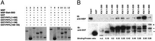
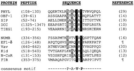
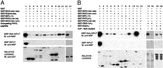
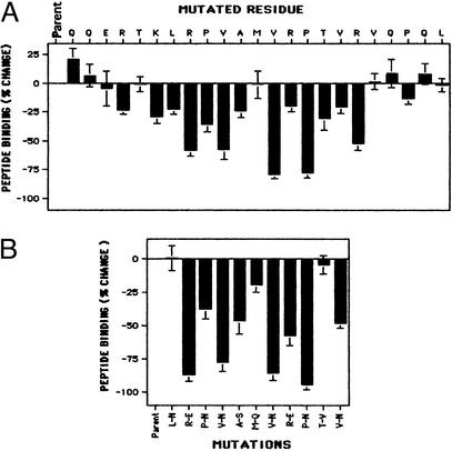
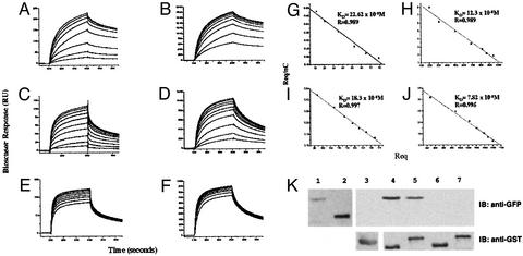
The Drosophila SINA (seven in absentia) protein and its mammalian orthologs (Siah, seven in absentia homolog) are RING domain proteins that function in E3 ubiquitin ligase complexes and facilitate ubiquitination and degradation of a wide range of cellular proteins, including beta-catenin. Despite these diverse targets, the means by which SINASiah recognize substrates or binding proteins has remained unknown. Here we identify a peptide motif (RPVAxVxPxxR) that mediates the interaction of Siah protein with a range of protein partners. Sequence alignment and mutagenesis scanning revealed residues that are important to this interaction. This consensus sequence correctly predicted a high-affinity interaction with a peptide from the cytoskeletal protein plectin-1 (residues 95-117). The unusually high-affinity binding obtained with a 23-residue peptide (K(Dapp) = 29 nM with SINA) suggests that it may serve as a useful dominant negative reagent for SINASiah proteins.


Figures

Figure 1
Figure 1
(A) Binding of PHYL to Siah maps to amino acids 108–130 of PHYL. Interaction between GST-PHYL fragments and a soluble fusion protein MBP-Siah-SBD was investigated in an in vitro GST pull-down. PHYL constructs were mixed with MBP alone (lanes 2–6) and MBP-Siah-SBD (lanes 8–12). Lane 1 shows the MBP alone input, and lane 7 shows the MBP-Siah-SBD input. The gels were stained with Coomassie blue. The MBP-Siah-SBD bound to the PHYL fragments is highlighted with an asterisk. (B) Binding of GST-Kid C-terminal deletion mutants to MBP-Siah SBD. Experiment was as in A, although bound proteins were detected by Western blotting using anti-MBP antibody. MBP alone did not bind Kid fragments (data not shown). The full-length GST-Kid is highlighted with an asterisk. The binding/protein ratio was determined by using densitometry of the bands, excluding the nonspecific bands highlighted by dashes.
Figure 2
Figure 2
Presence of a common motif in many Siah-SBD binding proteins. Alignment of PHYL108–130 with fragments of proteins previously reported to interact with Siah-SBD. Totally conserved residues are highlighted in black and residues conserved in >60% of sequences are in gray. ¶, Y. Hu and D.D.L.B., unpublished data.
Figure 3
Figure 3
Consensus motif confers binding of protein partners to Siah SBD. (A) Relative binding of Siah SBD to fragments of DCC (1203–1364), Kid (404–665), SIP (1–77), PHYL (108–130), and DCC (1324–1347) was assessed in an in vitro GST pull-down assay, in the absence and presence of 20 μM free PHYL108–130 peptide. Note that the loading on to the gel for the Western detection of the PHYL interaction was 10% of that for the other proteins, as this interaction was avid and gave an extremely strong signal. For this reason the GST-PHYL108–130 protein was undetectable when probed with anti-GST to show relative loading of solid-phase proteins. The amount of GST-PHYL108–130 fusion protein used in the experiment was approximately equal to that of the GST-Kid (404–665). (B) Effect of mutation of the VxP motif to NxN in a number of GST-fused interacting proteins, assessed by the binding of these proteins to MBP-Siah-SBD. Loading of the PHYL interaction products is 10% of that loaded for the other interactions. The relative loading was assessed by anti-GST immunoblot or Coomassie staining (TIEG-1FL and KidFL). In both panels, the migration of the solid-phase binding proteins are highlighted by an asterisk in the loading control Westerns.
Figure 4
Figure 4
Scanning mutagenesis of PHYL peptide identifies core residues required for Siah-SBD binding. Immobilized PHYL mutant peptides were tested for their ability to bind MBP-Siah-SBD in an ELISA-based assay. Binding is compared with the PHYL108–130 parent peptide. The results are averages of five separate experiments, showing SEM. (A) Alanine mutagenesis of individual residues in the PHYL108–130 peptide. (B) Altering the charge and hydrophobicity characteristics of individual residues in the core of the PHYL peptide.
Figure 5
Figure 5
Biosensor analysis of the interactions of MBP-SINA-SBD, MBP-Siah-SBD, and Siah-SBD with immobilized PHYL108–130 and plectin-195–117 peptides. Biotinylated peptides were immobilized onto a neutravidin sensor surface as described in Materials and Methods. Varying concentrations of MBP-SINA-SBD (25–800 nM) were injected over immobilized PHYL108–130 (A) and immobilized plectin-195–117 (B). MBP-Siah-SBD (50–1000 nM) was injected over immobilized PHYL108–130 (C) and immobilized plectin-195–117 (D). Siah-SBD (300–1,000 nM) was injected over immobilized PHYL108–130 (E) and immobilized plectin-195–117 (F). The sensorgrams shown have controls subtracted, in which the sample was passed over a control neutravidin surface. Not all traces are shown, to aid clarity. An equilibrium binding analysis of the interaction between MBP-Siah-SBD and Siah-SBD and immobilized peptides PHYL108–130 and plectin-195–117 was performed for the following interactions: MBP-Siah-SBD and PHYL108–130 (G), MBP-Siah-SBD and plectin-195–117 (H), Siah-SBD and PHYL108–130 (I), Siah-SBD and plectin-195–117 (J). KD was obtained from the reciprocal of the slope of the graph (KA) obtained by plotting the biosensor data in Scatchard format [(Req/nC) versus Req, where Req is the biosensor response at equilibrium, n is the valency, and C is the concentration). The KD values are tabulated in Table 1. (K) Plectin exon1 binds Siah in vitro. GST fusions of Siah1 and Siah2 were used to pull-down recombinant plectin exon 1 or exon 1c-GFP. Bound protein was detected by anti-GFP Western blotting. Lane 1, plectin exon1-GFP; lane 2, plectin exon1c-GFP; lane 3, GST plus exon1-GFP; lane 4, GST-Siah1a plus plectin exon1-GFP; lane 5, GST-Siah2 plus plectin exon1-GFP; lane 6, GST-Siah1a plus plectin exon1c-GFP; lane 7, GST-Siah plectin plus plectin exon1c-GFP. Loading controls are shown in Lower.

References

    1. Ciechanover A. EMBO J. 1998;17:7151–7160. - PMC - PubMed
    1. Zheng N, Wang P, Jeffrey P D, Pavletich N P. Cell. 2000;102:533–539. - PubMed
    1. Zheng N, Schulman B A, Song L, Miller J J, Jeffrey P D, Wang P, Chu C, Koepp D M, Elledge S J, Pagano M, et al. Nature. 2002;416:703–709. - PubMed
    1. Lorick K L, Jensen J P, Fang S, Ong A M, Hatakeyama S, Weissman A M. Proc Natl Acad Sci USA. 1999;96:11364–11369. - PMC - PubMed
    1. Carthew R W, Rubin G M. Cell. 1990;63:561–577. - PubMed

Publication types

MeSH terms

LinkOut - more resources