Review
. 2003 Mar 18;42(10):2759-67.
doi: 10.1021/bi027224+.

Sequence analyses of G-protein-coupled receptors: similarities to rhodopsin

Affiliations
Review

Sequence analyses of G-protein-coupled receptors: similarities to rhodopsin

Tara Mirzadegan et al. Biochemistry. .

Abstract

G-protein-coupled receptors (GPCRs) constitute a large superfamily of receptor proteins responsible for signal transduction (see http://www.gpcr.org/7tm). Throughout all higher organisms, these receptors mediate recognition of environmental stimuli like light, odor, and taste, but also hormonal and other types of communications across plasma membranes (). They are also important targets for pharmacological intervention via activating or blocking their action (). Three families of GPCRs were identified, with family A being by far the largest (reviewed in refs -). Its members are more closely related to each other within a few functional domains than those of the other families. In addition, numerous diseases have been linked to specific mutations within the genes encoding GPCRs, also making these receptors targets for specific therapeutic interventions including gene transfer (-).


Figures

F<sc>igure</sc> 1
Figure 1
Comparison of the structural models of rhodopsin. (A) Comparison of 1F88 and 1HZX, and 1HZX and 1L9H coordinates. 1F88 ribbon is in orange and treated as a reference structure. 1HZX ribbon is colored orange to violet. Color as well as the width of 1HZX ribbon change is proportional to distances between identical amino acids (orange—no difference, violet—maximal difference). (B) Superimposed structures of 1HZX (orange) and 1L9H (orange to violet). Color as well as the width of 1L9H ribbon change proportionally to distances between identical amino acids. Spots with maximal discrepancy between the two structures are denoted as I, II, and III and magnified in panel C. (C) Comparison of the most different parts of 1HZX (orange) and 1L9H (violet) within selected regions. Both structures were superimposed as a whole, and no fitting for particular parts of the structure was made.
F<sc>igure</sc> 2
Figure 2
Space filling representation of the crystal structure of bovine rhodopsin showing the organization of helices viewed from cytoplasmic (A) and extracellular sides (B). Loops are shown as silver lines, and the chromophore is a stick model in magenta. A break in loop C-III was filled with suitable amino acids and optimized. Figure drawn using MolMol ().
F<sc>igure</sc> 3
Figure 3
Sequence length analysis of the extracellular domains of family A GPCRs. The horizontal axis shows the number of amino acids in each domain. The vertical axis shows the number of receptors, 270 receptors in total. The loop size for rhodopsin is shown in all the graphs as a red diamond.
F<sc>igure</sc> 4
Figure 4
Extracellular domains of seven-transmembrane receptors. (A) Rhodopsin (1HZX). (B) Glutamate receptor (1EWK). (C) Drosophila Methuselah (1FJR). (D) Frizzled receptor (1IJY). Figure drawn using MolMol ().
F<sc>igure</sc> 5
Figure 5
Compositions of helices I and II in family A GPCRs (270 receptors). (A) Helix I. The first and last residue numbers (using rhodopsin residue numbers) are 35–60. The color indicates conservation in each position in the helix: green (0–20%), yellow (20–40%), orange (40–60%), red (60–80%), and brown (80–100%). (B) Helix II. Composition of helix II in family A GPCRs. The first and last residue numbers (using rhodopsin residue numbers) are 71–100. The color coding is an indication of residue conservation in each position in the helix: green (0–20%), yellow (20–40%), orange (40–60%), red (60–80%), and brown (80–100%).
F<sc>igure</sc> 6
Figure 6
Sequence length analysis of the intracellular domains of family A GPCRs. The horizontal axis shows the number of amino acids in each domain. The vertical axis shows the number of receptors, 270 receptors in total. The loop size for rhodopsin is shown in all the graphs by a red diamond.
F<sc>igure</sc> 7
Figure 7
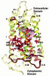
Crystal structure of bovine rhodopsin. The conserved residues are color coded (no conservation, pale yellow; 10–20% conservation, light yellow; 20–40%, light green; 40–60%, orange; 60–80%, red; and 80–100%, brown) based on the percent similarity among family A GPCRs (270 receptors). The conserved extracellular loop (E-I) and cytoplasmic loops (C-I and C-II) are colored brown. Residues that are colored red are as follows: helix I—54, 57, 58; helix II—76, 80, 82, 84, 85; helix III—124, 128, 131; helix IV—170; helix V—212, 215; helix VI—249, 252, 264, 268; helix VII—294, 298, 299, 302; and helix VIII—313, 314. Residues that are colored brown are as follows: helix I—55; helix II—79, 83; helix III—110, 134, 135, 136; helix IV—161; helix V—223; helix VI—261, 265, 267; and helix VII—303, 306.

References

    1. Horn F, Weare J, Beukers MW, Horsch S, Bairoch A, Chen W, Edvardsen O, Campagne F, Vriend G. Nucleic Acids Res. 1998;26:275–279. - PMC - PubMed
    1. Sautel M, Milligan G. Curr. Med. Chem. 2000;7:889–896. - PubMed
    1. Ballesteros JA, Shi L, Javitch JA. Mol. Pharmacol. 2001;60:1–19. - PubMed
    1. Gether U. Endocr. Rev. 2000;21:90–113. - PubMed
    1. Ballesteros J, Palczewski K. Curr. Opin. Drug Discovery Dev. 2001;4:561–574. - PMC - PubMed

Publication types