

Inactivation of Exonuclease 1 in mice results in DNA mismatch repair defects, increased cancer susceptibility, and male and female sterility
- PMID: 12629043
- PMCID: PMC196005
- DOI: 10.1101/gad.1060603
Inactivation of Exonuclease 1 in mice results in DNA mismatch repair defects, increased cancer susceptibility, and male and female sterility
Abstract
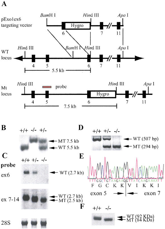
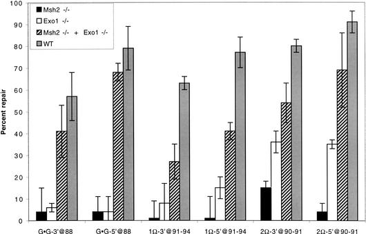
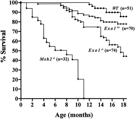
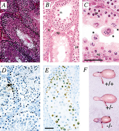
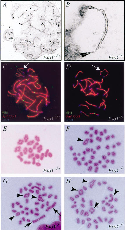
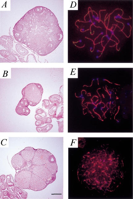
Exonuclease 1 (Exo1) is a 5'-3' exonuclease that interacts with MutS and MutL homologs and has been implicated in the excision step of DNA mismatch repair. To investigate the role of Exo1 in mammalian mismatch repair and assess its importance for tumorigenesis and meiosis, we generated an Exo1 mutant mouse line. Analysis of Exo1(-/-) cells for mismatch repair activity in vitro showed that Exo1 is required for the repair of base:base and single-base insertion/deletion mismatches in both 5' and 3' nick-directed repair. The repair defect in Exo1(-/-) cells also caused elevated microsatellite instability at a mononucleotide repeat marker and a significant increase in mutation rate at the Hprt locus. Exo1(-/-) animals displayed reduced survival and increased susceptibility to the development of lymphomas. In addition, Exo1(-/-) male and female mice were sterile because of a meiotic defect. Meiosis in Exo1(-/-) animals proceeded through prophase I; however, the chromosomes exhibited dynamic loss of chiasmata during metaphase I, resulting in meiotic failure and apoptosis. Our results show that mammalian Exo1 functions in mutation avoidance and is essential for male and female meiosis.
Figures
Similar articles
-
Exonuclease 1 and its versatile roles in DNA repair.Crit Rev Biochem Mol Biol. 2016 Nov/Dec;51(6):440-451. doi: 10.1080/10409238.2016.1215407. Epub 2016 Aug 5. Crit Rev Biochem Mol Biol. 2016. PMID: 27494243 Review.
-
Meiotic recombination intermediates and mismatch repair proteins.Cytogenet Genome Res. 2004;107(3-4):232-48. doi: 10.1159/000080601. Cytogenet Genome Res. 2004. PMID: 15467368 Review.
-
Mammalian Exo1 encodes both structural and catalytic functions that play distinct roles in essential biological processes.Proc Natl Acad Sci U S A. 2013 Jul 2;110(27):E2470-9. doi: 10.1073/pnas.1308512110. Epub 2013 Jun 10. Proc Natl Acad Sci U S A. 2013. PMID: 23754438 Free PMC article.
-
Comparative analysis of meiotic progression in female mice bearing mutations in genes of the DNA mismatch repair pathway.Biol Reprod. 2008 Mar;78(3):462-71. doi: 10.1095/biolreprod.107.065771. Epub 2007 Dec 5. Biol Reprod. 2008. PMID: 18057311
-
A possible mechanism for exonuclease 1-independent eukaryotic mismatch repair.Proc Natl Acad Sci U S A. 2009 May 26;106(21):8495-500. doi: 10.1073/pnas.0903654106. Epub 2009 May 6. Proc Natl Acad Sci U S A. 2009. PMID: 19420220 Free PMC article.
Cited by
-
hMRE11 deficiency leads to microsatellite instability and defective DNA mismatch repair.EMBO Rep. 2005 May;6(5):438-44. doi: 10.1038/sj.embor.7400392. EMBO Rep. 2005. PMID: 15864295 Free PMC article.
-
Male infertility: a risk factor for testicular cancer.Nat Rev Urol. 2009 Oct;6(10):550-6. doi: 10.1038/nrurol.2009.179. Epub 2009 Sep 1. Nat Rev Urol. 2009. PMID: 19724246 Review.
-
Tocotrienol Rich Fraction Supplementation Modulate Brain Hippocampal Gene Expression in APPswe/PS1dE9 Alzheimer's Disease Mouse Model.J Alzheimers Dis. 2019;70(s1):S239-S254. doi: 10.3233/JAD-180496. J Alzheimers Dis. 2019. PMID: 30507571 Free PMC article.
-
Coordinating Multi-Protein Mismatch Repair by Managing Diffusion Mechanics on the DNA.J Mol Biol. 2018 Oct 26;430(22):4469-4480. doi: 10.1016/j.jmb.2018.05.032. Epub 2018 May 21. J Mol Biol. 2018. PMID: 29792877 Free PMC article. Review.
-
Mouse models in colon cancer, inferences, and implications.iScience. 2023 May 25;26(6):106958. doi: 10.1016/j.isci.2023.106958. eCollection 2023 Jun 16. iScience. 2023. PMID: 37332609 Free PMC article. Review.
References
-
- Baker SM, Bronner CE, Zhang I, Plug A, Robatzek M, Warren G, Elliott EA, Yu J, Ashley T, Arnheim N, et al. Male mice defective in the DNA mismatch repair gene PMS2 exhibit abnormal chromosome synapsis in meiosis. Cell. 1995;82:309–320. - PubMed
-
- Baker SM, Plug AW, Prolla TA, Bronner CE, Harris AC, Yao X, Christie DM, Monell C, Arnheim N, Bradley A, et al. Involvement of mouse Mlh1 in DNA mismatch repair and meiotic crossing over. Nat Genet. 1996;13:336–342. - PubMed
Publication types
MeSH terms
Substances
Grants and funding
LinkOut - more resources
Full Text Sources
Other Literature Sources
Medical
Molecular Biology Databases
Miscellaneous