. 2003 Mar 1;17(5):629-37.
doi: 10.1101/gad.1062903.

Suppression of Ras-stimulated transformation by the JNK signal transduction pathway

Affiliations

Suppression of Ras-stimulated transformation by the JNK signal transduction pathway

Norman J Kennedy et al. Genes Dev. .

Abstract

The c-Jun NH(2)-terminal kinase (JNK) phosphorylates and activates members of the activator protein-1 (AP-1) group of transcription factors and is implicated in oncogenic transformation. To examine the role of JNK, we investigated the effect of JNK deficiency on Ras-stimulated transformation. We demonstrate that although JNK does play a role in transformation in vitro, JNK is not required for tumor development in vivo. Importantly, the loss of JNK expression resulted in substantial increases in the number and growth of tumor nodules in vivo. Complementation assays demonstrated that this phenotype was caused by JNK deficiency. These data demonstrate that, in contrast to expectations, the normal function of JNK may be to suppress tumor development in vivo. This conclusion is consistent with the presence in human tumors of loss-of-function mutations in the JNK pathway.


Figures

Figure 1
Figure 1
Characterization of wild-type and Jnk-null fibroblasts. (A) Wild-type (WT) and Jnk-null cells were transduced with a retrovirus vector that expresses activated Ras (Leu-61) or with the empty vector (Control). The cells obtained were pools of at least 100 individual clones. MAP kinase activity was examined by in vitro protein kinase assays using the substrate c-Jun, MBP, and ATF2 for JNK, ERK, and p38, respectively. The cells were exposed in the absence or presence of 60 J/m2 UV-C radiation 45 min prior to harvesting. The protein kinase activity was detected by autoradiography and was quantitated by PhosphorImager analysis (the relative activity is indicated below the autoradiograph). The expression of MAP kinases was examined by immunoblot analysis. (B) The fibroblasts were examined by immunoblot analysis of cell lysates for the expression of JNK, p53, p21, PUMA, ARF, and α-tubulin. Activated Ras was isolated from the lysates by incubation with Raf beads (Upstate Biotechnology), and the bound (activated) Ras was detected by immunoblot analysis. The level of total Ras detected by immunoblot analysis of lysates was approximately twofold greater in Ras-transformed cells than in the nontransformed cells (data not shown). (C) Wild-type and p53−/− primary murine embryo fibroblasts (MEF) and wild-type and Jnk-null immortalized fibroblasts were exposed to ionizing radiation (8 Gy). The cells were incubated (15 h) and then pulse-labeled by incubation with 10 μM BrdU (3 h). DNA content and BrdU incorporation were examined by flow cytometry.
Figure 2
Figure 2
JNK is required for Ras-stimulated apoptosis in vitro. (A) Nucleosomal fragmentation of chromosomal DNA was examined in cultures of exponentially growing wild-type and Jnk-null fibroblasts. Some cultures were exposed to 60 J/m2 UV-C radiation 16 h prior to harvesting. The data are presented as the normalized mean optical density (OD) ± S.D. (n = 3). The data presented are representative of three independent experiments. The UV-stimulated apoptosis of wild-type 3T3 cells is reduced compared with the robust response of wild-type primary murine embryo fibroblasts (MEF). However, both Jnk-null MEF (Tournier et al. 2000) and Jnk-null 3T3 cells (the present study) are resistant to UV-C-stimulated apoptosis. (B) The number of cells with activated caspase was measured in cultures of wild-type and Jnk-null fibroblasts. The effect of Ras transformation was examined. The data are presented as the percent of total cells with activated caspases ± S.D. (n = 3). (C) Wild-type and Jnk-null fibroblasts were transduced with a bicistronic retrovirus that expresses GFP and an estrogen receptor/c-Myc (ER/Myc) fusion protein. c-Myc-dependent apoptosis was examined by incubation (24 h) of the cells with 1 μM 4-hydroxytamoxifen (Tamoxifen) in serum-free media. Nucleosomal fragmentation of chromosomal DNA was examined. The data are presented as the normalized mean OD ± S.D. (n = 3). The data presented are representative of three independent experiments. The basal level of apoptosis observed in serum-free medium (C) was greater than the basal apoptosis detected in the presence of serum (A).
Figure 3
Figure 3
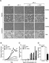
Analysis of wild-type and Jnk-null fibroblasts transformed with Ras in vitro. (A) Cell morphology in sparse and confluent cultures was examined by phase contrast microscopy. The morphology of wild-type cells, Jnk-null cells, and Jnk-null cells complemented with Jnk1 or Jnk2 is shown. (B) Cell proliferation in medium supplemented with 10% fetal calf serum was examined by crystal violet staining (mean OD at 590 nm ± S.D.; n = 3) following the addition of 1 × 104 cells to 20-mm tissue culture dishes. The data are expressed as relative cell number. (C) The saturation growth density in different concentrations of serum was examined by crystal violet staining (mean OD at 590 nm ± S.D.; n = 3). Relative cell numbers were measured at day 0 (D = 0) and after culture for 10 d (D = 10). (D) Wild-type and Jnk-null fibroblasts were plated in soft agar, then incubated for 14 d, and the number of colonies was measured. The data presented are the mean ± S.D. of triplicate data obtained in three independent experiments.
Figure 4
Figure 4
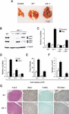
JNK suppresses tumor development in vivo. (A,B) Wild-type and Jnk-null cells (5 × 105) were injected into the tail vein of 12-week-old male athymic nude mice (Charles River). The mice were injected with 400 μg of BrdU on day 13 and euthanized on day 14. (A) Representative Ras-induced tumor nodules in the lungs are illustrated. (B) Wild-type and Jnk-null cells were examined by immunoblot analysis using antibodies to JNK and α-tubulin. Complementation assays were performed using Jnk-null cells expressing Jnk1 or Jnk2. (C) The lung mass as a percentage of total body mass (mean ± S.D.; n = 5) is presented as relative lung mass. The data presented are representative of three independent experiments. (DF) Dose-response analysis of tumor formation by Ras-transformed wild-type and Jnk-null cells. The effects of injecting different numbers of transformed cells on the tumor burden (D), the number of tumor nodules (E), and the number of tumor nodules with a surface greater than 2 mm2 (F) are shown. The data presented represent the mean mass ± S.D. (n = 5). (G) The wild-type and Jnk-null tumor nodules were examined by immunocytochemistry. Representative images of sections stained with hematoxylin and eosin (H&E), with an antibody to BrdU to detect proliferating cells, by TUNEL assay for apoptotic cells, and with an antibody to the endothelial cell protein PECAM-1.

References

    1. Behrens A, Jochum W, Sibilia M, Wagner EF. Oncogenic transformation by ras and fos is mediated by c-Jun N-terminal phosphorylation. Oncogene. 2000;19:2657–2663. - PubMed
    1. Bennett BL, Sasaki DT, Murray BW, O’Leary EC, Sakata ST, Xu W, Leisten JC, Motiwala A, Pierce S, Satoh Y, et al. SP600125, an anthrapyrazolone inhibitor of Jun N-terminal kinase. Proc Natl Acad Sci. 2001;98:13681–13686. - PMC - PubMed
    1. Bos JL. Ras oncogenes in human cancer: A review. Cancer Res. 1989;49:4682–4689. - PubMed
    1. Bost F, McKay R, Bost M, Potapova O, Dean NM, Mercola D. The Jun kinase 2 isoform is preferentially required for epidermal growth factor-induced transformation of human A549 lung carcinoma cells. Mol Cell Biol. 1999;19:1938–1949. - PMC - PubMed
    1. Clark GJ, Cox AD, Graham SM, Der CJ. Biological assays for Ras transformation. Methods Enzymol. 1995;255:395–412. - PubMed

Publication types

MeSH terms

Substances