. 2003 Mar 1;17(5):664-75.
doi: 10.1101/gad.1052003.

Protein:protein interactions and the pairing of boundary elements in vivo

Affiliations

Protein:protein interactions and the pairing of boundary elements in vivo

Jason Blanton et al. Genes Dev. .

Abstract

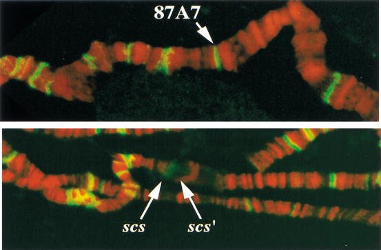
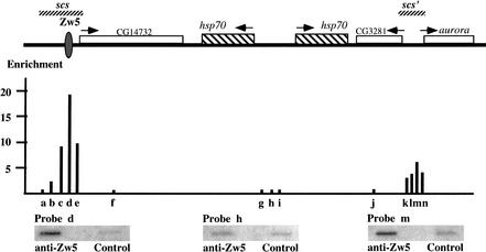
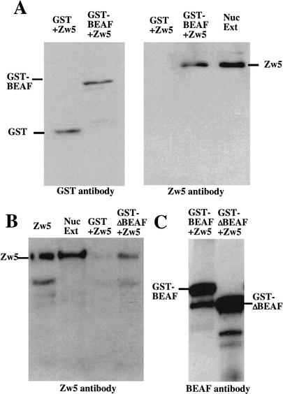
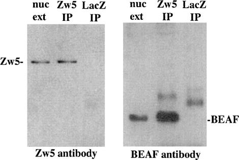
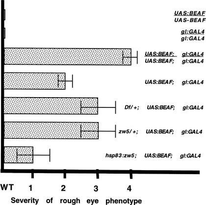
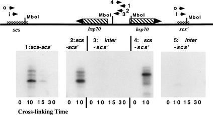
Although it is now well-established that boundary elements/insulators function to subdivide eukaryotic chromosomes into autonomous regulatory domains, the underlying mechanisms remain elusive. One idea is that boundaries act as barriers, preventing the processive spreading of "active" or "silenced" chromatin between domains. Another is that the partitioning into autonomous functional units is a consequence of an underlying structural subdivision of the chromosome into higher order "looped" domains. In this view, boundaries are thought to delimit structural domains by interacting with each other or with some other nuclear structure. The studies reported here provide support for the looped domain model. We show that the Drosophila scs and scs' boundary proteins, Zw5 and BEAF, respectively, interact with each other in vitro and in vivo. Moreover, consistent with idea that this protein:protein interaction might facilitate pairing of boundary elements, we find that that scs and scs' are in close proximity to each other in Drosophila nuclei.


Figures

Figure 1
Figure 1
Zw5 localizes to scs in polytene chromosomes. A Zw5 polyclonal antibody was used to probe fixed salivary gland polytene chromosomes prepared from control larvae grown at 18°C, and larvae heat-shocked for 30 min at 37°C. The DNA was counterstained with propidium iodide, and the preparations were then visualized by confocal microscopy. Zw5 is in green, DNA is in red. More than 100 different sites are labeled with the Zw5 antibody in polytene chromosomes from non-heat-shocked larvae. (Top) One of these sites on the third chromosome is the condensed 87A7 heat-shock locus. In polytene chromosomes from heat-shock larvae, the two 87A7 hsp70 genes are induced, and the locus forms a large puff. (Bottom) scs is on the proximal side of the 87A7 puff; scs‘ is on the distal side. Zw5 staining is seen on the proximal, scs, side of the puff. No staining is observed on the distal, scs‘, side of the puff.
Figure 2
Figure 2
Distribution of Zw5 across the 87A7 heat-shock locus. The chromatin immunoprecipitation (ChIP) procedure of Orlando and Paro (1993; see Materials and Methods) was used to examine the distribution of Zw5 across the 87A7 locus. Affinity-purified rabbit anti-Zw5 antibody (Gaszner et al. 1999) or preimmune rabbit serum was used to immunoprecipitate the CsCl-purified cross-linked chromatin. DNA recovered from each pellet was amplified by LM-PCR (Zhao et al. 1995), slot blotted, and then probed with short fragments (an) derived from different sequences in the 87A7 locus. The approximate position of each fragment used as a probe is indicated below the map of 87A7 in the diagram and is given in base pairs in the Materials and Methods section. The degree of enrichment in the Zw5 immunoprecipitate was calculated relative to preimmune serum control and is plotted in the diagram. The signal for probes a, f, and gj in the Zw5 immunoprecipitate was less than that of the preimmune serum, while all other probes gave signals in the Zw5 immunoprecipitate that were greater than the preimmune serum. The highest degree of enrichment in this experiment is for probe d, which is nearly 20-fold greater in the Zw5 immunoprecipitate than in the immunoprecipitate from the control preimmune serum. Probe d spans the Zw5 binding site. The degree of enrichment for probes around scs‘ was between three- and sixfold. Probes k and m contain BEAF binding sites.
Figure 3
Figure 3
Zw5 and BEAF interact with each other in vitro. (A) Recombinant Zw5 associates with GST-BEAF-32A but not GST. Zw5 was mixed with either GST-BEAF 32A or GST alone in a 1:10 ratio. The proteins were allowed to bind for 30 min at room temperature before subsequently being added to 400 μL of PBS containing ∼25 μL of glutathione beads. After rocking overnight at 4°C, the beads were washed, and the proteins were eluted from the beads by boiling in 2× sample loading buffer. The proteins were separated by SDS-PAGE and transferred to PVDF membranes. The membrane on the left was probed with anti-GST antibody. GST+Zw5, Zw5 mixed with GST alone; GST-BEAF+Zw5, Zw5 mixed with GST-BEAF-32A. The membrane on the right was probed with Zw5 antibody. GST+Zw5, GST mixed with Zw5. Zw5 does not associate with GST. GST-BEAF+Zw5, Zw5 mixed with GST-BEAF-32A. Zw5 forms a stable complex with GST-BEAF 32A. Nuc Ext, nuclear extract used as a positive control for Zw5. (B) Recombinant Zw5 associates specifically with GST-ΔN BEAF-32A in a GST pull-down assay. Recombinant Zw5 was mixed with either GST-ΔN BEAF or GST alone in a 1:10 ratio. The proteins were allowed to interact for 30 min at room temperature before being added to 400 μL of PBS containing ∼25 μL of glutathione beads. After rocking overnight at 4°C, the beads were washed, and the proteins were eluted from the beads by boiling in 2× sample loading buffer. The proteins were separated by SDS-PAGE and transferred to a PVDF membrane. The blot was probed with anti-Zw5 antibody. Zw5, recombinant Zw5 protein; Nuc Ext, nuclear extract; GST+Zw5, Zw5 mixed with GST alone; GST-ΔN BEAF+Zw5, Zw5 mixed with GST-ΔN BEAF-32A. (C) Zw5 immunoprecipitation. GST-BEAF-32A or GST-ΔN BEAF-32A were mixed with Zw5. After incubating at room temperature, the mixture was diluted in PBS containing 25 μL of Zw5 antibody. The samples were rocked overnight at 4°C, the beads were washed, and the proteins were eluted from the beads by boiling in 2× sample loading buffer. The proteins were separated by SDS-PAGE and transferred to a PVDF membrane. The blot was probed with BEAF antibody.
Figure 4
Figure 4
Zw5 and BEAF are in an immunoprecipitable complex in Drosophila embryos. Anti-Zw5 antibody cross-linked to protein A/G beads was mixed with embryonic nuclear extract. After rocking overnight at 4°C, the beads were washed, and the proteins were eluted from the beads by boiling in 2× sample loading buffer. The proteins were separated by SDS-PAGE and transferred to PVDF membranes. The blot on the left was probed with Zw5 antibody. In the lane labeled nuc ext, the starting nuclear extract was loaded and used as the positive control for Zw5. The lane labeled Zw5 IP contains the proteins immunoprecipitated from nuclear extracts with Zw5 antibody. As evident from a comparison with the “nuc ext” lane, Zw5 protein is present in the immunoprecipitate. The lane labeled LacZ IP contains the proteins immunoprecipitated with the control β-galactosidase antibody. Note that Zw5 is not detected in the β-galactosdiase immunoprecipitate. There is, however, a weakly labeled band of ∼40 kD. The blot on the right was probed with BEAF antibody. In the lane labeled nuc ext, nuclear extract was loaded and used as the positive control for BEAF. In the Zw5 IP lane, proteins isolated from the Zw5 immunoprecipitate were loaded. As can be seen from comparison with the “nuc ext” lane, BEAF is present in the Zw5 immunoprecipitate. In the lane labeled LacZ IP, proteins isolated from the β-galactosidase immunoprecipitate were loaded. BEAF is not present; however, there is a larger protein species of ∼40 kD. As noted above, a protein species of this size was also detected when the β-galactosidase immunoprecipitate was probed with Zw5 antibody. Consequently, we suspect that it corresponds to protein that associates with the β-galactosidase and is recognized by the secondary antibody.
Figure 5
Figure 5
Genetic interactions between zw5 and beaf. Yamaguchi et al. (2001) showed that inducing BEAF-32A expression from a UAS–BEAF transgene using a gl:GAL4 driver has deleterious effects on eye development. We scored female flies from each cross on a scale of 0–4 based on the severity of the phenotypic effects on eye development. Wild-type females had a score of 0. The most severe rough eye phenotype was observed for females carrying two copies of both the UAS:BEAF expression construct and the gl:GAL4 driver, GMR-GAL4, and this phenotype was assigned a score of 4. An intermediate rough eye phenotype was observed for females carrying a single copy each of the UAS:BEAF and gl:GAL4 transgenes, and this phenotype was assigned a score of 2. The eye phenotype observed for each experimental cross was then compared with these three controls and assigned a score based on this comparison. Each of the experimental crosses was also scored independently by another investigator who had no knowledge of the genotypes being examined. As reported by Yamaguchi et al. (2001), animals homozygous for either the UAS:BEAF expression construct or the gl:GAL4 driver (lines 1,2) had a wild-type phenotype (#0), while animals carrying two copies of the expression construct and driver (line 3) had a severe (#4) eye phenotype. This rough eye phenotype was comparatively homogenous within the population. Females hemizygous for the expression construct and driver (line 4) were generated by mating females carrying two copies of the expression construct and the driver to w‘ males. The hemizygous females had an intermediate rough eye phenotype (#2). To obtain the female progeny heterozygous for Df(1)936 that also carry single copy of both transgenes (line 5), we crossed Df(1)935/Bal females to males hemizygous for the X-linked gl:GAL4 driver, GMR-GAL4, and homozygous for the UAS:BEAF-32A transgene. Nonbalancer females were scored. In our hands, the Df(l)935 deletion, which uncovers zw5, enhances the rough eye phenotype associated with overexpressed BEAF 32A, contrary to the findings published by Yamaguchi et al. (2001). The reason for this discrepancy is uncertain. To obtain the female progeny heterozygous for zw562j1 that also carry a single copy of both transgenes (line 6) we crossed zw562j/FM7 females to males that were hemizygous for the X-linked gl:GAL4 driver and homozygous for the UAS–BEAF-32A transgene. Female progeny lacking the FM7 balancer were examined. This strong zw5 loss-of-function allele enhances the rough eye phenotype compared with that observed for otherwise wild-type flies hemizygous for the two transgenes. The disruptions in eye development are not, however, as severe as that observed for females carrying two copies of both the expression construct and the driver. Similar results were obtained for a second independent zw5 allele, zw590. Finally, to increase the level of zw5 protein (line 7), females carrying an hsp83:zw5 cDNA transgene (line 17.1.1) over the Cyo balancer were crossed to males hemizygous for the gl:GAL4 driver, and homozygous for the UAS:BEAF-32A transgene. Females lacking the Cyo balancer were examined. They had a weaker rough eye phenotype than the single copy control. All crosses were done at 29°C.
Figure 6
Figure 6
scs and scs‘ are paired in Drosophila embryos. The chromosome conformation capture procedure of Dekker et al. (2002) was used to determine whether scs and scs‘ are in close proximity to each other in vivo. Formaldehyde cross-linked Drosophila embryonic nuclei were restricted with MboI and then ligated. After ligation, the cross-linking was reversed and the DNA was PCR-amplified with primer combinations derived from scs‘ and either scs or the 5′ ends of the two 87A7 hsp70 genes. The PCR products were then detected with either ethidium bromide staining or by hybridization with a probe derived from scs‘. The primer combinations for each experiment were as follows. (1) scs-scs‘: samples in each lane were cross-linked for the times indicated and then PCR-amplified for 40 cycles using scs-i and scs‘-i as primers. (2) scs-scs‘: samples were cross-linked for the times indicated and then PCR-amplified in two steps. In the first step, the samples were PCR-amplified for 20 cycles with scs-o and scs‘-o as primers. In the second step, an aliquot of from the first step was PCR-amplified for 20 cycles with scs-i and scs‘-i as primers. (3) scs‘-inter: samples were cross-linked for the times indicated and then PCR-amplified in two steps. In the first step, the samples were amplified for 20 cycles with inter-1 and scs‘-o as primers. In the second step, an aliquot from the first step was PCR-amplified for 20 cycles with inter-2 and scs‘-i as primers. (4) scs-scs‘: samples were cross-linked for the times indicated and PCR-amplified in two steps. In the first step, the samples were amplified for 20 cycles with scs-o and scs‘-o as primers. In the second step, an aliquot from the first step was PCR-amplified for 20 cycles with scs-o and scs‘-i as primers. (5) scs‘-inter: samples were cross-linked for the times indicated and PCR-amplified in two steps. In the first step, the samples were amplified for 20 cycles with inter-3 and scs‘-o as primers. In the second step, an aliquot from the first step was PCR-amplified for 20 cycles with inter-3 and scs‘-i as primers. As a positive control, we also ligated a mixture of Sau3a-digested plasmids containing scs, scs‘, and the 87A7 intergenic spacer. We then PCR-amplified using scs-scs‘ or scs‘-inter primer pairs and detected the mixed ligation products by ethidium bromide staining or by probing filters with the scs‘. The expected hybrid scs‘-scs and scs‘-inter amplification products were observed.

References

    1. Bell AC, West AG, Felsenfeld G. Insulators and boundaries: Versatile regulatory elements in the eukaryotic genome. Science. 2001;291:447–450. - PubMed
    1. Bellard M, Kuo MT, Dretzen G, Chambon P. Differential nuclease sensitivity of the ovalbumin and β-globin chromatin regions in erythrocytes and oviduct cells of laying hen. Nucleic Acids Res. 1980;8:2737–2750. - PMC - PubMed
    1. Cai HN, Shen P. Effects of cis arrangement of chromatin insulators on enhancer-blocking activity. Science. 2001;291:493–495. - PubMed
    1. Chung JH, Whiteley M, Felsenfeld G. A 5′ element of the chicken β-globin domain serves as an insulator in human erythroid cells and protects against position effect in Drosophila. Cell. 1993;74:505–514. - PubMed
    1. Cuvier O, Hart CM, Laemmli UK. Identification of a class of chromatin boundary elements. Mol Cell Biol. 1998;18:7478–7486. - PMC - PubMed

Publication types