. 2003 Mar 17;160(6):909-18.
doi: 10.1083/jcb.200212064. Epub 2003 Mar 10.

Skeletal muscle repair by adult human mesenchymal stem cells from synovial membrane

Affiliations

Skeletal muscle repair by adult human mesenchymal stem cells from synovial membrane

Cosimo De Bari et al. J Cell Biol. .

Abstract

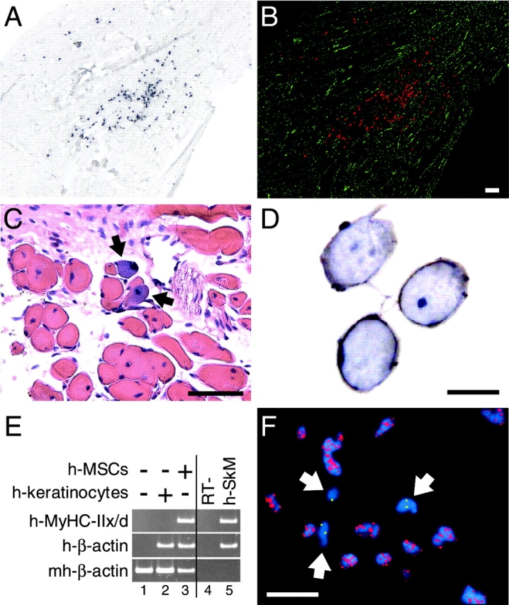
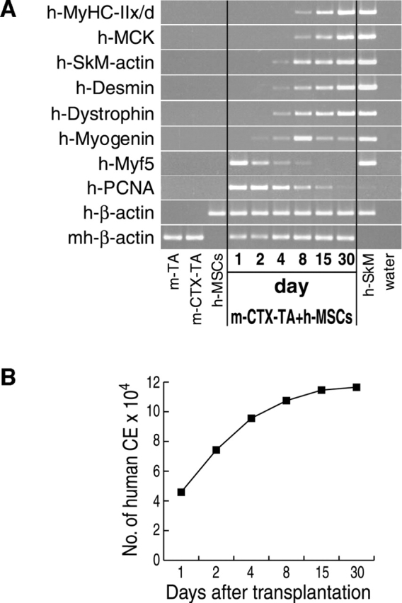
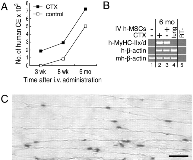
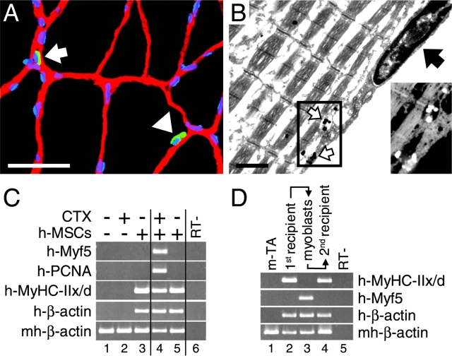
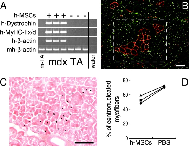
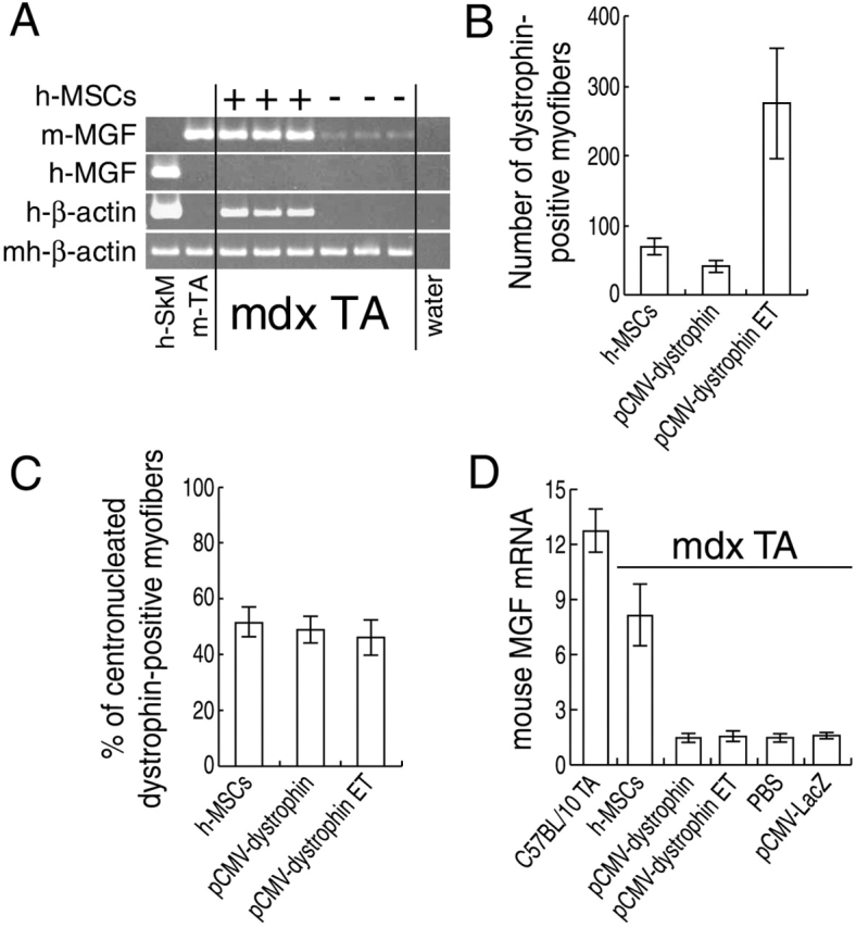
We have demonstrated previously that adult human synovial membrane-derived mesenchymal stem cells (hSM-MSCs) have myogenic potential in vitro (De Bari, C., F. Dell'Accio, P. Tylzanowski, and F.P. Luyten. 2001. Arthritis Rheum. 44:1928-1942). In the present study, we have characterized their myogenic differentiation in a nude mouse model of skeletal muscle regeneration and provide proof of principle of their potential use for muscle repair in the mdx mouse model of Duchenne muscular dystrophy. When implanted into regenerating nude mouse muscle, hSM-MSCs contributed to myofibers and to long term persisting functional satellite cells. No nuclear fusion hybrids were observed between donor human cells and host mouse muscle cells. Myogenic differentiation proceeded through a molecular cascade resembling embryonic muscle development. Differentiation was sensitive to environmental cues, since hSM-MSCs injected into the bloodstream engrafted in several tissues, but acquired the muscle phenotype only within skeletal muscle. When administered into dystrophic muscles of immunosuppressed mdx mice, hSM-MSCs restored sarcolemmal expression of dystrophin, reduced central nucleation, and rescued the expression of mouse mechano growth factor.


Figures

Figure 1.
Figure 1.
Contribution of hSM-MSCs to skeletal muscle regeneration in vivo. (A and B) Localization of the human nuclei within the mouse skeletal muscle. (A) ISH for human Alu repeats on a representative longitudinal cryosection from a nude mouse TA muscle at 4 wk after hSM-MSC transplantation. Human nuclei are stained black. In B, brightfield and fluorescence (DAPI counterstaining) images were given artificial colors and superimposed. The Alu-positive nuclei are shown in red, and the Alu-negative, DAPI-stained nuclei are shown in green. Bar, 200 μm. (C) X-gal staining with hematoxylin-eosin counterstaining at 3 wk after transplantation of hSM-MSCs transduced with AdCMV-LacZ. Arrows point to muscle fibers with diffuse β-gal expression. Bar, 50 μm. (D) Immunohistochemistry for human β2M (brown). Nuclei are counterstained with hematoxylin (blue). Bar, 20 μm. (E) SQ–RT-PCR for human MyHC-IIx/d. CTX-treated TA muscle was injected with either human keratinocytes (lane 2) or hSM-MSCs (lane 3); CTX-treated muscle injected with PBS was used as tissue-negative control (lane 1). Lane 4, RT-negative control of lane 3; lane 5, human skeletal muscle (h-SkM) as a positive control. cDNAs were equalized for the expression of human β-actin. At 3 wk after transplantation, human MyHC-IIx/d was detected only in the muscle injected with SM-MSCs. (F) Double genomic FISH on a cryosection from a TA muscle at 4 wk after hSM-MSC transplantation using a probe for human CEN18 (green) and a probe for mouse centromeric satellite DNA (red). Nuclei were counterstained with DAPI (blue). Arrows point to human nuclei. Bar, 50 μm.
Figure 2.
Figure 2.
Muscle differentiation of hSM-MSCs recapitulates embryonic myogenesis. (A) hSM-MSCs were injected into regenerating (CTX-treated) TA muscles of nude mice. Mice were killed at different time points as indicated. The dissected muscles were subjected to SQ–RT-PCR analysis. TA muscles containing human cells were equalized for the expression of human β-actin. TA muscles with no human cells served as controls for species specificity of the primers for human cDNAs. For each time point, a PBS-injected regenerating (CTX-treated) mouse TA muscle was included for a negative control. m-TA, uninjected mouse TA muscle; m-CTX-TA, mouse CTX-treated TA muscle. (B) Number of human CE per TA muscle over time as determined by Q–RT-PCR (see Results for details).
Figure 3.
Figure 3.
Differentiation is sensitive to environmental cues. (A) Preferential homing to the damaged skeletal muscle of systemically delivered hSM-MSCs. As determined by Q–RT-PCR, the number of human CE increased in parallel over time in both CTX-injured and uninjured host TA muscles, remaining constantly higher in the CTX-injured muscles. Two mice were killed at each time point examined with similar results. (B) SQ–RT-PCR analysis at the 6 mo time point. cDNAs were equalized for the expression of mouse/human β-actin. Human MyHC-IIx/d was detected in both the CTX-treated and -untreated TA muscles (lanes 2 and 3). Lungs (lane 4) expressed human β-actin at levels at least comparable to TA muscles, but MyHC-IIx/d was not detectable. Controls were mouse TA muscle (lane 1) and RT-negative control of lane 2 (lane 5). (C) ISH for human Alu repeats on a longitudinal section from a TA muscle at 6 mo after systemic administration of hSM-MSCs. Counterstaining with eosin. Bar, 50 μm.
Figure 4.
Figure 4.
Contribution to functional satellite cells. (A) Double IF staining for laminin (red) and human β2M (green) at 6 mo after hSM-MSC transplantation into mouse TA muscle. The nuclei were revealed by DAPI staining (blue). A mononuclear cell staining positive for human β2M (arrowhead) is shown between the laminin-positive basal lamina and a distinct (β2M-negative) myofiber. The arrow points to another mononuclear cell of human origin lying outside of the basal lamina. Bar, 50 μm. (B) TEM of a hSM-MSC–derived satellite cell at 6 mo after hSM-MSC transplantation. The high magnification (bar, 1 μm) of a satellite cell shows a plasma membrane positive for human β2M (white arrows), separating the satellite cell from its adjacent β2M-negative myofiber, the continuous basal lamina (black arrow) surrounding the satellite cell and myofiber, and the heterochromatic appearance of the nucleus. Inset shows an inverted, magnified view of the staining for human β2M. (C) Satellite cell-like response of hSM-MSCs in vivo upon muscle injury. 6 mo after hSM-MSC bilateral implantation into regenerating TA muscles, CTX was injected into the right TA muscle and PBS into the contralateral muscle. Animals were killed 12 h later. SQ–RT-PCR analysis was performed after equalization of cDNAs for human β-actin. High levels of human Myf5 and human PCNA were observed in the CTX-treated TA muscle (lane 4) compared with the contralateral muscle (lane 5), indicating the presence of hSM-MSC–derived functional satellite cells. The following were controls: mouse TA muscle (lane 1); mouse TA muscle at 12 h after CTX treatment (lane 2); mouse CTX-treated TA muscle implanted with hSM-MSCs and harvested after 6 mo as external control (lane 3); and RT-negative control of lane 4 (lane 6). (D) Human mononuclear cells recovered from first recipient mice retain in vivo myogenic activity when transplanted into a second recipient. 6 mo after hSM-MSC transplantation, first recipient mice were killed, and cultures of primary myoblasts were established from the injected TA muscles. At 70% confluence, these cells were injected into regenerating TA muscles of second recipient mice, which were killed 1 mo later. Lane 1, control mouse TA muscle; lane 2, TA muscle from first mouse recipient; lane 3, first recipient primary myoblasts; lane 4, TA muscle from second mouse recipient; lane 5, RT-negative control of lane 4.
Figure 5.
Figure 5.
Restoration of dystrophin in mdx muscle fibers by hSM-MSCs. (A) SQ–RT-PCR for human dystrophin. cDNAs were equalized for mouse/human β-actin expression. In all mdx mice, TA muscles injected with hSM-MSCs expressed human dystrophin and human MyHC-IIx/d, whereas the contralateral muscles did not. (B) IF staining for human dystrophin (red) of a TA muscle transverse cryosection from an mdx mouse at 4 wk after hSM-MSC transplantation. Nuclei are shown in green. Clusters of muscle fibers display peripheral localization of the human dystrophin protein. Bar, 100 μm. (C) ISH for human Alu repeats on a parallel, nonconsecutive section, showing human nuclei in the squared area of (B). Counterstaining with eosin. Bar, 100 μm. (D) The percentage of CN human-dystrophin–positive fibers was significantly lower in the hSM-MSC–injected TA muscles than in the contralateral PBS-injected TA muscle fibers in three mdx mice (P < 0.05).
Figure 6.
Figure 6.
Rescue of mouse MGF in mdx muscle by hSM-MSCs. (A) SQ–RT-PCR for mouse MGF. cDNAs were equalized for mouse/human β-actin expression. In the first lane, human skeletal muscle was added for mouse primer specificity. Mouse MGF expression was reproducibly restored in the mdx TA muscles injected with hSM-MSCs to levels comparable to the TA muscle from a wild-type C57BL/10 mouse. (B) Maximal numbers of human-dystrophin–positive fibers in TA muscles of mdx mice injected with either hSM-MSCs or pCMV-dystrophin. In three mice, ET was applied after plasmid DNA injection. Animals were assayed for human-dystrophin expression at 4 wk after injections. For each treatment group, three TA muscles were serially cross sectioned and stained for human dystrophin. The sections containing the highest number of human- dystrophin–positive fibers were selected. Data are mean ± SD of maximal numbers of human-dystrophin–positive fibers per treatment group. (C) Percentage of CN human-dystrophin–positive fibers. We observed no significant difference comparing the three methods of dystrophin delivery. (D) Real-time Q–RT-PCR for mouse MGF normalized for mouse/human β-actin. The expression levels of mouse MGF in mdx TA muscles injected with hSM-MSCs were significantly higher (P < 0.05) than those found in mdx TA muscles injected with pCMV-dystrophin (with or without ET), with PBS, or with pCMV-LacZ. Normal TA muscles were from three age-matched C57BL/10 mice.

Comment in

References

    1. Asakura, A., P. Seale, A. Girgis-Gabardo, and M.A. Rudnicki. 2002. Myogenic specification of side population cells in skeletal muscle. J. Cell Biol. 159:123–134. - PMC - PubMed
    1. Bischoff, R. 1994. The satellite cell and muscle regeneration. Myogenesis. A.G. Engel and C. Franszini-Armstrong, editors. McGraw-Hill, New York. 97–118.
    1. Blau, H.M., G.K. Pavlath, E.C. Hardeman, C.P. Chiu, L. Silberstein, S.G. Webster, S.C. Miller, and C. Webster. 1985. Plasticity of the differentiated state. Science. 230:758–766. - PubMed
    1. Braun, S., C. Thioudellet, P. Rodriguez, D. Ali-Hadji, F. Perraud, N. Accart, J.M. Balloul, C. Halluard, B. Acres, B. Cavallini, and A. Pavirani. 2000. Immune rejection of human dystrophin following intramuscular injections of naked DNA in mdx mice. Gene Ther. 7:1447–1457. - PubMed
    1. Cooper, R.N., S. Tajbakhsh, V. Mouly, G. Cossu, M. Buckingham, and G.S. Butler-Browne. 1999. In vivo satellite cell activation via Myf5 and MyoD in regenerating mouse skeletal muscle. J. Cell Sci. 112:2895–2901. - PubMed

Publication types

MeSH terms