. 2003 Mar 17;197(6):735-42.
doi: 10.1084/jem.20021153. Epub 2003 Mar 10.

Role of adhesin release for mucosal colonization by a bacterial pathogen

Affiliations

Role of adhesin release for mucosal colonization by a bacterial pathogen

Loic Coutte et al. J Exp Med. .

Abstract

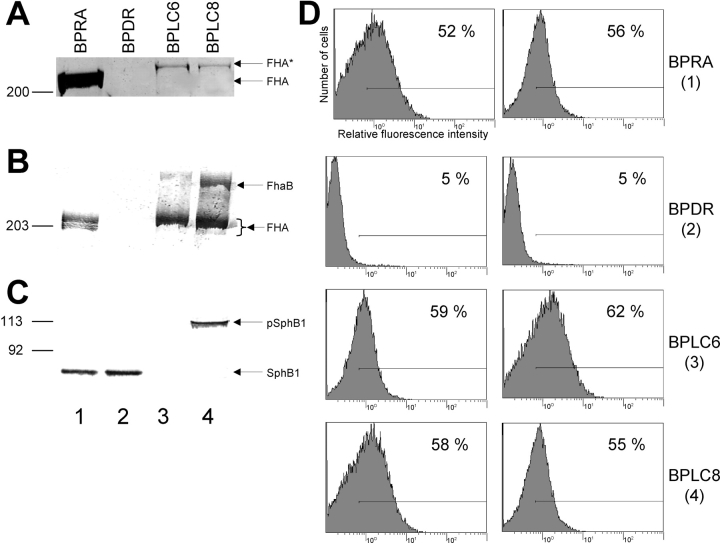
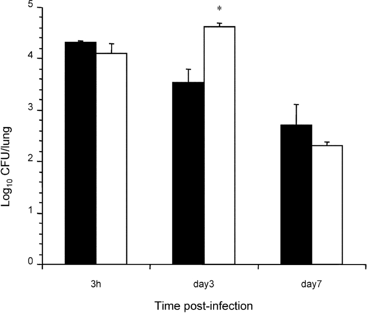
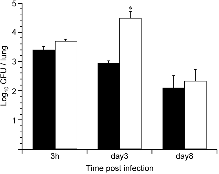
Pathogen attachment is a crucial early step in mucosal infections. This step is mediated by important virulence factors called adhesins. To exert these functions, adhesins are typically surface-exposed, although, surprisingly, some are also released into the extracellular milieu, the relevance of which has previously not been studied. To address the role of adhesin release in pathogenesis, we used Bordetella pertussis as a model, since its major adhesin, filamentous hemagglutinin (FHA), partitions between the bacterial surface and the extracellular milieu. FHA release depends on its maturation by the specific B. pertussis protease SphB1. We constructed SphB1-deficient mutants and found that they were strongly affected in their ability to colonize the mouse respiratory tract, although they adhered even better to host cells in vitro than their wild-type parent strain. The defect in colonization could be overcome by prior nasal instillation of purified FHA or by coinfection with FHA-releasing B. pertussis strains, but not with SphB1-producing FHA-deficient strains, ruling out a nonspecific effect of SphB1. These results indicate that the release of FHA is important for colonization, as it may facilitate the dispersal of bacteria from microcolonies and the binding to new sites in the respiratory tract.


Figures

Figure 1.
Figure 1.
Secretion of FHA and detection of cell-associated FHA and SphB1 in B. pertussis parental and mutant strains. (A) Identical volumes of nonconcentrated supernatants from each culture at the late logarithmic phase of growth were analyzed by SDS-PAGE using an 8% polyacrylamide gel. The gel was stained with Coomassie Blue. Only the gel portion corresponding to high molecular masses is shown. BPRA (FHA+ SphB1+), BPDR (FHA SphB1+), BPLC6 (FHA+ SphB1), and BPLC8 (FHA+ S412→A SphB1). FHA indicates the position of the mature protein and FHA* that of a slightly larger FHA-related polypeptide. (B) Cellular lysates were subjected to SDS-PAGE using a 6% polyacrylamide gel and immunoblotting with anti-FHA antibodies. FhaB and FHA represent the precursor and mature forms of FHA, respectively. (C) Cellular lysates were subjected to SDS-PAGE using an 8% polyacrylamide gel and immunoblotting with an anti-SphB1 antiserum. pSphB1 and SphB1 are the precursor and mature forms of the protease SphB1, respectively. (D) BPRA (1), BPDR (2), BPLC6 (3), and BPLC8 (4) were incubated with monoclonal anti-FHA antibodies 55.4G9 (left row) or 55.6.C4 (right row), followed by anti–mouse FITC conjugate. The fluorescent cells were detected by flow cytometry, with 20,000 events counted for each sample. The fluorescence threshold (left end of the horizontal bar) was set such that 98% of nonlabeled cells had intensities of autofluorescence below the threshold value. A representative experiment is shown, with percentages of fluorescent cells indicated in each panel.
Figure 2.
Figure 2.
Adherence of B. pertussis to cell lines in vitro. Human pulmonary epithelial cells A549 (A) and murine alveolar macrophage-like MH-S cells (B) were incubated with the indicated 35S-labeled bacteria at a multiplicity of infection of 20. After washing, adherence was assessed by scintillation counting. The results are expressed as percentages of counts per minute relative to the counts per minute present in the inoculum. Black bars, BPRA, gray bars, BPDR, white bars, BPLC6 and stippled bars, BPLC8. The data represent averages and standard deviations for triplicate experiments. *P < 0.01 relative to values obtained for BPRA.
Figure 3.
Figure 3.
Lung colonization by SphB1 mutant strains. Balb/c mice were infected intranasally with ∼105 CFU of B. pertussis BPRA (black bars), BPLC6 (white bars), BPLC8 (stippled bars), or BPDR (gray bars). At the indicated time points, mice were killed, and the viable bacteria present in the lungs were counted. Four mice were analyzed per time point for each group. *P < 0.05 relative to the BPRA values.
Figure 4.
Figure 4.
Lung colonization by B. pertussis BPLC9 in coinfection. Balb/c mice were infected intranasally with ∼104 CFU of B. pertussis BPLC9 (SphB1; black bars), or coinfected with 104 CFU of B. pertussis BPLC9 and 106 CFU of B. pertussis BPDR (FHA SphB1+). The white bars represent the numbers of CFU of BPLC9 (as distinguished by their resistance to gentamycin) in the lungs of coinfected mice, and the striped bars represent the numbers of BPDR in the coinfected mice.
Figure 5.
Figure 5.
Effect of an intranasal treatment with FHA on colonization by BPLC6. Balb/c mice were treated by an intranasal instillation of either 20 μl of PBS containing 0.5 M NaCl (PBS/NaCl) and 5 μg of purified FHA or 20 μl of PBS/NaCl, and then immediately infected intranasally with ∼5 × 10⁴ CFU of B. pertussis BPLC6 (SphB1). At the indicated time points, mice were killed, and the viable bacteria present in the lungs were counted. Black bars, numbers of BPLC6 after mock treatment, white bars, numbers of BPLC6 after the FHA treatment. *P < 0.05 relative to the numbers of BPLC6 CFU administered without FHA.
Figure 6.
Figure 6.
Lung colonization by B. pertussis BPLC9 in coinfection. Balb/c mice were infected intranasally with ∼104 CFU of B. pertussis BPLC9 (SphB1, black bars), or coinfected with 104 CFU of B. pertussis BPLC9 and 105 CFU of B. pertussis BPRA (FHA+ SphB1+). The white bars represent the numbers of CFU of BPLC9 (as distinguished by their resistance to gentamycin) in the lungs of coinfected mice. *P < 0.05 relative to the number of CFU of BPLC9 administered alone.

Similar articles

Cited by

References

    1. Beachey, E.H. 1981. Bacterial adherence: adhesin-receptor interactions mediating the attachement of bacteria to mucosal surfaces. J. Infect. Dis. 143:325–345. - PubMed
    1. Isberg, R.R., and G. Tran Van Nhieu. 1994. Binding and internalization of microorganisms by integrin receptors. Trends Microbiol. 2:10–14. - PubMed
    1. Rostand, K.S., and J.D. Esko. 1997. Microbial adherence to and invasion through proteoglycans. Infect. Immun. 65:1–8. - PMC - PubMed
    1. Wadström, T., and A. Ljungh. 1999. Glycosaminoglycan-binding microbial proteins in tissue adhesion and invasion: key events in microbial pathogenicity. J. Med. Microbiol. 48:223–233. - PubMed
    1. Menozzi, F.D., K. Pethe, P. Bifani, F. Soncin, M.J. Brennan, and C. Locht. 2002. Enhanced bacterial virulence through exploitation of host glycosaminoglycans. Mol. Microbiol. 43:1379–1386. - PubMed

Publication types

MeSH terms

Substances