. 2003 Mar 1;23(5):1593-604.
doi: 10.1523/JNEUROSCI.23-05-01593.2003.

Afterhyperpolarization regulates firing rate in neurons of the suprachiasmatic nucleus

Affiliations

Afterhyperpolarization regulates firing rate in neurons of the suprachiasmatic nucleus

Robin K Cloues et al. J Neurosci. .

Abstract

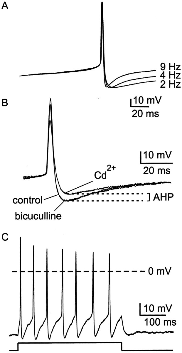
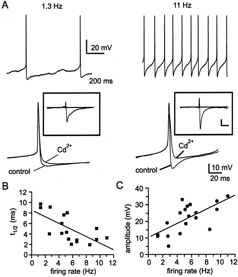
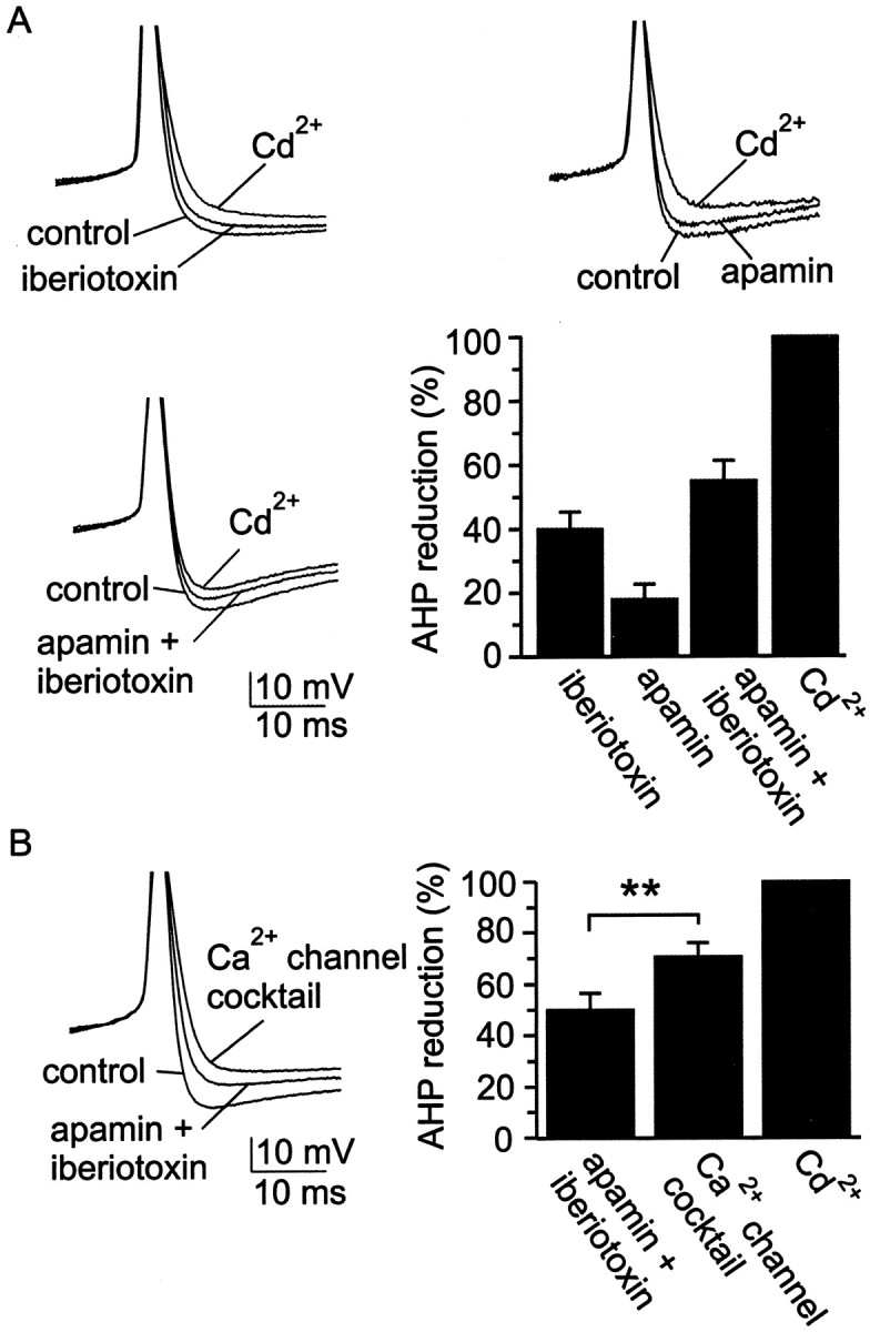
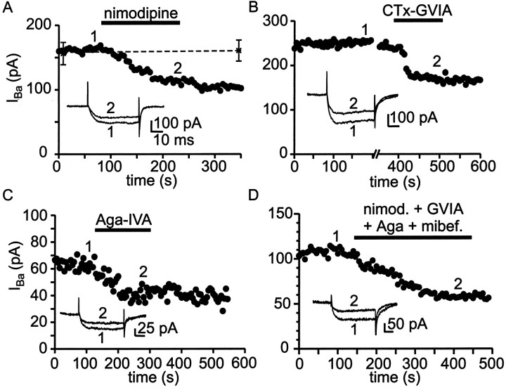
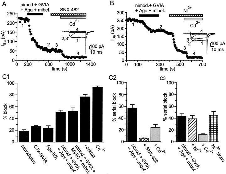
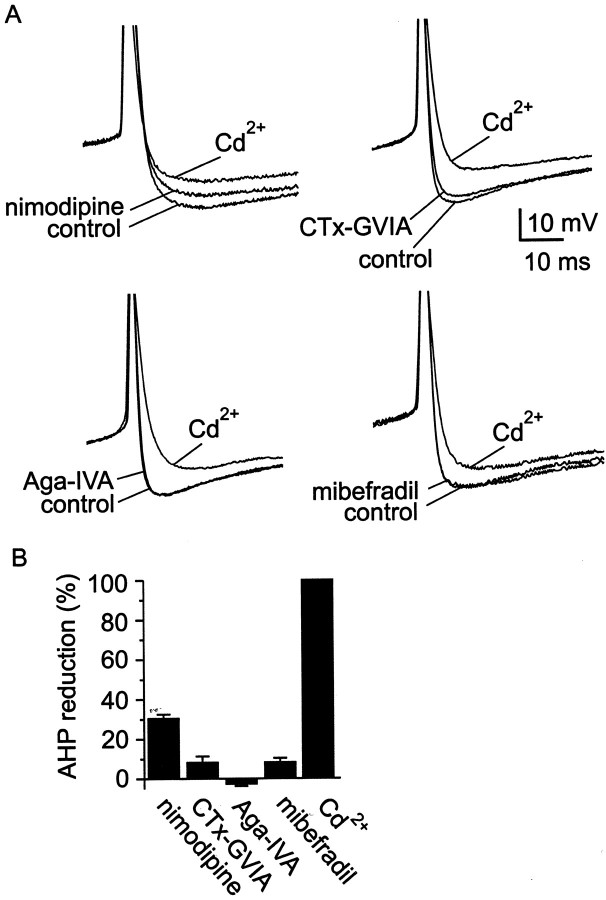
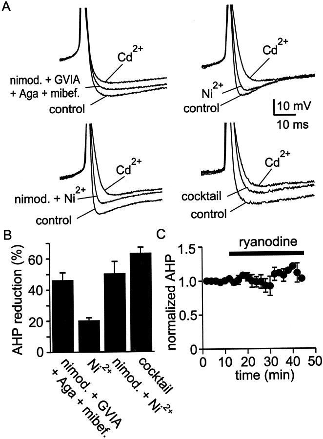
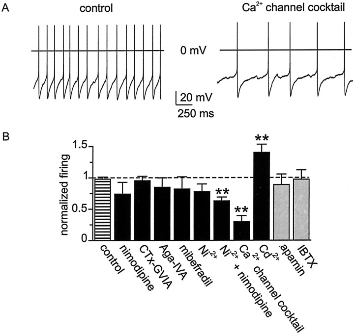
Cluster I neurons of the suprachiasmatic nucleus (SCN), which are thought to be pacemakers supporting circadian activity, fire spontaneous action potentials that are followed by a monophasic afterhyperpolarization (AHP). Using a brain slice preparation, we have found that the AHP has a shorter duration in cells firing at higher frequency, consistent with circadian modulation of the AHP. The AHP is supported by at least three subtypes of K(Ca) channels, including apamin-sensitive channels, iberiotoxin-sensitive channels, and channels that are insensitive to both of these antagonists. The latter K(Ca) channel subtype is involved in rate-dependent regulation of the AHP. Voltage-clamped, whole-cell Ca(2+) channel currents recorded from SCN neurons were dissected pharmacologically, revealing all of the major high-voltage activated subtypes: L-, N-, P/Q-, and R-type Ca(2+) channel currents. Application of Ca(2+) channel antagonists to spontaneously firing neurons indicated that predominantly L- and R-type currents trigger the AHP. Our findings suggest that apamin- and iberiotoxin-insensitive K(Ca) channels are subject to diurnal modulation by the circadian clock and that this modulation either directly or indirectly leads to the expression of a circadian rhythm in spiking frequency.


Figures

Fig. 1.
Fig. 1.
Firing properties of cluster I SCN neurons under whole-cell current clamp. A, Action potential waveforms, averaged from four to six spikes, from three different cells firing at the rates indicated. Note that in cells with faster firing rates, the duration of the AHP was briefer. Records of action potentials were shifted along the voltage axis to superimpose action potential threshold. B, Single action potentials recorded in control conditions, in the presence of 30 μm bicuculline methiodide and in the presence of 30 μmCd2+ are superimposed. Cd2+ was used as a measure of the Ca2+-dependent AHP. Bicuculline did not cause a reduction in the amplitude of the AHP.C, Positive current injection (20 pA, as indicated below current-clamp record), evoked a train of action potentials that did not exhibit spike frequency adaptation. In many, but not all neurons, there was a progressive diminution in the amplitude of action potentials that followed the initial spike.
Fig. 2.
Fig. 2.
Changes in the Ca2+-dependent AHP over a range of firing frequencies. A, Comparison of two cells, one firing at 1.3 Hz (left panel) and one firing at 11 Hz (right panel). After several minutes of spontaneous firing in control conditions, 30 μm Cd2+ was applied to slices. Ensemble-averaged action potential waveforms (4–6 action potentials for each condition) are shown superimposed. Insets show subtraction of waveforms recorded in 30 μmCd2+ from control waveforms (Cd2+-subtracted AHP). Solid line in the inset indicates 0 mV. Calibration: 10 mV, 10 msec.B, Half-time for decay of the Cd2+-subtracted AHP as a function of spontaneous firing frequency. For each neuron analyzed, decay half-time (t1/2) was measured as the time from maximum hyperpolarization to 50% of that value, and the measuredt1/2 value was plotted versus the spontaneous firing frequency in that particular neuron. Thet1/2 versus frequency data were fit with a linear regression, using a maximum likelihood estimate. Correlation coefficient was r = −0.67. C, Amplitude of the Cd2+-subtracted AHP as a function of spontaneous firing frequency. Amplitude of the subtracted AHP was measured from maximum hyperpolarization to 0 mV. Correlation coefficient for the regression fit was r = 0.66.
Fig. 3.
Fig. 3.
SCN neurons possess at least three classes of KCa channels. A, Example records and group data from experiments using blockers of KCa channels. Spontaneous action potentials were recorded under control conditions and in the presence of iberiotoxin (100 nm), apamin (100 nm), or both of these channel blockers. Cd2+ (30 μm) was added at the end of each experiment to fully block the Ca2+-dependent AHP, thus establishing a baseline from which to estimate the contribution to the AHP of KCa channels that were sensitive to apamin, sensitive to iberiotoxin, or insensitive to both blockers. Superimposed records were always obtained from the same neuron, but each panel of records was recorded from a different neuron. Iberiotoxin reduced AHP amplitude by 40 ± 5%, with a range of 18–55% (n = 6); apamin reduced AHP amplitude by 18 ± 5%, with a range of 8–37% (n = 6); and the combination of apamin and iberiotoxin reduced AHP amplitude by 54 ± 10%, with a range of 32–91% (n = 5).B, Further diminution in the AHP by the Ca2+ channel blocker mixture after application of apamin and iberiotoxin. The Ca2+ channel mixture contained 10 μm nimodipine, 3 μmω-CTx-GVIA, 200 nm ω-Aga-IVA, 30 μmNi2+, and 500 nm mibefradil, which will block most of the Ca2+ entry into SCN neurons (see also Fig. 5). Example action potentials in the various conditions are superimposed on the left, and a bar chartshowing the percentage reduction in AHP amplitude is shown on theright. The reductions in AHP amplitude produced by the two sets of blockers (apamin + iberiotoxin,Ca2+channel cocktail) were statistically different from one another at the p < 0.05 level.
Fig. 4.
Fig. 4.
Pharmacological identification of Ca2+ channel currents in SCN neurons.A, Plot of peak inward Ba2+ current versus time for a single SCN neuron recorded in a hypothalamic slice. The L channel antagonist nimodipine (10 μm) was applied during the period indicated by the black bar. Nimodipine blocked 29% of total Ba2+ current in this neuron (filled circles). Asterisksconnected by a dotted line mark the mean Ba2+ current recorded from seven neurons to which no antagonist was applied. Inset shows current records obtained at the times indicated by the numbers. The slow time course of the tail currents reflects in part the speed of voltage clamp for SCN neurons in slices. B, Application of the N channel antagonist ω-CTx-GVIA (3 μm) to a neuron in another slice blocked 30% of total Ba2+ current in this example. C, Application of the P/Q channel antagonist ω-Aga-IVA (200 nm) to a neuron in another slice blocked 47% of total Ba2+ current in this example. D, A combination of nimodipine (nimod.) (10 μm), ω-CTx-GVIA (3 μm), ω-Aga-IVA (200 nm), and mibefradil (mibef.) (500 nm) was added to block L-, N-, P/Q-, and T-type Ca2+ channels, revealing a prominent component of antagonist-resistant current (R-type Ca2+ current). In all examples currents were activated every 5 sec by 50 msec voltage steps from −80 to −10 mV (voltage at which peak inward current was obtained).
Fig. 5.
Fig. 5.
SCN neurons possess a large R-type Ca2+ current. A, Plot of peak inward Ba2+ current versus time for an experiment designed to test the sensitivity of R-type Ca2+ current to the α1E channel blocker SNX-482. A combination of nimodipine (10 μm), ω-CTx-GVIA (3 μm), ω-Aga-IVA (200 nm), and mibefradil (500 nm) was added to block L-, N-, P/Q-, and T-type Ca2+channels. After a washout period, SNX-482 (200 nm) was applied but had no effect on the current. At the end of the experiment, complete block of Ca2+ channels was produced by applying 30 μm Cd2+. Example records obtained at various times during the experiment are illustrated in theinset. B, In a similar experiment, the R current antagonist Ni2+ (30 μm) was applied, which blocked 75% of the current remaining after application of blockers of L-, N-, P/Q-, and T-type channels. C, Summary of average percentage block by Ca2+ channel antagonists (same concentrations as described in A). InC1, the various antagonists were applied either alone or in various combinations. As a percentage of total Ba2+ current, nimodipine blocked 18 ± 3%, with a range of 0–33% (n= 18); ω-CTx-GVIA blocked 27 ± 1%, with a range of 24–30% (n= 6); ω-Aga-IVA blocked 24 ± 4%, with a range of 15–47% (n= 8). Combined application of nimodipine, ω-CTx-GVIA, ω-Aga-IVA, and mibefradil blocked 51 ± 4%, with a range of 31–77% (n = 12); substitution of ω-Aga-IVA with ω-CTx-MVIIC (3 μm) blocked 52 ± 6%, with a range of 36–62% (n = 4). Combined application of nimodipine, ω-CTx-GVIA, ω-Aga-IVA, mibefradil, and Ni2+ (cocktail) blocked 76 ± 4% (n= 6) of total Ba2+ current. At 30 μm, Cd2+ applied alone blocked 93 ± 2% (n= 10) of total Ba2+current in SCN neurons. In C2, nimodipine, ω-CTx-GVIA, ω-Aga-IVA, and mibefradil were applied together to neurons first, followed by SNX-482 and then Cd2+. The combination of nimodipine, ω-CTx-GVIA, ω-Aga-IVA, and mibefradil blocked 57 ± 6%, with a range of 44–77% (n= 6) of total Ba2+ current. After application of this combination of blockers, addition of SNX-482 blocked only 6 ± 2% (n = 6) of total Ba2+ current, and subsequent application of 30 μm Cd2+ blocked the remaining current (25 ± 5%; n = 6). In C3, the experiment was conducted in an identical manner, with the exception that application of the L-, N-, P/Q-, and T-type blockers was followed by Ni2+ and then Cd2+. The combination of nimodipine, ω-CTx-GVIA, ω-Aga-IVA, and mibefradil blocked 44 ± 7%, with a range of 31–50% (n= 6) of total Ba2+ current. After application of this combination of blockers, addition of Ni2+ blocked 52 ± 3% (n = 6) of total Ba2+ current, and subsequent application of 30 μm Cd2+ blocked the remaining current (15 ± 5%; n = 6). Ni2+ added alone to SCN neurons blocked 45 ± 6%, with a range of 29–66% (n=6).
Fig. 6.
Fig. 6.
Action of Ca2+ channel blockers on AHP amplitude. A, Ca2+ channel antagonists were applied to spontaneously firing neurons in SCN slices. Examples from individual experiments are shown, superimposing three records from each experiment (control, block by an isoform-specific antagonist, and block byCd2+). B, A summary of the results from these experiments is presented as a bar chart. Doses used for individual antagonists were 10 μm nimodipine, 3 μm ω-CTx-GVIA, 200 nm ω-Aga-IVA, and 500 nmmibefradil. Nimodipine reduced AHP amplitude by 31 ± 2%, with a range of 17–51% (n = 6); the blockers ω-CTx-GVIA, ω-Aga-IVA, and mibefradil reduced AHP amplitude by <10% (n = 6 for each).
Fig. 7.
Fig. 7.
Participation of R-type current in AHP activation.A, Coapplication of antagonists for L-, N-, P/Q-, and T-type Ca2+ channels blocked ∼50% of the AHP, suggesting that R-type Ca2+ channels must also participate in activation of KCa channels. Application of Ni2+ (30 μm) reduced AHP amplitude and was additive with nimodipine (10 μm). A mixture of antagonists that included nimodipine, ω-CTx-GVIA, ω-Aga-IVA, mibefradil, and Ni2+ blocked the AHP by a similar amount as Ni2+ and nimodipine applied together.B, Summary of AHP reduction by combinations of Ca2+ channel blockers. A combination of nimodipine (10 μm), ω-CTx-GVIA (3 μm), ω-Aga-IVA (200 nm), and mibefradil (500 nm) inhibited the AHP by 46 ± 5%, with a range of 31–56% (n= 5). Ni2+ added alone (30 μm) reduced AHP amplitude by 20 ± 2%, with a range of 10–25% (n= 6). Inhibition of the AHP by coapplication of nimodipine plus Ni2+ (50 ± 8%; range, 17–90%; n = 8) was comparable with application of a mixture containing nimodipine, ω-CTx-GVIA, ω-Aga-IVA, mibefradil, and Ni2+ (63 ± 5%; range, 49–78%; n = 7). C, Plot of normalized AHP amplitude versus time for four cells in which ryanodine (10 μm), a blocker of Ca2+-induced Ca2+ release, was applied for >30 min. Long-term recordings were made using the perforated-patch method to prevent rundown of the AHP.
Fig. 8.
Fig. 8.
Effect on firing rate of Ca2+channel and KCa channel antagonists. A, Records (2 sec duration) of spontaneous firing in an SCN neuron under control conditions and after application of a mixture of Ca2+ channel blockers in the bath solution. The Ca2+ channel mixture contained nimodipine (10 μm), ω-CTx-GVIA (3 μm), ω-Aga-IVA (200 nm), mibefradil (500 nm), and Ni2+ (30 μm). B, Normalized firing rate in the presence of various channel blockers.Black bars represent results for Ca2+channel antagonists, and gray bars represent results for KCa channel antagonists. All firing rates with antagonist were compared with a baseline firing rate in that neuron measured just before antagonist application. In control recordings, firing frequency did not significantly change over the first 15 min of whole-cell recording; at 15 min, firing frequency was the same as that measured just after establishment of whole-cell recording (striped bar labeled control; p > 0.1; n = 5). The duration of experiments testing antagonist action on firing frequency was in all cases <15 min. Individual selective Ca2+ channel antagonists were applied at a dose equal to that used in the Ca2+channel blocker mixture. Apamin and iberiotoxin were applied at 100 nm. Cd2+ was applied at 30 μm. Firing rate experiments were performed in the presence of 30 μm bicuculline methiodide. Sample size ranged from n = 6–10, except for experiments with Cd2+, for which n = 16. Paired Student's t test; **p < 0.01.

References

    1. Aizenman CD, Linden DJ. Regulation of the rebound depolarization and spontaneous firing patterns of deep nuclear neurons in slices of rat cerebellum. J Neurophysiol. 1999;82:1697–1709. - PubMed
    1. Akasu T, Shoji S, Hasuo H. Inward rectifier and low-threshold calcium currents contribute to the spontaneous firing mechanism in neurons of the rat suprachiasmatic nucleus. Pflügers Arch. 1993;425:109–116. - PubMed
    1. Akita T, Kuba K. Functional triads consisting of ryanodine receptors, Ca2+ channels and Ca2+-activated K+ channels in bullfrog sympathetic neurons: plastic modulation of action potential. J Gen Physiol. 2000;116:697–720. - PMC - PubMed
    1. Anderson CS, MacKinnon R, Smith C, Miller C. Charybdotoxin block of single Ca2+-activated K+ channels. Effects of channel gating, voltage, and ionic strength. J Gen Physiol. 1988;91:317–333. - PMC - PubMed
    1. Barrett EF, Barrett JN. Separation of two voltage-sensitive potassium currents, and demonstration of a tetrodotoxin-resistant calcium current in frog motoneurones. J Physiol (Lond) 1976;255:737–774. - PMC - PubMed

Publication types

MeSH terms

LinkOut - more resources