. 2003 Mar 1;23(5):1638-48.
doi: 10.1523/JNEUROSCI.23-05-01638.2003.

Role of integrin-linked kinase in nerve growth factor-stimulated neurite outgrowth

Affiliations

Role of integrin-linked kinase in nerve growth factor-stimulated neurite outgrowth

Julia Mills et al. J Neurosci. .

Abstract

The role of integrin-linked kinase (ILK), a kinase that is involved in various cellular processes, including adhesion and migration, has not been studied in primary neurons. Using mRNA dot blot and Western blot analysis of ILK in rat and human brain tissue, we found that ILK is expressed in various regions of the CNS. Immunohistochemical and immunocytochemical techniques revealed granular ILK staining that is enriched in neurons and colocalizes with the beta1 integrin subunit. The role of ILK in neurite growth promotion by NGF was studied in rat pheochromocytoma cells and dorsal root ganglion neurons using a pharmacological inhibitor of ILK (KP-392) or after overexpression of dominant-negative ILK (ILK-DN). Both molecular and pharmacological inhibition of ILK activity significantly reduced NGF-induced neurite outgrowth. Survival assays indicate that KP-392-induced suppression of neurite outgrowth occurred in the absence of cell death. ILK kinase activity was stimulated by NGF. NGF-mediated stimulation of phosphorylation of both AKT and the Tau kinase glycogen synthase kinase-3 (GSK-3) was inhibited in the presence of KP-392 and after overexpression of ILK-DN. Consequently, ILK inhibition resulted in an increase in the hyperphosphorylation of Tau, a substrate of GSK-3. Together these findings indicate that ILK is an important effector in NGF-mediated neurite outgrowth.


Figures
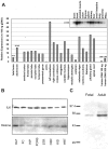
Fig. 1.
Fig. 1.
ILK transcript and protein expression in rat and human tissue. A, Human multiple tissue expression array. The signals obtained by hybridization of an ILK-specific probe to the multiple tissue expression membrane were quantified by phosphoimage analysis, as described in Materials and Methods. The data are represented as fold increases over the signal obtained from 100 ng of genomic DNA. Inset, Specificity of the ILK probe was assured by Northern blot analysis. B, Top, Rat homogenates from neural (olfactory bulb, lane 1; frontal cortex, lane 2; hippocampus, lane 3; pons, lane 4; striatum, lane 5; cerebellum, lane 6) and non-neuronal (kidney,lane 7; heart, lane 8) tissues were immunoblotted and probed with an ILK polyclonal antibody from Upstate Biotechnology. Bottom, Western blots were also probed with a histone H1 monoclonal antibody as a loading control.C, Representative Western blot, performed in triplicate, showing ILK expression in adult or fetal (7 week) human brain tissue. ILK was visualized using a rabbit polyclonal antibody (Upstate Biotechnology).
Fig. 2.
Fig. 2.
Immunohistochemical staining of ILK is enhanced in neurons from the human hippocampus and cerebellum. Coronal sections through the hippocampus (top left panel) or cerebellum (bottom left panel) showing high somatodendritic ILK immunoreactivity in both CA1 pyramidal cells and Purkinje cells, respectively. IgG controls for both sections are shown in the adjacent panels. Scale bar, 50 μm.
Fig. 3.
Fig. 3.
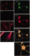
Immunocytochemical localization of ILK and β1 integrin in neuronal cells. Labeling of cells with ILK reveal both a punctate and continuous staining pattern with concentrations at the axon, neurite tip, and periphery of the cell soma. A, Punctate immunoreactivity was evident in differentiated PC12 cells overexpressing gp140trk and plated onto collagen I after incubation with a polyclonal ILK antibody (A) but was not present in the corresponding IgG controls (B). Immunoreactivity to an anti-ILK antibody was continuous at the axons of DRG neurons plated onto laminin (C, D) but appeared more patchy and granular at the growth cone (E). Codistribution of ILK and the β1 integrin subunit occurred throughout the neuronal cell body and processes of cerebellar granule neurons (F–H). Area outlined by the box(H) is shown here at a higher magnification (I). Scale bar, 20 μm.
Fig. 4.
Fig. 4.
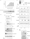
ILK inhibition reduces AKT and GSK-3 phosphorylation in PC12 cells after NGF receptor stimulation.A, NGF increases ILK activity in a concentration-dependent manner. PC12 cells, plated onto laminin, were exposed to increasing concentrations of NGF for 30 min. After immunoprecipitation, ILK activity was measured by AKT substrate radiolabeling. Top, Average CPM of AKT substrate radiolabeling; n = 3 ± SEM.Bottom, Representative in vitro kinase assay. B, NGF increases ILK phosphorylation of AKT Ser-473. ILK kinase activity was measured in PC12 cells plated onto collagen I after a 10 min exposure to NGF (100 ng/ml). ILK activity was measured using a His-tagged fusion protein corresponding to human AKT as an exogenous substrate. Phosphorylation of the substrate by ILK is detected using a phosphospecific anti-AKT Ser-473 antibody. The antibody control (lane 3) corresponds to lysate immunoprecipitated with an irrelevant antibody (anti-flag monoclonal antibody; Upstate Biotechnology). C, KP-392 inhibits NGF stimulation of AKT and GSK phosphorylation in a dose-dependent manner. Western blots of lysates from PC12 cells grown on collagen I and treated with NGF (50 ng/ml) for 4 hr in the presence of vehicle control or increasing concentrations of KP-392. Representative Western blots of cell lysates, run in parallel and probed with antibodies for phospho-AKT, phospho-GSK-3, or phospho-p38 MAPK; n = 3–5. These same blots were subsequently stripped and reprobed for AKT, GSK-3, or p38 MAPK as a control for protein expression levels. D, KP-392 does not inhibit phosphorylation of PI-3 kinase or TrkA. Left, Representative Western blot of PC12 cell lysates from PC12 cells treated for 4 hr with NGF in the presence or absence of KP-392 (100 μm) and probed with a phosphospecific antibody for TrkA (Tyr490). Right, Levels of the p85 regulatory subunit of PI-3 kinase were analyzed in anti-phosphotyrosine immunoprecipitates by Western blotting. E, Long-term effects of KP-392 (100 μm) on cell signaling in PC12 cells grown on collagen I (Col I), collagen IV (Col IV), or laminin (L) and incubated in the presence or absence of NGF (50 ng/ml) for 48 hr. Representative Western blots of cell lysates, run in parallel and probed with antibodies for phospho-AKT, phospho-GSK-3, or phospho-ERK; n = 3. After membrane stripping, Western blots were subsequently reprobed for AKT, GSK-3, and ERK.F, Overexpression of ILK-DN decreases NGF-induced stimulation of AKT and GSK-3 phosphorylation. PC12 cells were transfected with ILK-DN:V5 or EmptyV5. Twenty-four hours after transfection, cells were replated onto collagen I and were serum-starved overnight. Representative Western blots of phospho-AKT and phospho-GSK-3 after a 15 min exposure to NGF (50 ng/ml); n = 3. These same Western blots were subsequently stripped and reprobed with an anti-V5 or anti-GSK-3 antibody, respectively.
Fig. 5.
Fig. 5.
NGF increases phosphorylation of AKT Ser-473: effect of ILK-DN and ILK-WT overexpression in primary neurons.Top, ILK in vitro kinase activity was measured in rat cerebrocortical cultures after a 15 min exposure to NGF. NGF increased ILK phosphorylation of AKT Ser-473. Growth factor stimulation of AKT phosphorylation was inhibited after overexpression of ILK-DN (lanes 2 and 6, respectively). Competition of ILK-WT was observed with increasing concentrations of ILK-DN (the ratio of ILK-WT:ILK-DN is indicated above).Bottom, Immunoblots were stripped and subsequently reprobed with an anti-AKT antibody as a control for His-tagged, AKT fusion protein loading.
Fig. 6.
Fig. 6.
ILK inhibition increases Tauhyperphosphorylation in PC12 cells after integrin and NGF receptor stimulation. A representative Western blot of the effect of KP-392 (100 μm) on levels of PHF-1 in PC12 cells grown on collagen I (Col I), collagen IV (Col IV), or laminin (L) after a 48 hr incubation in media lacking serum with or without NGF (50 ng/ml);n = 3. The bottom panel shows Tau-1-immunoreactive bands from the same blot as a control for protein expression levels.
Fig. 7.
Fig. 7.
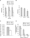
Inhibition of ILK attenuates NGF-induced neurite outgrowth in PC12 cells and DRG cells. A, NGF-induced neurite outgrowth was assessed in PC12 cells grown for 48 hr on various ECM [collagen I (Col I), collagen IV (Col IV), or laminin (L)], in the presence or absence of KP-392 (100 μm) or drug vehicle. The number of neurite-bearing PC12 cells after NGF treatment was significantly reduced in the presence of KP-392. Data represent four independent experiments ± SEM and have been normalized to PC12 cells grown on collagen I. *p < 0.05, different from control cells plated onto the same extracellular matrix.B, NGF-induced neurite outgrowth was measured in PC12 cells plated onto collagen I and transiently cotransfected with ILK-WT, ILK-DN, or empty vector together with EGFP. The percentage of neurite-bearing cells was determined from cells positive for EGFP expression. Data represent four independent experiments ± SEM. *p < 0.05, different from control cells plated on collagen I. C, Average length of axon front cultured in the presence of KP-392 for 24 hr was significantly decreased compared with the drug vehicle. D, Assessment of the total percentage of neurite bundles reaching a distance of 500, 1000, or 1500 μm shows a significant decrease at each distance in the presence of KP-392. Data represent 18 explants from four independent experiments ± SEM. *p < 0.05, different from treatment groups lacking inhibitor grown on the same extracellular matrix.
Fig. 8.
Fig. 8.
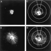
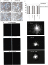
Representative explants of DRG neurons with neurofilament (1:500, Sigma-Aldrich) labeled neurites after 24 hr in the presence of KP-392 (A, C) or DMSO (B, D). Outgrowth was assayed using a series of concentric circles at radia of 500, 1000, or 1500 μm (C, D) measured from the edge of the explant. The center of the circles were placed around the edge of the entire explant to obtain an accurate assessment of the total number of axon bundles crossing each distance.
Fig. 9.
Fig. 9.
Inhibition of ILK does not decrease neuronal cell survival in the presence of NGF. A–D, Phase-contrast and fluorescence photomicrographs of PC12 cells plated onto laminin (A, B) or collagen IV (C, D) and exposed to NGF with or without KP-392. After 2 d, cells were fixed and stained with propidium iodide and Hoechst 33342. Inhibition of neurite outgrowth by KP-392 was independent from propidium iodide staining.E–G, Representative fluorescence photomicrographs of PC12 cells plated onto collagen I and exposed to NGF with or without KP-392. After 2 d cells were labeled for TUNEL (right). DAPI staining corresponding to each of these fields is shown in the adjacent left panel. A positive control, performed according the manufacturer's instructions (In Situ Cell Death Detection Kit; Boehringer Mannheim) is included (G). H, To assess survival, the number of propidium iodide-negative cells or TUNEL-negative cells was determined and expressed as a percentage of total cell number (Hoechst-positive or DAPI-positive cells, respectively);n = 3 ± SD. I–K, Caspase inhibition did not block KP-392 effects on DRG neurite outgrowth. DRG neurons with neurofilament-labeled neurites after 24 hr in the presence of NGF alone (I), with KP-392 (J), or KP-392 combined with the caspase inhibitor BAF (K).
Fig. 10.
Fig. 10.
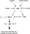
Schematic model of integrin and NGF-mediated ILK regulation of differentiation. ILK activity is regulated by integrins and growth factors in a PI-3 kinase-dependent manner. Active ILK phosphorylates AKT on Ser-473, resulting in its activation. Activated AKT in turn phosphorylates and inhibits GSK-3. ILK may also inhibit GSK-3 activity directly. Inhibition of ILK by KP-392 would thus increase GSK-3 activity and result in Tauhyperphosphorylation, microtubule instability, and decreased neurite growth.

Similar articles

Cited by

References

    1. Andjelkovic M, Alessi DR, Meier R, Fernandez A, Lamb NJ, Frech M, Cron M, Cohen P, Lucocq JM, Hemmings BA. Role of translocation in the activation and function of protein kinase B. J Biol Chem. 1997;272:31515–31524. - PubMed
    1. Andjelkovic M, Suidan HS, Meier R, Frech M, Alessi DR, Hemmings BA. Nerve growth factor promotes activation of the α, β and γ isoforms of protein kinase B in PC12 pheochromocytoma cells. Eur J Biochem. 1998;251:195–200. - PubMed
    1. Arregui CO, Carbonetto S, McKerracher L. Characterization of neural cell adhesion sites: point contacts are the sites of interaction between integrins and the cytoskeleton in PC12 cells. J Neurosci. 1994;14:6967–6977. - PMC - PubMed
    1. Ashcroft M, Stephens RM, Hallberg B, Downward J, Kaplan DR. The selective and inducible activation of endogenous PI 3-kinase in PC12 cells results in efficient NGF-mediated survival but defective neurite outgrowth. Oncogene. 1999;18:4586–4597. - PubMed
    1. Attwell S, Roskelley C, Dedhar S. The integrin-linked kinase (ILK) suppresses anoikis. Oncogene. 2000;19:3811–3815. - PubMed

Publication types

MeSH terms

LinkOut - more resources