. 2003 Mar 1;23(5):1649-58.
doi: 10.1523/JNEUROSCI.23-05-01649.2003.

The E2F-Cdc2 cell-cycle pathway specifically mediates activity deprivation-induced apoptosis of postmitotic neurons

Affiliations

The E2F-Cdc2 cell-cycle pathway specifically mediates activity deprivation-induced apoptosis of postmitotic neurons

Yoshiyuki Konishi et al. J Neurosci. .

Abstract

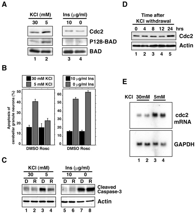
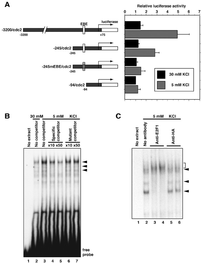
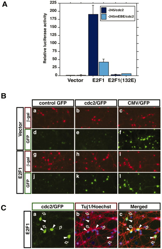
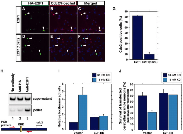
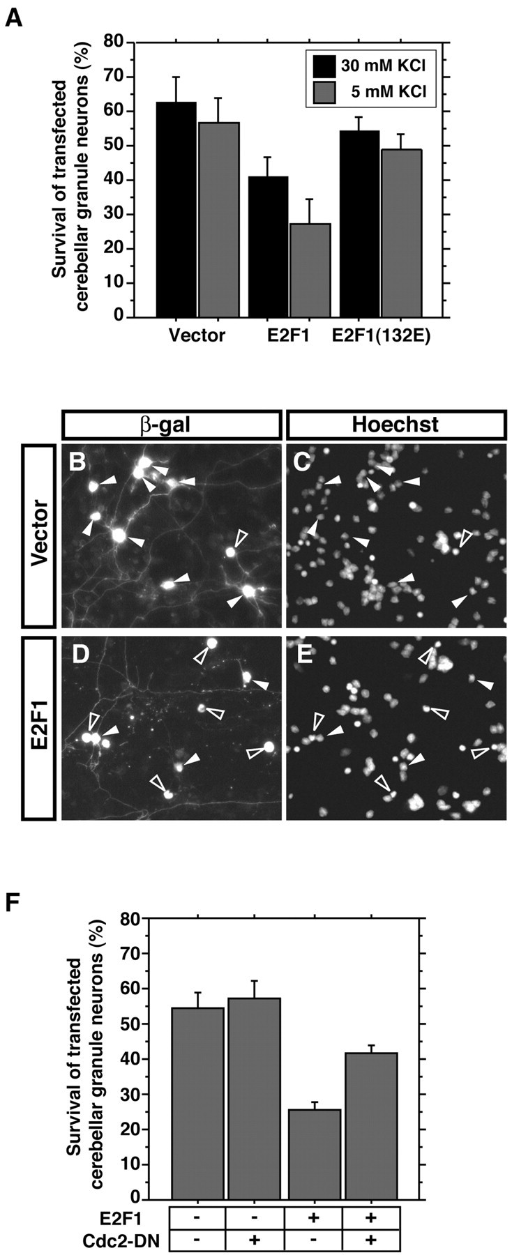
Neuronal apoptosis plays a critical role in the normal development of the mammalian brain and is thought to contribute to the pathogenesis of several neurologic disorders. However, the intracellular mechanisms underlying apoptosis of neurons remain incompletely understood. In the present study, we characterized a cell-cycle-based mechanism by which neuronal activity deprivation induces apoptosis of postmitotic neurons. Activity deprivation, but not growth factor withdrawal, was found to induce Cdc2 expression and consequent Cdc2-mediated apoptosis in granule neurons of the developing rat cerebellum. We found that activity deprivation induces cdc2 transcription in neurons via an E2F-binding element (EBE) within the cdc2 promoter. The transcription factor E2F1 that is expressed in granule neurons was found in DNA binding assays to bind to the EBE of the cdc2 gene. In chromatin immunoprecipitation analysis, endogenous E2F1 forms a complex with the promoter of the endogenous cdc2 gene in granule neurons, indicating that endogenous E2F1 is poised to activate transcription of the endogenous cdc2 gene in neurons. Consistent with this conclusion, a dominant interfering form of E2F, when expressed in granule neurons, blocked activity deprivation-induced cdc2 transcription. In other experiments, we found that the expression of E2F1 in granule neurons induces Cdc2 expression and promotes neuronal apoptosis via the activation of Cdc2. Remarkably, in contrast to inducing the E2F-mediated expression and activation of Cdc2 in granule neurons, activity deprivation fails to stimulate the expression of E2F-target genes that trigger DNA synthesis and replication. Together, our findings define a novel apoptotic mechanism whereby E2F selectively couples an activity deprivation-induced signal to cdc2 transcription in the absence of stimulating DNA synthesis and thus culminating in Cdc2-mediated apoptosis of postmitotic neurons.


Figures

Fig. 1.
Fig. 1.
Cdc2 specifically mediates activity deprivation-induced apoptosis of cerebellar granule neurons.A, Lysates of granule neuron cultures (P6 plus 2 DIV) that were in survival medium [30 mm KCl plus serum,lane 1; 30 mm KCl plus insulin (Ins), lane 3)], that were deprived of KCl (5 mm KCl plus serum, lane 2), or that were deprived of growth factors (30 mm KCl, lane 4) for 24 hr were immunoblotted with a mouse monoclonal antibody that recognizes Cdc2 (top), the rabbit phospho128 (P128)-BAD antibody (middle), or an antibody that recognizes BAD regardless of its phosphorylation (bottom). Activity withdrawal significantly induced the expression of Cdc2 (1.4 ± 0.1 fold; n = 3;p < 0.001; ANOVA) and the phosphorylation of BAD at serine 128 (1.9 ± 0.1 fold; n = 3; ANOVA;p < 0.001), but insulin withdrawal failed to induce Cdc2 expression (0.8 ± 0.1 fold; n = 3) or to induce the BAD serine 128 phosphorylation (1.1 ± 0.2 fold; n = 3). B, Cerebellar granule neurons (P6 plus 2 DIV) were kept in survival medium (30 mmKCl plus serum, left; 30 mm KCl plus insulin, right) or were deprived for 48 hr of KCl (5 mm KCl plus serum, left) or insulin (30 mm KCl, right) in the presence of the Cdc2 inhibitor roscovitine (Rosc; 10 μm) or its vehicle [dimethylsulfoxide (DMSO)]. Roscovitine inhibited neuronal activity deprivation-induced apoptosis (mean ± SEM;n = 3; ANOVA; p < 0.01) but not growth factor withdrawal-induced apoptosis. C, Lysates of cerebellar granule neurons (P6 plus 2 DIV) that were treated as in B in the presence of DMSO (D) or roscovitine (R) were immunoblotted using an antibody to the cleaved form of caspase-3 (top) or an antibody that recognizes actin (bottom) to serve as control for equal protein loading.D, Lysates of cerebellar granule neurons that were placed in full medium (30 mm KCl plus serum) or deprived of KCl (5 mm KCl plus serum) for the indicated times were immunoblotted with the antibody to Cdc2 (top) or the antibody to actin (bottom). E, Northern blot analysis of total RNA from cerebellar granule neuron cultures that had either been maintained in full medium (30 mm KCl plus serum, lanes 1 and 2) or deprived of activity (5 mm KCl plus serum, lanes 3 and4) for 24 hr. RNA was subjected to Northern blot analysis using a cdc2 probe (top) or glyceraldehyde-3-phosphate dehydrogenase (GAPDH;bottom).
Fig. 2.
Fig. 2.
Activity deprivation induces cdc2transcription via an EBE within the cdc2 promoter.A, Schematic representation of cdc2promoter–luciferase reporter constructs and their activity in cerebellar granule neurons. Cerebellar granule neurons (P6 plus 2 DIV) were transfected with the indicated cdc2 firefly-luciferase reporter plasmid and reporter gene that is controlled by the elongation factor promoter (EF-renilla) and incubated for 4–5 hr in conditioned full medium (30 mm KCl plus serum). Transfected neurons were then placed in full medium (30 mm KCl plus serum;black bars) or deprived of neuronal activity (5 mm KCl plus serum; gray bars) for 36 hr and then subjected to dual-luciferase assay (Promega). The normalized firefly-luciferase activity of each cdc2 luciferase reporter gene is shown relative to the activity of −94/cdc2luciferase gene in membrane-depolarized granule neuron cultures. Values shown are mean ± SEM (n = 4). Activity deprivation significantly increased the expression of the −3200/cdc2 and −245/cdc2 reporter genes (ANOVA;p < 0.05) but not of the −245mEBE/cdc2or the −94/cdc2 reporter genes. B, Nuclear extracts prepared from cerebellar granule neuron cultures (P6 plus 2 DIV) maintained in full survival medium (30 mm KCl plus serum) or deprived of neuronal activity (5 mm KCl plus serum) for 24 hr were subjected to an EMSA. The radiolabeled probe contains the EBE within the cdc2 promoter. For competition analysis, excess amount of wild-type cold probe (lanes 4and 5) or a probe that contains a point mutation within the EBE (lanes 6 and 7) was added to the reaction. C, A rabbit antibody to E2F1 (2 μg,lane 3; 5 μg, lane 4) or a control antibody (anti-HA antibody, 2 μg, lane 5; 5 μg, lane 6) was added to the EMSA reaction. Three protein–DNA complexes that are shown as specific EBE–protein complexes in B (arrowheads) were blocked or super-shifted (bracket) by the addition of E2F1 antibody but not by control antibody.
Fig. 3.
Fig. 3.
E2F1 activates cdc2 promoter-mediated transcription in postmitotic cerebellar granule neurons.A, Cerebellar neuron cultures (P6 plus 2 DIV) were transfected with the −245/cdc2 or −245/mEBE/cdc2 luciferase reporter plasmids together with the EF-renilla reporter gene and an expression plasmid encoding E2F1, an E2F1 mutant in which the DNA-binding domain is mutated [E2F1(132E)], or their control vector. One day after transfection, transfected cells were harvested and subjected to a dual-luciferase assay. Normalized reporter activities are shown relative to the reporter activity of the −245/cdc2 luciferase gene that was cotransfected with the vector control plasmid. Values shown are mean ± SEM (n = 4). The activity of the −245/cdc2 reporter gene was significantly increased by E2F1 (ANOVA; p < 0.001) but not by E2F1(132E). Mutation within the EBE (−245/mEBE/cdc2) significantly decreased E2F1 induction of cdc2 transcription (ANOVA;p < 0.001). B, Cerebellar granule neurons were transfected with the control GFP, cdc2/GFP reporter gene, or CMV/GFP reporter gene together with an expression plasmid encoding E2F1 or its control vector and an expression plasmid encoding β-galactosidase (β-gal). Two days after transfection, cells were subjected to indirect immunofluorescence with an antibody to β-galactosidase, and GFP reporter gene activity was monitored by visualizing GFP in transfected cells. E2F1 induced thecdc2/GFP reporter. C, Cerebellar granule neuron cultures were transfected with the E2F1-expression vector together with the cdc2/GFP reporter gene. Transfected cultures were subjected to indirect immunofluorescence with an antibody to β-tubulin type III (Tuj1) that is expressed in postmitotic neurons, and GFP visualization was used to monitor thecdc2/GFP reporter activity. Cell bodies (arrowheads) and neurites (open arrows) of all GFP-positive cells also stained with the Tuj1 antibody.
Fig. 4.
Fig. 4.
E2F1 mediates activity deprivation-inducedcdc2 transcription and apoptosis in cerebellar granule neurons. A–F, Cerebellar granule neurons (P6 plus 2 DIV) were transfected with an expression plasmid encoding HA-tagged E2F1 or HA-tagged E2F1(132E). Two days after transfection, cultures were subjected to indirect immunofluorescence using a rabbit anti-HA antibody and the mouse monoclonal antibody to Cdc2 together with the DNA dye bisbenzimide (Hoechst 33258) to reveal cell nuclei.Arrowheads indicate HA–E2F1- or HA–E2F1(132E)-expressing neurons. G, Quantification of experiments in A–F showing the percentages of E2F1- or E2F1(132E)-expressing neurons that exhibit high endogenous Cdc2 immunoreactivity. The percentage of transfected neurons with high Cdc2 immunoreactivity was significantly higher in E2F1-expressing neurons than in E2F1(132E)-expressing neurons (mean ± SEM;n = 3; p < 0.001;t test). H, Chromatin immunoprecipitation from cerebellar granule neuron cultures using no antibody (lane 1), a rabbit antibody to HA to serve as a control (lane 2), and an antibody against E2F1 (lane 3). Precipitated chromatin was subjected to PCR analysis using a set of primers encompassing the EBE within the cdc2 promoter (as shown in the figure). Endogenous cdc2 promoter was specifically coprecipitated with immunoprecipitated E2F1 (lane 3). I, Cerebellar granule neurons (P6 plus 2 DIV) were transfected with the −3200/cdc2–luciferase reporter plasmid together with a control vector or an expression plasmid encoding the dominant interfering form of E2F (E2F-Rb), in which the E2F1 DNA-binding region (1–368) was fused to Rb (379–972). Transfected cultures were processed as in Figure2A. Activity deprivation induced cdc2promoter-mediated transcription in vector-transfected (mean ± SEM; n = 4; ANOVA; p < 0.0001) but not in E2F-Rb-expressing granule neurons. J, Cerebellar granule neurons (P6 plus 2 DIV) were transfected with the E2F-Rb expression plasmid or the control vector together with an expression plasmid encoding β-galactosidase. One day after transfection, granule neurons were placed in full medium (30 mm KCl plus serum) or deprived of neuronal activity (5 mm KCl plus serum) for 2 d. Transfected neurons were subjected to indirect immunofluorescence using the monoclonal antibody to β-galactosidase and the DNA dye bisbenzimide (Hoechst 33258). Cell survival and death were measured in β-galactosidase-expressing neurons based on the integrity of neurites and nucleus. KCl withdrawal significantly reduced the survival of vector-transfected granule neurons (n = 3; ANOVA; p < 0.05) but not of E2F-Rb-expressing granule neurons.
Fig. 5.
Fig. 5.
Cdc2 mediates E2F1-induced neuronal apoptosis.A, Cerebellar granule neurons (P6 plus 2 DIV) were transfected with the expression plasmid encoding E2F1, E2F1(132E), or their control vector together with an expression plasmid encoding β-galactosidase. One day after transfection, granule neurons were placed in full medium (30 mm KCl plus serum) or deprived of neuronal activity (5 mm KCl plus serum) for 24 hr. Transfected neurons were subjected to indirect immunofluorescence and cell survival assays as in Figure 4J. The expression of E2F1 significantly reduced the survival of cerebellar granule neurons [mean ± SEM; n = 3; ANOVA;p < 0.05 (30 mm KCl),p < 0.005 (5 mm KCl)]. However, E2F1(132E) had little effect on the survival of cerebellar granule neurons.B–E, Representative images of cerebellar granule neurons transfected with the control vector (B,C) or the E2F1 expression plasmid (D,E). In the vector control, several healthy neurons appear (closed arrowheads), whereas several E2F1-transfected neurons are undergoing apoptosis (open arrowheads). F, Cerebellar granule neurons were transfected with the E2F1 expression plasmid together with Cdc2-DN or its vector control together with the β-galactosidase expression plasmid. Transfected neurons were deprived of neuronal activity (5 mm KCl plus serum) for 24 hr and subjected to indirect immunofluorescence and cell survival assay as in Figure4J. Cdc2-DN significantly reduced E2F1-induced apoptosis (mean ± SEM; n = 4; ANOVA;p < 0.01).
Fig. 6.
Fig. 6.
Activity deprivation does not stimulate DNA synthesis in cerebellar granule neurons. A–D, Cerebellar granule neuron cultures (P6 plus 2 DIV) were labeled with BrdU for 24 hr in cultures that were in full medium (30 mmKCl plus serum) or that were deprived of activity (5 mm KCl plus serum) in the presence or absence of the antimitotic agent AraC. Incorporated BrdU was detected by immunofluorescence using a mouse monoclonal antibody that recognizes BrdU. Postmitotic granule neurons in culture were identified using a rabbit antibody that recognizes microtubule-associated protein 2 (MAP2). MAP2-negative non-neuronal cells incorporated BrdU in the absence of AraC, whereas postmitotic granule neurons did not incorporate BrdU in the presence or absence of AraC. E, Lysates of cerebellar granule neurons (P6 plus 2 DIV) that were placed in full medium or deprived of activity for 24 or 48 hr were subjected to immunoblotting using an antibody to PCNA, Cdk2, Cdc2, or actin. Activity deprivation induced the level of Cdc2 protein but failed to induce the expression of PCNA or Cdk2.

Similar articles

Cited by

References

    1. Alessi DR, Andjelkovic M, Caudwell B, Cron P, Morrice N, Cohen P, Hemmings BA. Mechanism of activation of protein kinase B by insulin and IGF-1. EMBO J. 1996;15:6541–6551. - PMC - PubMed
    1. Altman J, Bayer S. Development of the cerebellar system: in relation to its evolution, structure, and functions. CRC; New York: 1997.
    1. Bonni A, Brunet A, West A, Datta SR, Takasu M, Greenberg ME. Cell survival promoted by the Ras-MAPK signaling pathway by transcription-dependent and -independent mechanisms. Science. 1999;286:1358–1362. - PubMed
    1. Busser J, Geldmacher DS, Herrup K. Ectopic cell cycle proteins predict the sites of neuronal cell death in Alzheimer's disease brain. J Neurosci. 1998;18:2801–2807. - PMC - PubMed
    1. Campanero MR, Armstrong M, Flemington E. Distinct cellular factors regulate the c-myb promoter through its E2F element. Mol Cell Biol. 1999;19:8442–8450. - PMC - PubMed

Publication types

MeSH terms

LinkOut - more resources